Anatomy 3
1/106
There's no tags or description
Looks like no tags are added yet.
Name | Mastery | Learn | Test | Matching | Spaced | Call with Kai |
|---|
No analytics yet
Send a link to your students to track their progress
107 Terms
how many bones?
skull: 22 —- cranial: 8, facial: 14
vertebral column: 26
thoracic cage: 25
ganglion
cluster of neuron cell bodies within PNS
nerve
bundle of axons within PNS
Nerve plexus
•network of nerves within PNS
Nuclei
cluster of neuron cell bodies within CNS
Tract
bundle of axons within CNS
Funiculus
group of tracts in a specific area of the spinal cord
Pathway
centers and tracts that connect the CNS with body organs and systems
Peduncle
stalk-like structure connecting two regions of the brain
Gray matter of the brain
•Cortex
•Motor neuron and interneuron cell bodies, dendrites, terminal arborizations, and unmyelinated axons
•Forms deep clusters of neuronal cell bodies called cerebral nuclei
White matter of the brain
•Deep to cortex
•Myelinated axons
cerebral hemispheres
•Cerebrum is composed of two cerebral hemispheres
•divided by longitudinal fissure
•connected by corpus callosum
cerebral lobes of each hemisphere
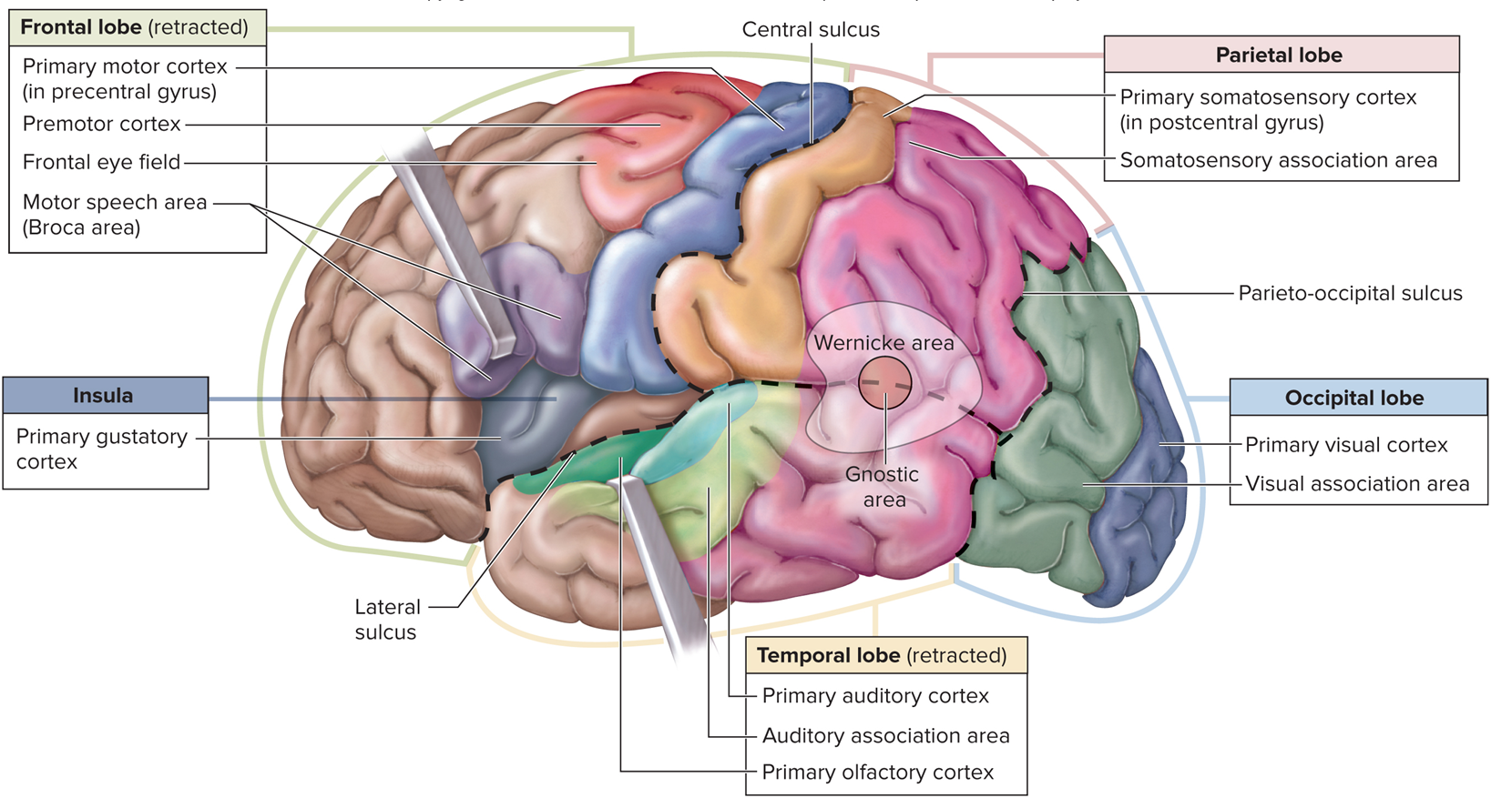
•Frontal lobe
•Parietal lobe
•Occipital lobe
•Temporal lobe
•Insula – deep within lateral sulcus
function of cerebral hemispheres
•Contralateral control
•Hemispheric lateralization
•Considerable overlap of functions in each region
•Separate except where tracts allow for communication
•Largest = corpus callosum
frontal lobe function
•Primary motor cortex: controls skeletal muscle movement, located in precentral gyrus
•Functions: voluntary muscle movement, concentration, verbal communication, decision making, planning, and personality
parietal lobe function
•Primary somatosensory cortex: receives somatic sensory information from touch, pain, pressure, and temperature receptors; located in postcentral gyrus
•Function: general sensory functions
temporal lobe function
•Primary auditory cortex: hearing
•Primary olfactory cortex: smell
•Function: involved with hearing and smell
occipital lobe function
•Primary visual cortex: vision
•Function: processes incoming visual information and stores visual memories
insula lobe function
•Primary gustatory cortex: taste
•Function: involved in emotional responses, empathy, and taste
Central White Matter tracts
•Association tracts
Connect regions of the cortex within the same hemisphere
•Commissural tracts
Extend between cerebral hemispheres
•Projection tracts
Link the cerebral cortex to the inferior brain regions and the spinal cord
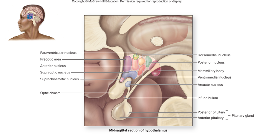
•Components of the Diencephalon
•Epithalamus
•Thalamus
•Hypothalamus
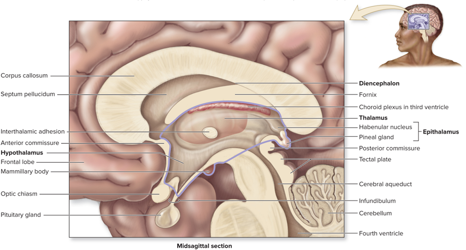
Epithalamus
•Pineal gland
•Melatonin à regulates circadian rhythm
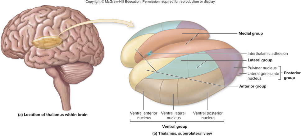
Thalamus
•Composed of thalamic nuclei
•Sensory impulses from all the conscious senses except olfaction converge on the thalamus and synapse in at least one of its nuclei
•“mailman”
hypothalamus
•Functions
•Autonomic integration center: Influences heart rate, blood pressure, digestive activities, and respirations
•Controls endocrine system
Brainstem
•Bidirectional passageway for tracts between cerebrum and spinal cord
•Contains autonomic centers
•Contains reflex centers
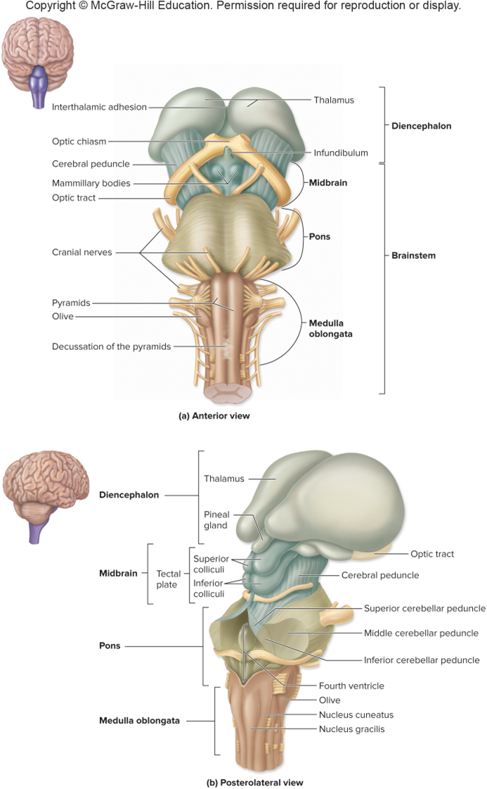
Midbrain
•Somatic motor axons descend from primary motor cortex through cerebral peduncles to spinal cord
•Superior cerebellar peduncles connect cerebellum to midbrain

Midbrain posterior view
Corpora quadrigemina
- Superior colliculi
- Visual reflex centers
- Visually track moving objects
- Turning eyes and head in response to a visual stimulus
Inferior colliculi
- Auditory reflex center
- Control reflexive turning of the head and eyes in the direction of a sound
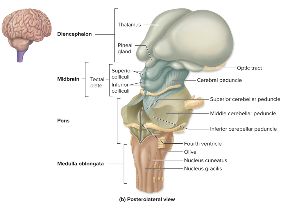
pons
•Middle cerebellar peduncles are transverse fibers that connect pons to cerebellum
•Contains autonomic nuclei in pontine (pneumotaxic) respiratory center, that help regulate breathing

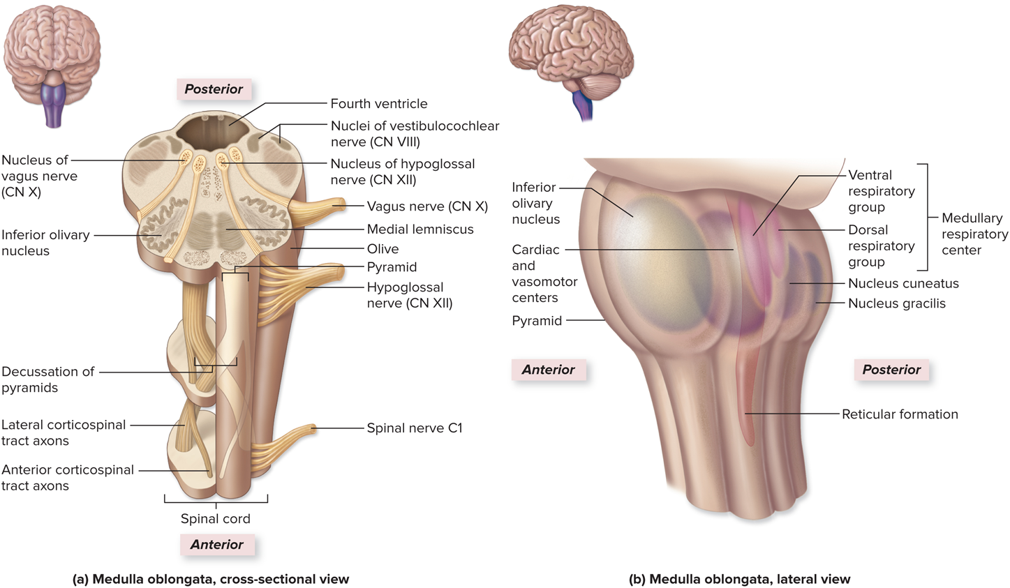
Medulla Oblongata
•Pyramids are composed of motor projection tracts called the corticospinal tracts
•Most axons in pyramids cross midline at decussation of the pyramids
•Inferior cerebellar peduncles connect medulla to cerebellum
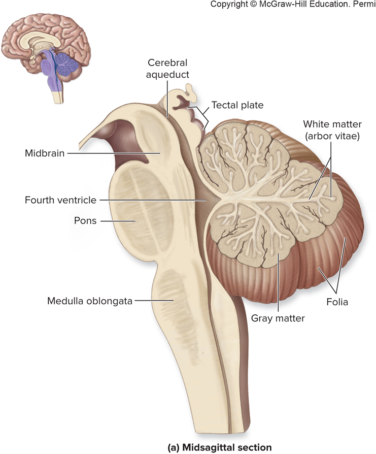

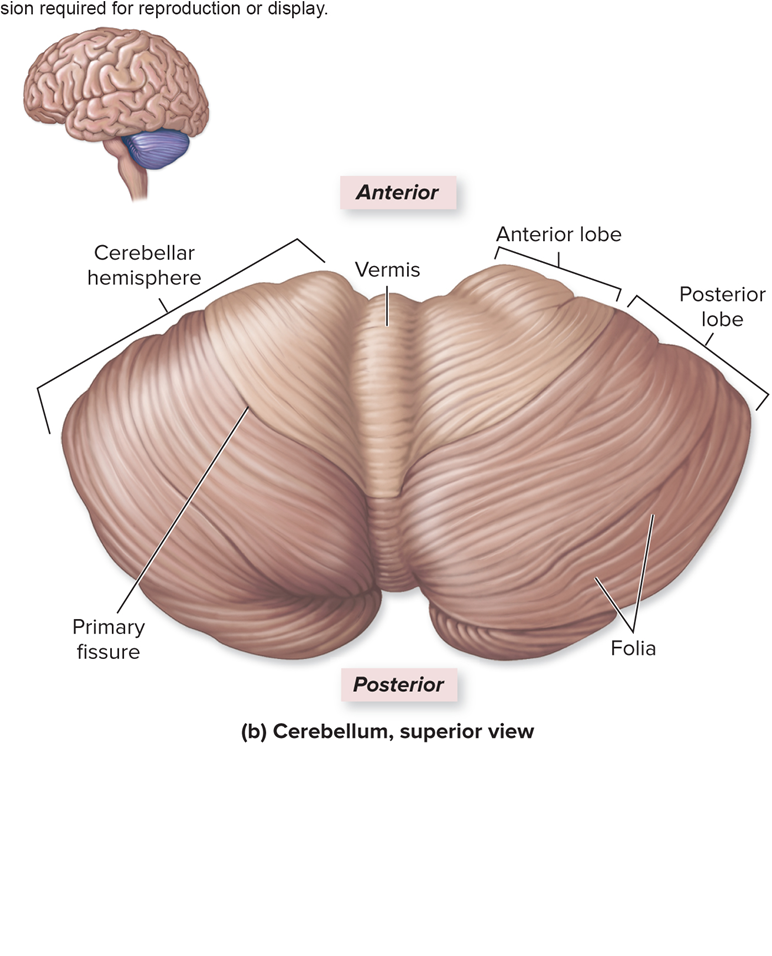
cerebellum
•Partitioned into three Regions:
1.Outer gray matter layer
2.Internal white matter
3.Cerebellar nuclei in deepest layer
•Cerebellum has left and right cerebellar hemispheres
• Anterior and posterior lobe
• Vermis sits in-between cerebellar hemispheres
•Folds of cerebellar cortex are called folia
cerebellum function
•Coordinates and “fine tunes” skeletal muscle movements
•Ensures that skeletal muscle contraction follows the correct pattern leading to smooth, coordinated movements
•Receives a “rough draft” from cerebrum
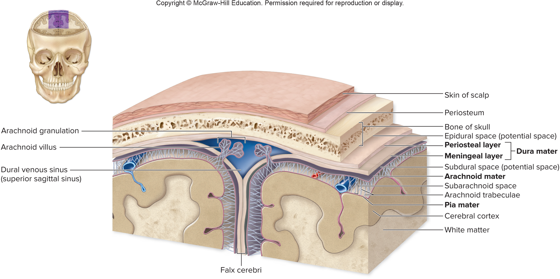
Cranial Meninges
•3 CT layers that separate the soft tissue of the brain from the cranium
•Pia mater
•Arachnoid mater
•Dura mater
•Enclose and protect blood vessel that supply the brain
•Contain and circulate cerebrospinal fluid
Cranial Dural Septa
•Falx cerebri
•Tentorium cerebelli
•Falx cerebelli
•Diaphragma sellae
Falx cerebri
project into longitudinal fissure, separates left and right cerebral hemisphere
Tentorium cerebelli
horizontal fold that separates cerebrum from cerebellum
Falx cerebelli
separates left and right cerebellar hemispheres
Diaphragma sellae
small septum between pituitary gland and hypothalamus
circular or rectangular sheet of dura mater that forms an incomplete roof over the sella turcica, covering the pituitary gland. It features a central aperture that allows the pituitary stalk to pass through and connects the pituitary gland to the hypothalamus.
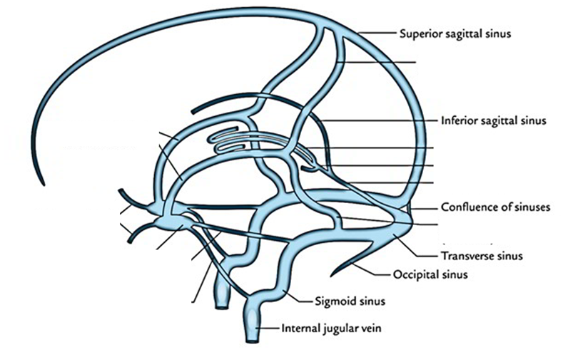
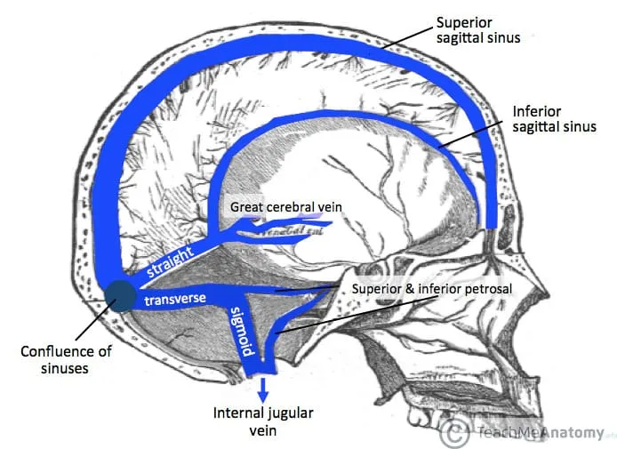
Dural Venous Sinuses
•No valves
•Drain blood from the brain to the internal jugular veins
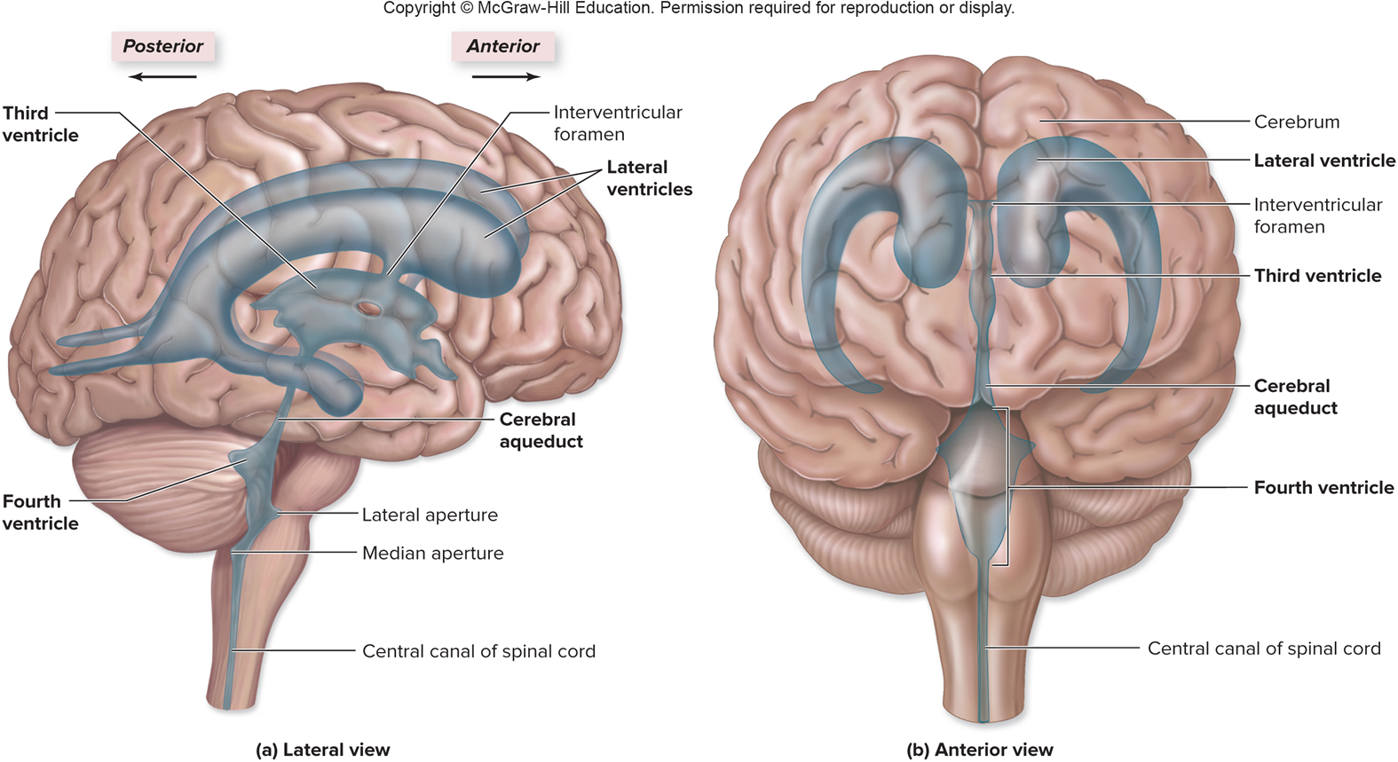
Brain Ventricles
•Cavities within the brain derived from the lumen of the neural tube
•Continuous with one another and the central canal of the spinal cord
Contain cerebrospinal fluid
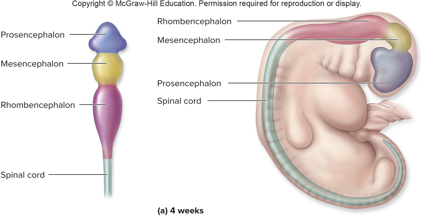
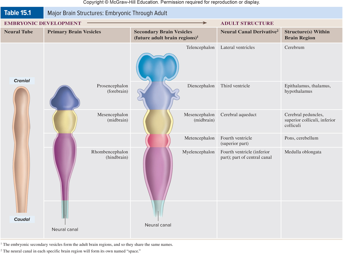
embryonic development of the brain
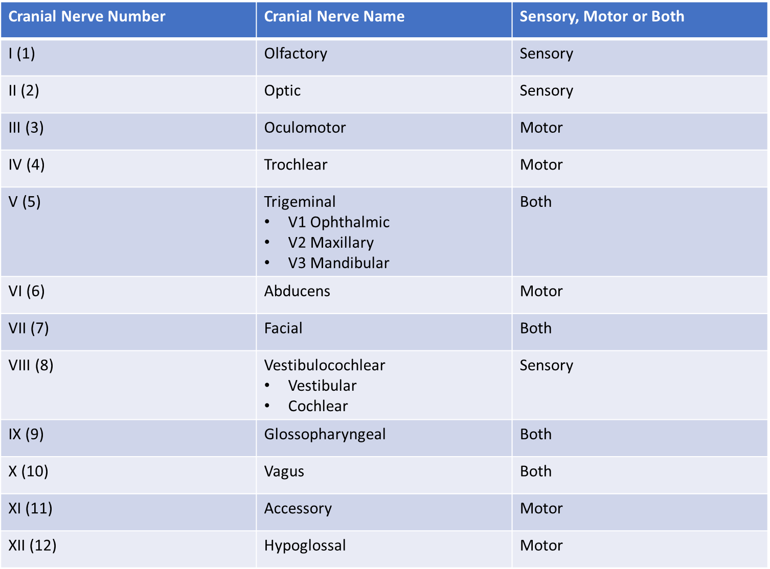
Cranial Nerves
•Part of PNS – nerve = PNS
•12 pairs —→ Each pair can be motor, sensory, or both
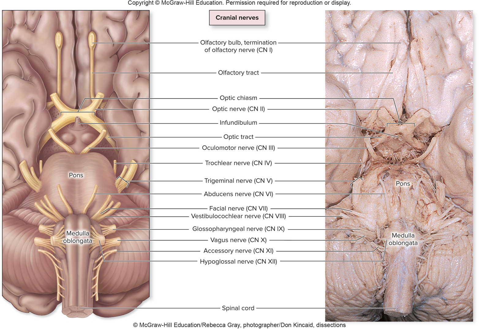
olfactory nerve
I, sensory, smell
optic nerve
II, sensory, vision
oculomotor nerve
III, motor, controls extrinsic and intrinsic eye muscles (moving the eye up, down, and inward), pupillary constriction, and eyelid elevation, near vision
Trochlear nerve
IV, motor, controls extrinsic eye muscles, innervates superior oblique muscle to facilitate eye movement (looking down and out)
Trigeminal nerve
V, both, sensory from face (touch, pain, temperature), motor innervation for jaw muscles and the soft palate to control muscles of mastication
trigeminal nerve divisions
v1 - ophthalmic - sensory fibers. sensation to forehead, scalp, upper eyelid, cornea, and nose.
v2 - maxillary - sensory fibers. sensation to cheek, lower eyelid, side of the nose, upper lip, upper teeth, palate, and nasal cavity.
v3 - mandibular - sensory and motor fibers, sensation to the lower lip, chin, lower teeth, tongue (anterior 2/3), and skin over the jaw, controls the muscles of mastication
Abducens nerve
VI, motor, controls extrinsic eye muscles, innervates lateral rectus muscle for abduction of eye
facial nerve
VII, both, taste (anterior two-thirds of the tongue), control of facial muscles, controls secretions from lacrimal and salivary glands
vestibulocochlear nerve
VIII, sensory, equilibrium and hearing
Glossopharyngeal nerve
IX, both, sensory and taste from tongue (posterior third of the tongue), innervates stylopharyngeus — triggers swallow reflex, innervation and sensation of pharynx/tonsils, controls secretions from salivary glands, blood pressure regulation via carotid artery/sinus
Vagus nerve
X, both, visceral and general sensory from structures inferior to the pharynx, motor innervation to control pharyngeal (involuntary swallow reflex) and laryngeal (sensory input for airway protection during swallowing), controls glands and visceral and cardiac muscles
speech, swallowing, and parasympathetic control of thoracoabdominal organs (heart, lungs, digestive tract).
Accessory nerve
XI, motor, controls trapezius and sternocleidomastoid, head turning and shoulder shrugging
hypoglossal nerve
XII, motor, innervates all intrinsic and extrinsic tongue muscles, facilitate tongue movement and speech articulation
all the 12 cranial nerves
brain ventricles direction of flow
• Lateral ventricles
• Septum pellucidum —- membrane in the midline of the brain from curpus callosum to fornix, separates the anterior horns of the left and right lateral ventricles.
—→ Interventricular foramen
• Third ventricle
—→ Cerebral aqueduct
• Fourth ventricle —→ merges with central canal of spinal cord
—→ Median and lateral apertures
• Subarachnoid space (between pia and arachnoid mater)
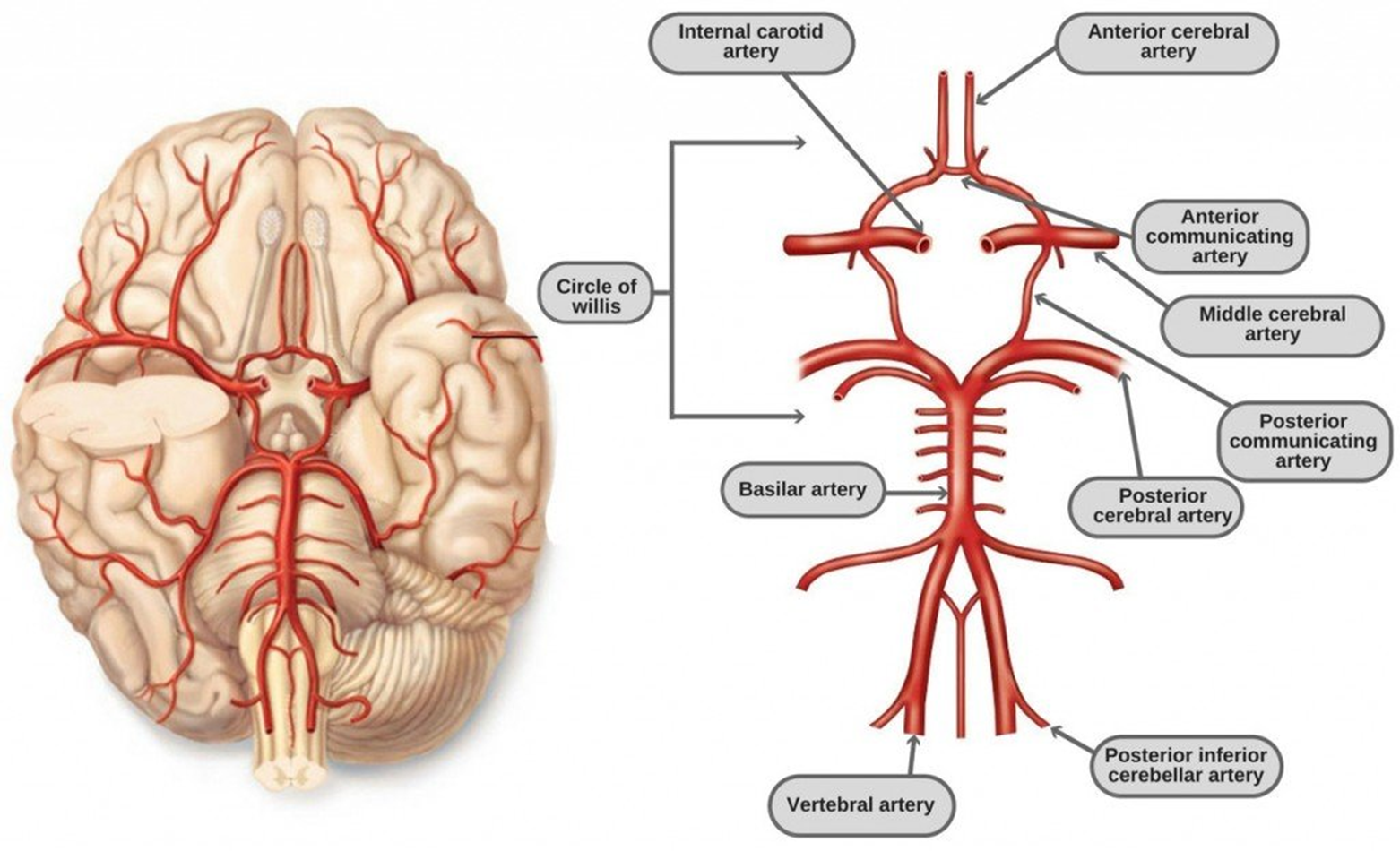
circle of willis
cerebral spinal fluid
•Formed by choroid plexus (located in the 4 ventricles)
•Ependymal cells + capillaries of pia mater
•Formed from blood plasma that filters from capillaries
•Further modified by ependymal cells
functions of csf
•Buoyancy – brain floats in CSF
•Protection – “movement buffer” – provides a liquid cushion
•Environmental stability – CSF transports nutrients and removes waste from the brain
meninges
dura mater, arachnoid mater, pia mater
dura mater
This is the tough, fibrous, and inextensible outermost layer that lines the inside of the skull. It consists of two layers (periosteal and meningeal) and creates folds like the falx cerebri and tentorium cerebelli to compartmentalize the brain, while also forming dural venous sinuses to drain blood from the brain.
layers of dura mater
periosteal layer is the superficial outer layer of the cranial dura mater that adheres to the inner surface of the cranium and fuses with bone at sutures and foramina.
meningeal layer is the deep inner layer that functions as the true dura mater, the brain and extending downward as the spinal dura mater through the foramen magnum.
**tightly fused in most areas but separate at specific locations to form the dural venous sinuses and dural reflections such as the falx cerebri, tentorium cerebelli, and falx cerebelli.
function of dura mater
The periosteal layer is responsible for anchoring the dura to the skull and contains large blood vessels
The meningeal layer contains more fibroblasts, forms the protective sheaths for cranial nerves, and creates the dural folds that compartmentalize the brain.
dural folds
limit the rotational displacement of the brain and stabilize its position within the cranial cavity during head movements.
These folds also divide the cranial cavity into freely communicating spaces, creating partitions that separate different brain regions to prevent direct contact and interference.
arachnoid mater
Located beneath the dura mater, this is a delicate, web-like, and avascular membrane that separates the brain from the skull's hard outer covering. It is separated from the dura by the subdural space and from the inner layer by the subarachnoid space, which is filled with cerebrospinal fluid (CSF) produced by the choroid plexuses.
•Arachnoid trabeculae —- spider web-like strands of connective tissue that span the subarachnoid space to connect the arachnoid mater and the pia mater
Subdural space
•Between arachnoid and dura
Potential space
Blood or fluid accumulation
pia mater
As the innermost layer, this thin, delicate matrix closely adheres to the brain's surface, following every fold and sulcus. It contains numerous blood vessels that supply the brain with nutrients and oxygen, and it is impermeable to fluid while allowing for the passage of CSF in the subarachnoid space.
features of subarachnoid space
•Arachnoid villi
Extensions of arachnoid mater into dural venous sinuses
fundamental, tiny projections of the arachnoid mater present from a young age, serving as the primary sites for cerebrospinal fluid (CSF) reabsorption into the blood.
•Arachnoid granulations
clusters of arachnoid villi that become visible to the naked eye, typically developing as early as age four and increasing in number and size with age or increased CSF pressure.
function of arachnoid villi and granulations
one-way valves that allow CSF to flow from the subarachnoid space into the dural venous sinuses while preventing backflow, a process driven by hydrostatic pressure. While villi are abundant and primarily located in the lateral lacunae of the superior sagittal sinus, granulations are fewer in number and often project directly into the inner table of the skull or the lumen of major venous sinuses.
CN 1 (olfactory) passes through
Cribriform foramina — Cribriform plate of the ethmoid
CN II (optic) passes through
optic canal
CN III, IV, V1*, VI pass through
•Superior orbital fissure
•Between greater and lesser wings of the sphenoid bone
*ophthalmic branch of trigeminal nerve
CN V2* passes through
foramen rotundum
*maxillary branch of trigeminal nerve
CN V3* passes through
foramen ovale
*mandibular branch of trigeminal nerve
CN VII – Facial nerve passes through
Stylomastoid foramen
CN XIII – Vestibulocochlear nerve — passes through
Internal Acoustic Meatus
CN IX, X, XI, jugular vein pass through
jugular foramen
IX —- glossopharyngeal nerve, X –- vagus nerve, XI – accessory nerve
CN XI*, vertebral arteries, spinal cord pass through
Foramen magnum
*spinal accessory
CN XII — hypoglossal nerve — passes through
hypoglossal canal
internal carotid artery passes through
carotid canal
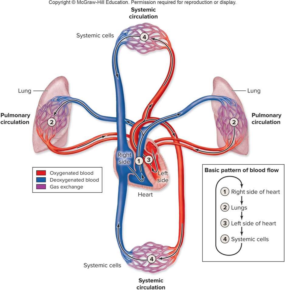
heart flow
• Connect to blood vessels to distribute blood between the heart and body tissues
Unidirectional flow —→ Valves prevent backflow
• 2 pump system (Pulmonary & Systemic)
• Develops blood pressure —→ Alternating contraction and relaxation
pulmonary circulation
heart —→ lungs —> heart

systemic circulation
heart —→ body tissues —→ heart
heart position
•Mediastinum: Between pleural cavities
•Apex: Anteroinferior and to the left
•Base: Sits on diaphragm
Surrounded by pericardium
fibrous Pericardium
•Restricts heart movements within thoracic cavity
•Prevents overfilling with blood
•Attached inferiorly to diaphragm up to base of great vessels
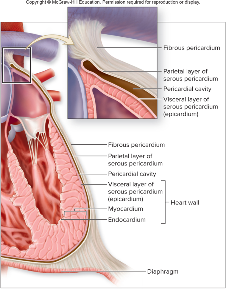
Serous pericardium
•Parietal and visceral pericardial layers
•Visceral also called epicardium
•Produce serous fluid to reduce friction
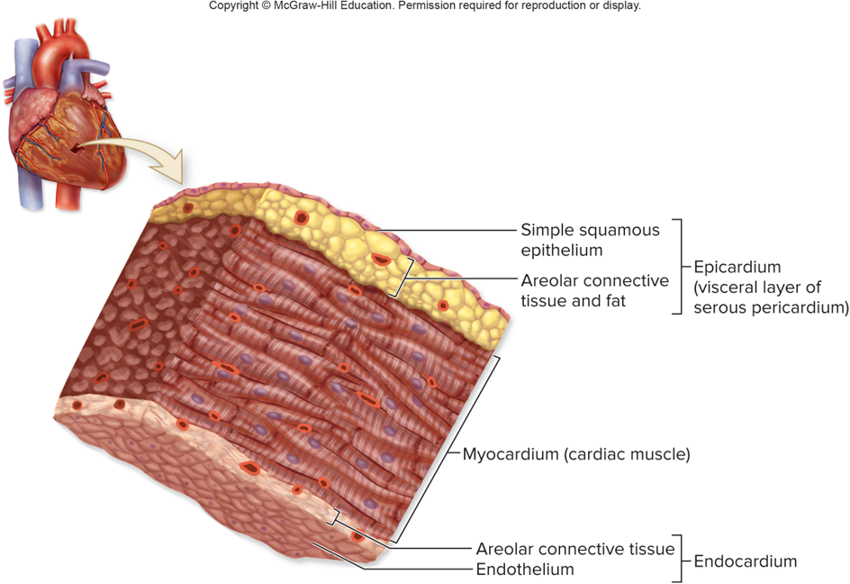
3 layers of Heart Wall
Epicardium: Simple squamous epithelium + areolar connective tissue and adipose
Myocardium: Cardiac muscle tissue
Endocardium: Endothelium + areolar connective tissue
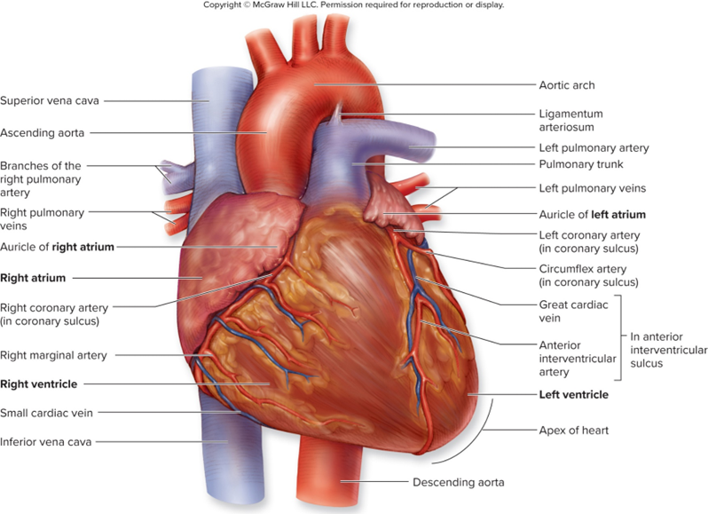
4 chambers of heart external anatomy
delineated by sulci (2 atriums and 2 ventricles)
Coronary/atrioventricular sulcus —→ Between atria and ventricles
Anterior interventricular sulcus —→ Right from left ventricle on anterior aspect
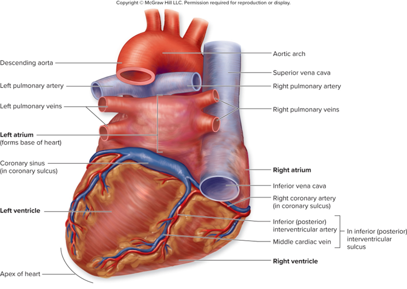
Posterior interventricular sulcus —→ Right from left ventricle on posterior aspect
auricles
One associated with each atrium (anterior)
small, ear-shaped muscular projections
expand the capacity of the atria to hold more blood and helping regulate blood flow into the ventricles during contraction.
wrinkled internal surface formed by pectinate muscles distinct from the smooth-walled main chambers of the atria.
vessels of heart
•Great vessels —→ Aorta, pulmonary trunk, superior vena cava, inferior vena cava
•Coronary vessels —→ Vessels that will supply blood to the heart tissues
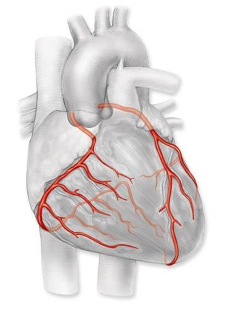
coronary circulation: arteries
•Right coronary artery (Right marginal a. and Posterior interventricular a.)
•Left coronary artery (Anterior interventricular a. and Circumflex a.)
left anterior descending (LAD) artery — largest in heart, widowmaker (frequent site of blockages)
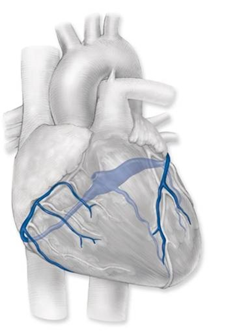
coronary circulation: veins
•not called coronary veins: cardiac
Great cardiac v., Middle cardiac v., Small cardiac v. — all drain into coronary sinus, which drains into right atrium of heart (through coronary sinus orifice)
Anterior cardiac veins
coronary sinus orifice
medial and to the left of the inferior vena cava and between the inferior vena cava and the tricuspid valve
Right atrium
pumps blood from the body via the superior vena cava, inferior vena cava, and the coronary sinus, before pumping it into the right ventricle through the tricuspid valve
houses the sinoatrial (SA) node, the heart's natural pacemaker, which initiates the electrical signals that drive the heartbeat.
•Interatrial septum (fibromuscular wall separates right atrium from left atrium)
•Pectinate muscle (internal ridges)
•Fossa ovalis (remnant of the foramen ovale from fetal development)
Right AV/Tricuspid valve (between the right atrium and the right ventricle, acting as a one-way gate for deoxygenated blood)
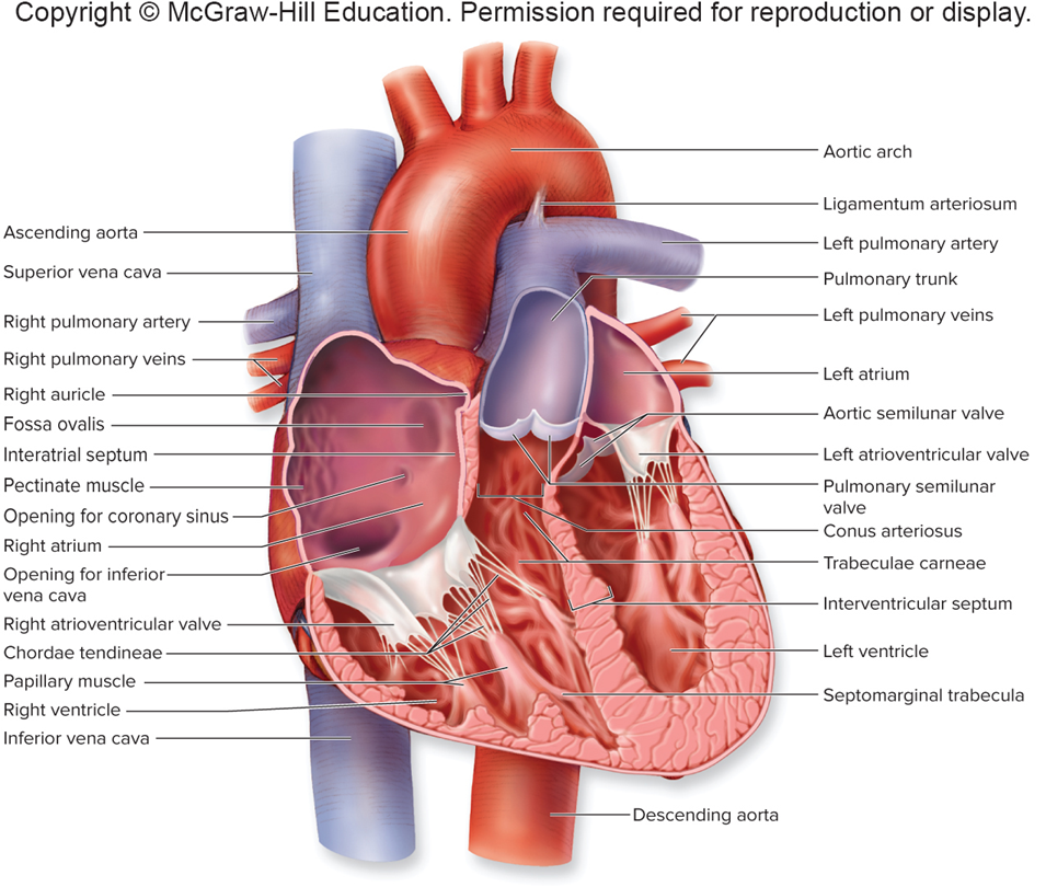
picture internal heart anatomy
foramen ovale
when baby is born, foramen ovale claps together so the lungs can be used. But if it doesn’t fully close, it is very dangerous.
‘hole in the heart’
Right ventricle
•Interventricular septum (the oblique wall that separates the left and right ventricles)
•Trabeculae carne (irregular muscular ridges and columns projecting from inner surface of myocardium)
•Papillary muscle (anterior, posterior, and septal muscles)
•Chordae tendineae (fibrocollagenous bands connect the free margins of the tricuspid valve leaflets to the papillary muscles: heart strings)
•Pulmonary semilunar valve (between right ventricle and pulmonary trunk, gateway for deoxygenated blood to exit heart toward lungs)
Left Atrium
Left AV/Bicuspid/Mitral valve (between left atrium and left ventricle, two cusps or leaflets regulate blood flow from left atrium to left ventricle while preventing backflow during ventricular contraction)
Left ventricle
•Thicker myocardium
• Aortic semilunar valve —→ between left ventricle and aorta, final gate for oxygenated blood to leave heart. allow blood flow from left ventricle to aorta during ventricular systole (contraction) and prevent backflow from the aorta into the ventricle during diastole (relaxation)
conus arteriosus
smooth-walled, conical outflow tract between the right ventricle and the pulmonary trunk, origin point for the pulmonary trunk
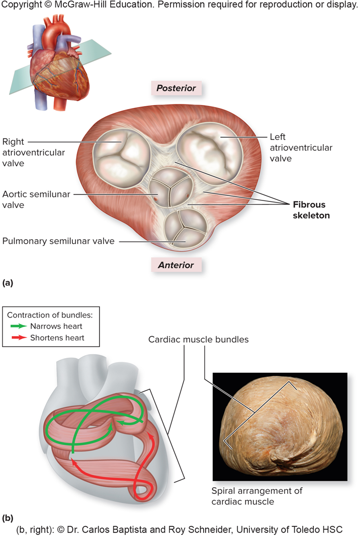
Fibrous skeleton
•Dense regular connective tissue
•Between the atria and ventricles