Respiratory Anatomy / Digestive Anatomy
1/95
There's no tags or description
Looks like no tags are added yet.
Name | Mastery | Learn | Test | Matching | Spaced | Call with Kai |
|---|
No analytics yet
Send a link to your students to track their progress
96 Terms

Contains Nose, Nasal Cavity, Sinuses, Pharynx, and Larynx (Divided by Larynx)
Upper Respiratory Tract
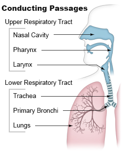
Contains Trachea, Bronchi, Bronchioles, and Alveoli (Divided by Larynx)
Lower Respiratory Tract
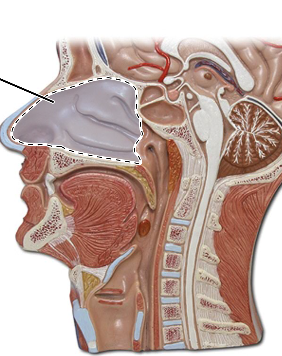
Nasal Cavity
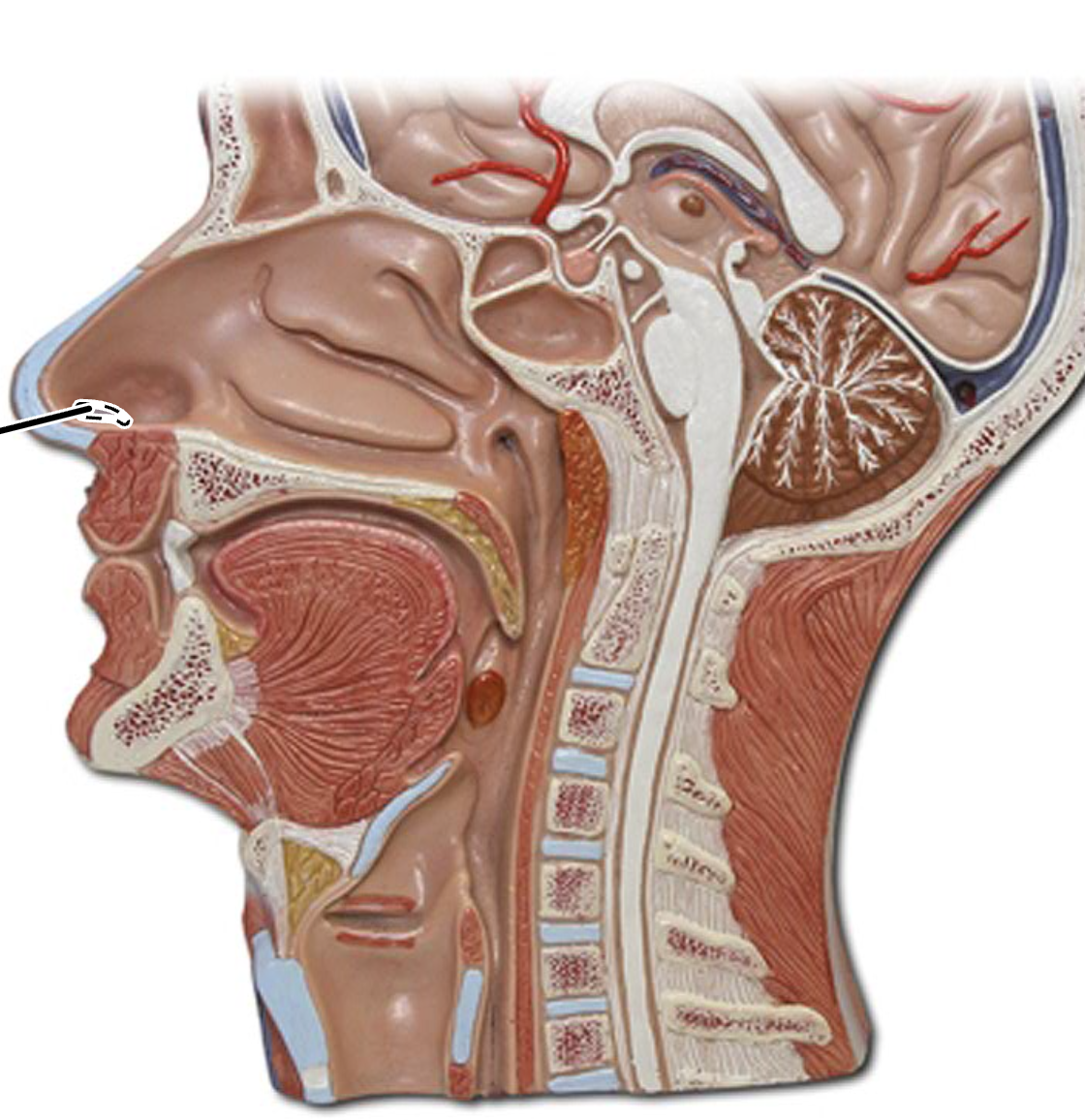
Anterior Nares
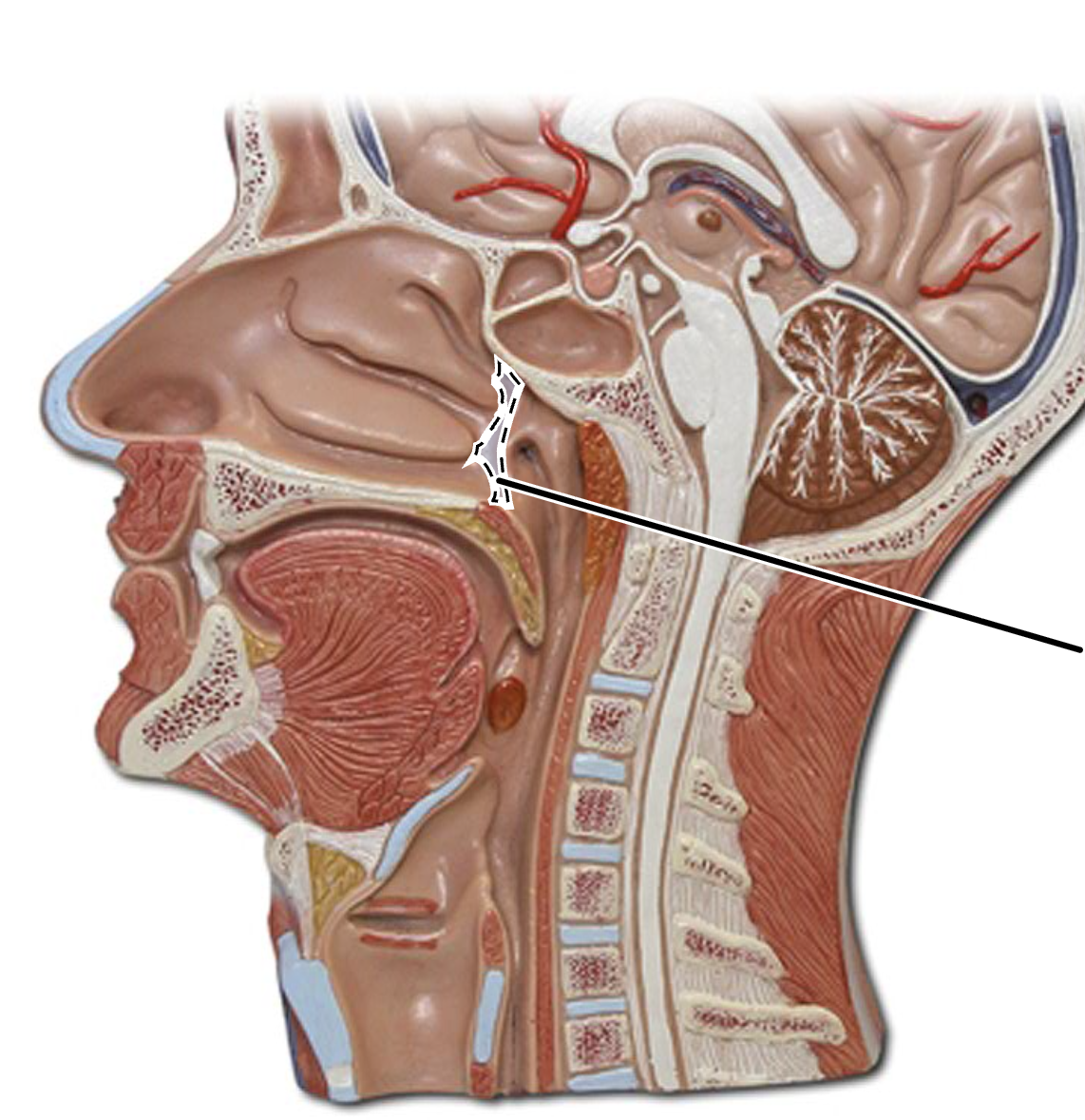
Posterior Nares
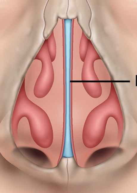
Nasal Septum
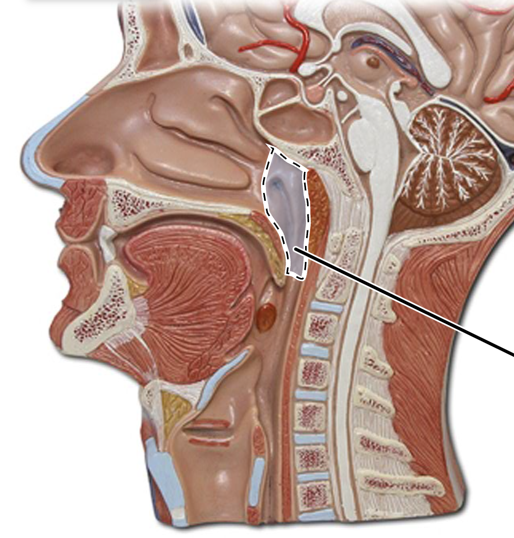
Nasopharynx
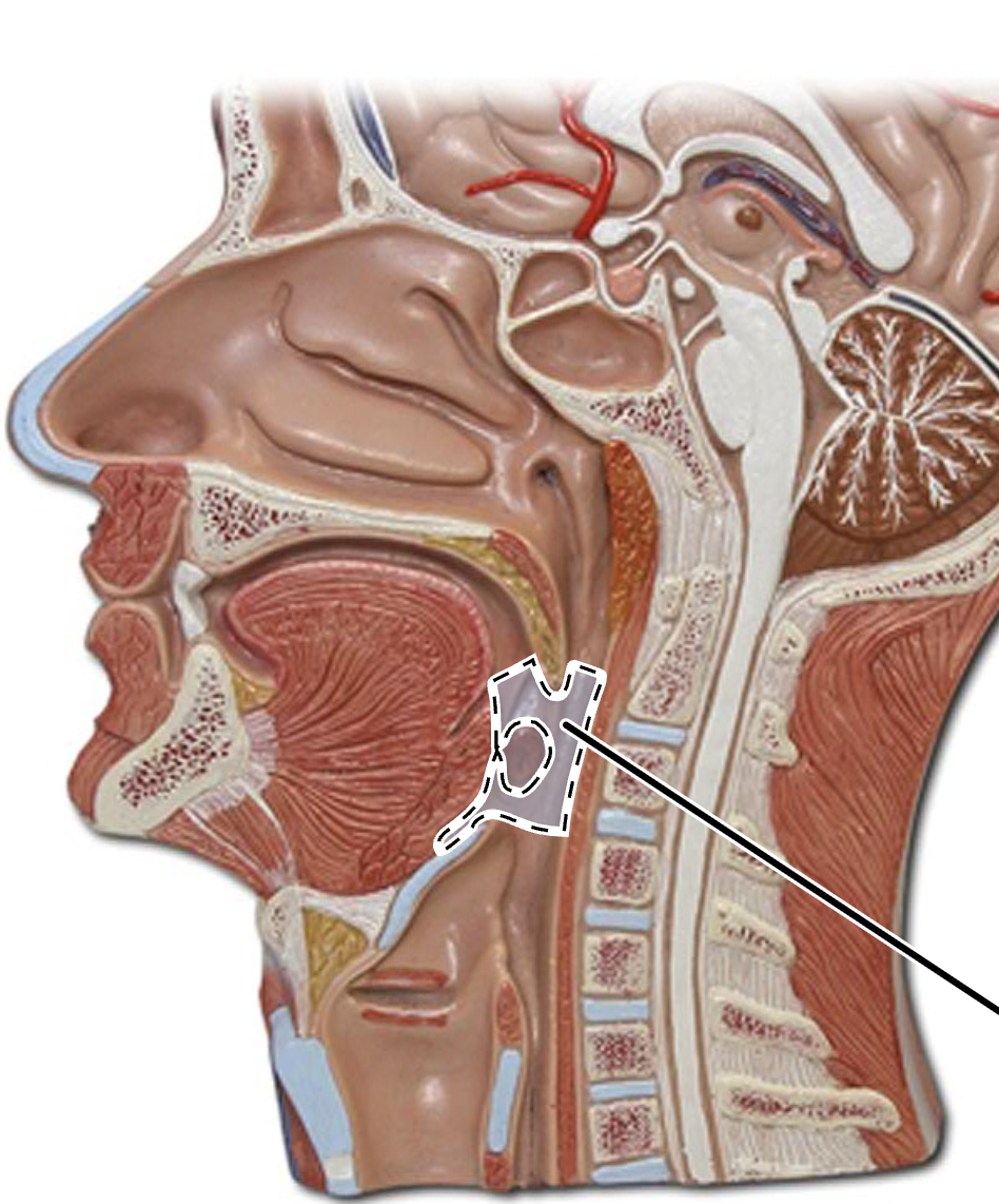
Oropharynx
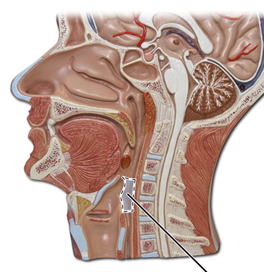
Laryngopharynx
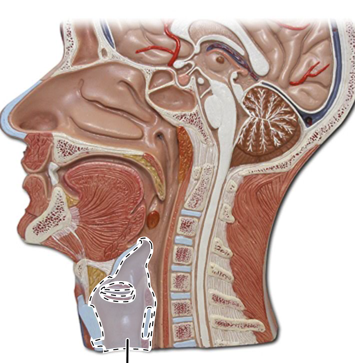
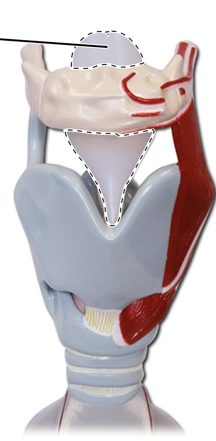
Larynx
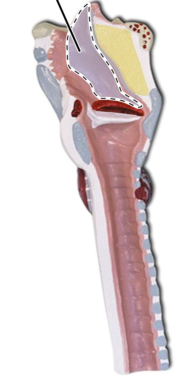
(Larynx Medial View)
Epiglottis
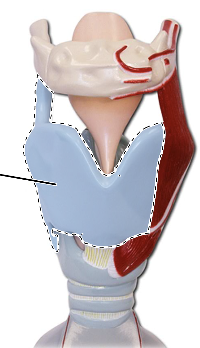
(Larynx Anterior View)
Thyroid Cartilage
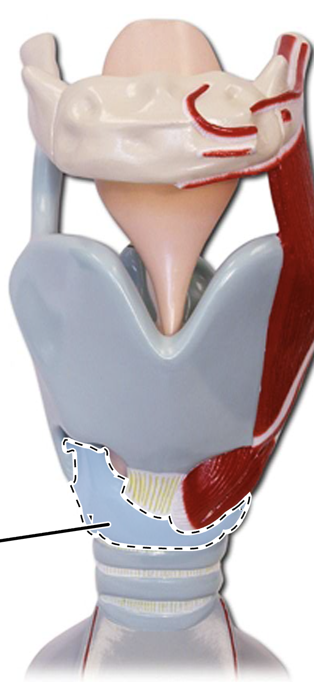
(Larynx Anterior View)
Cricoid Cartilage
(Larynx Anterior View)
Epiglottis (LAV)
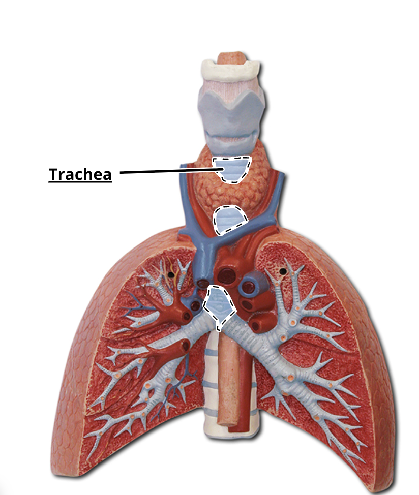
Trachea
Rings that support the trachea
Cartilage Rings

1
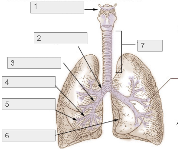
Larynx (1-7 Diagram)

2
Primary Bronchi (1-7 Diagram)

3
Secondary Bronchi (1-7 Diagram)

4
Tertiary Bronchi (1-7 Diagram)
5
Bronchioles (1-7 Diagram)

7
Trachea (1-7 Diagram)

6
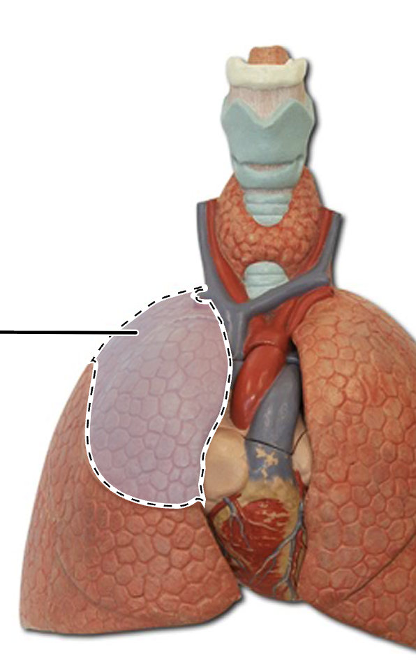
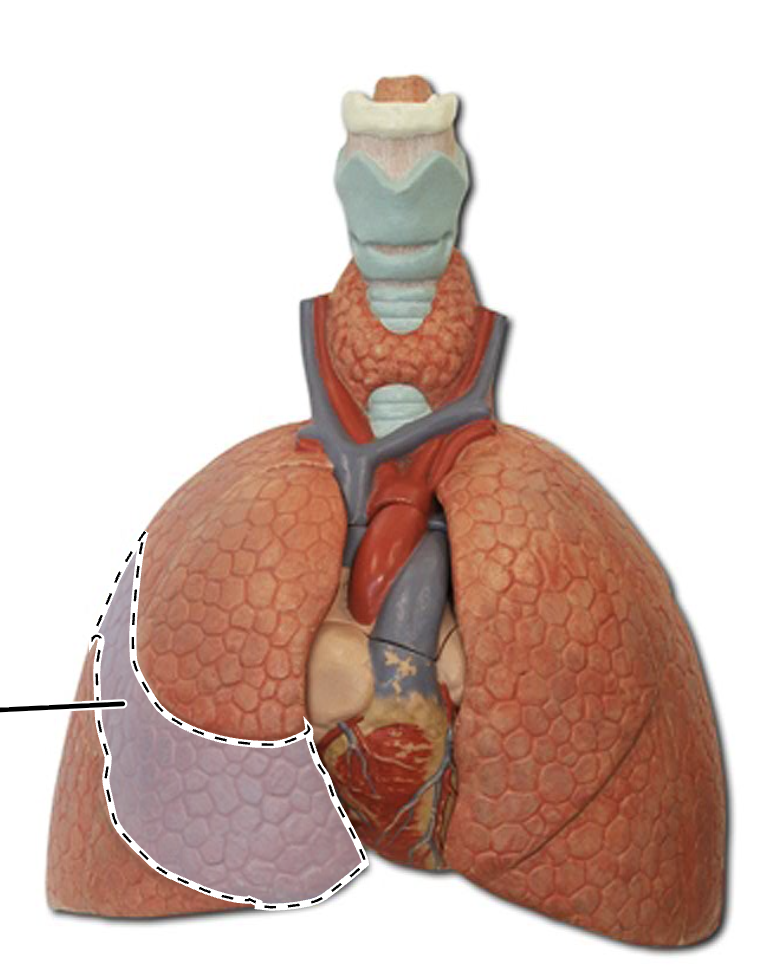
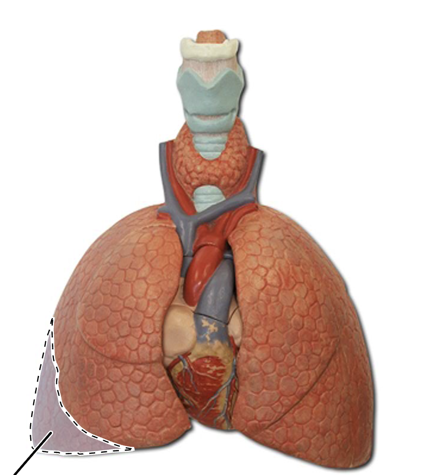
Cardiac Notch (1-7 Diagram)
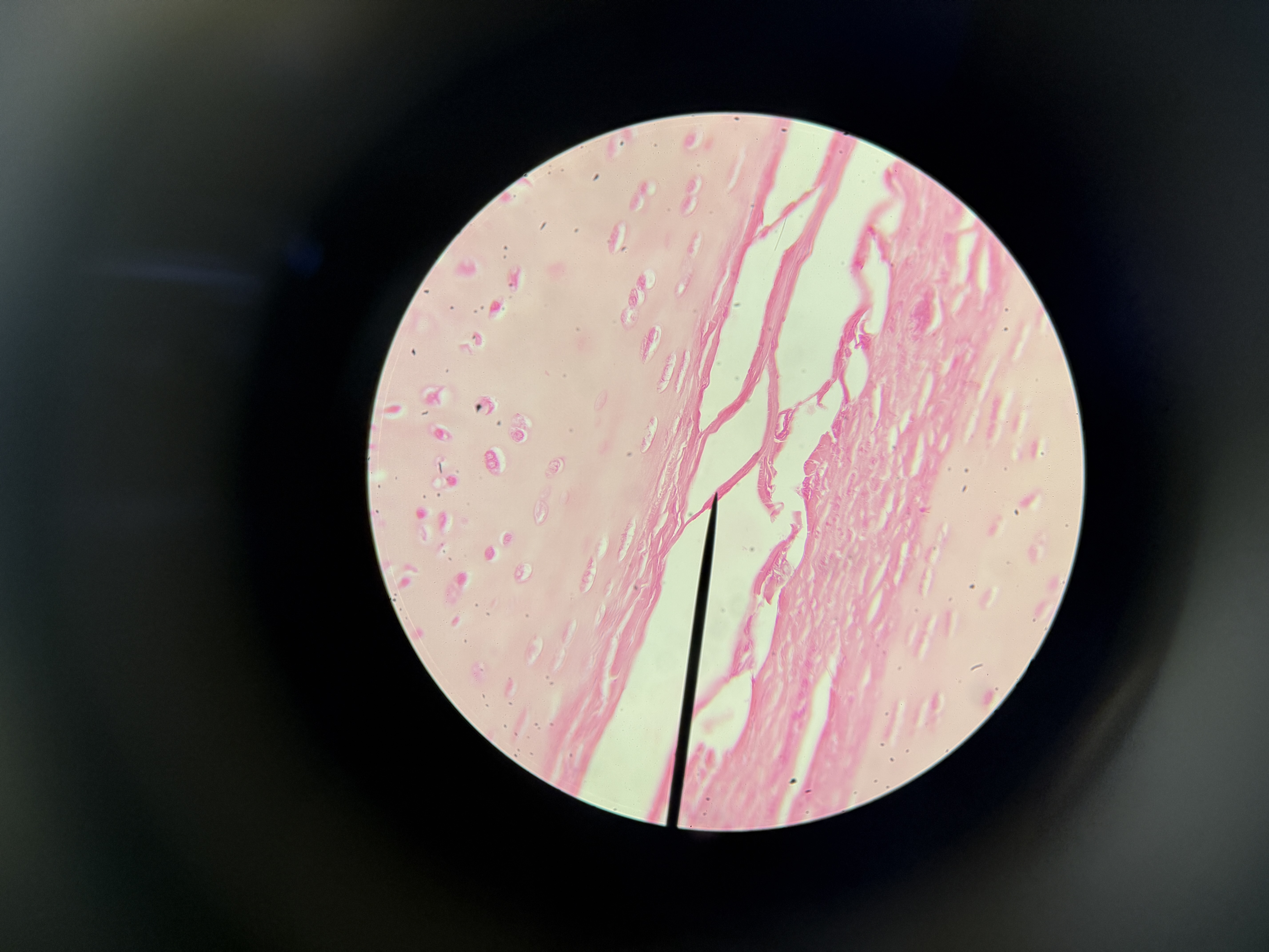
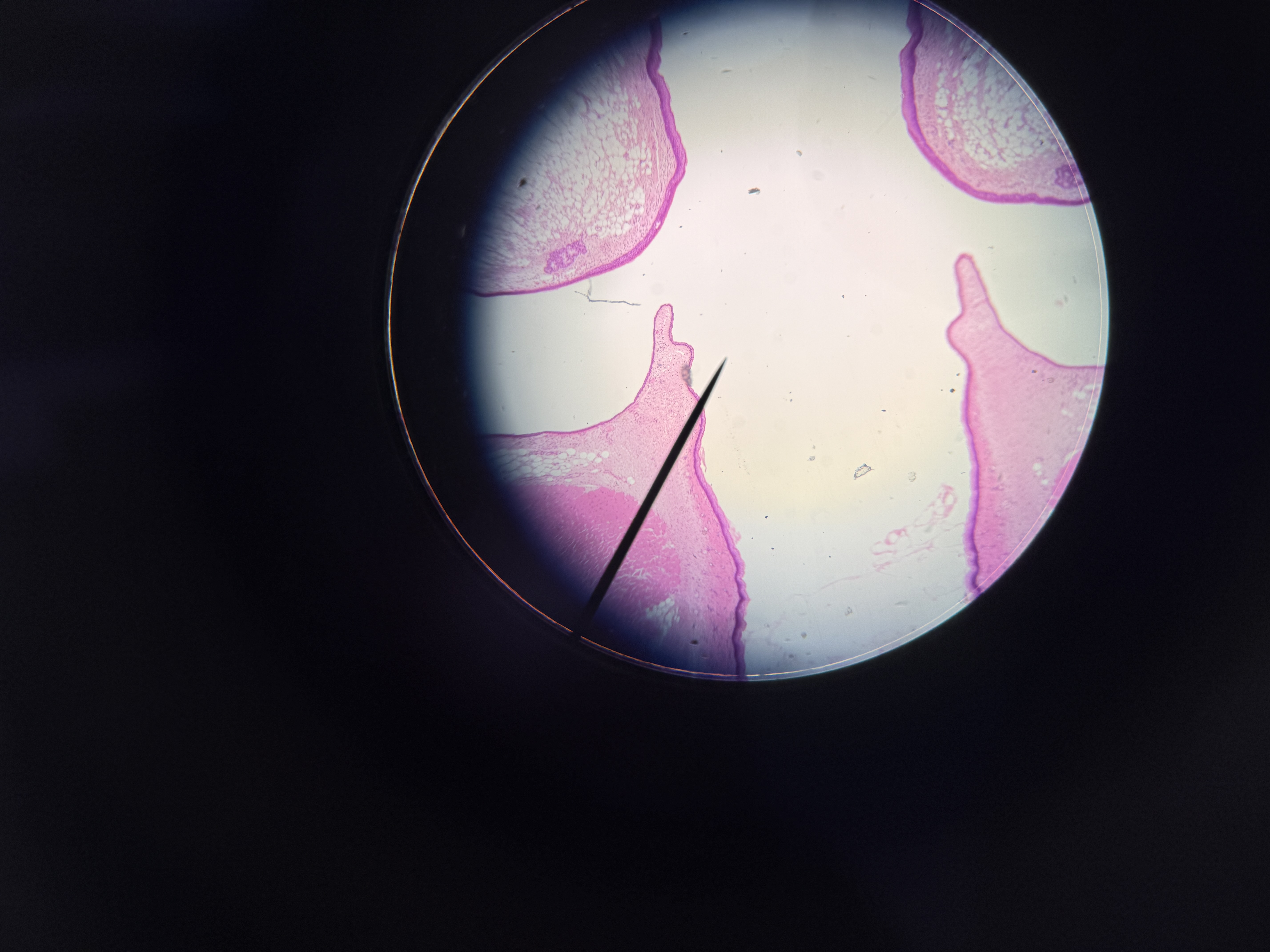
Trachea (Histology)
Larynx
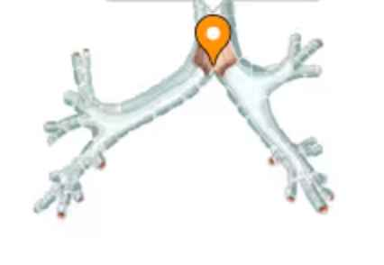
Carina
Superior Lobe of the Right Lung
Middle Lobe of the Right Lung
Inferior Lobe of the Right Lung
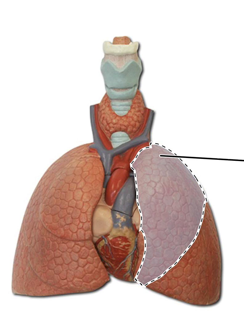
Superior Lobe of the Left Lung
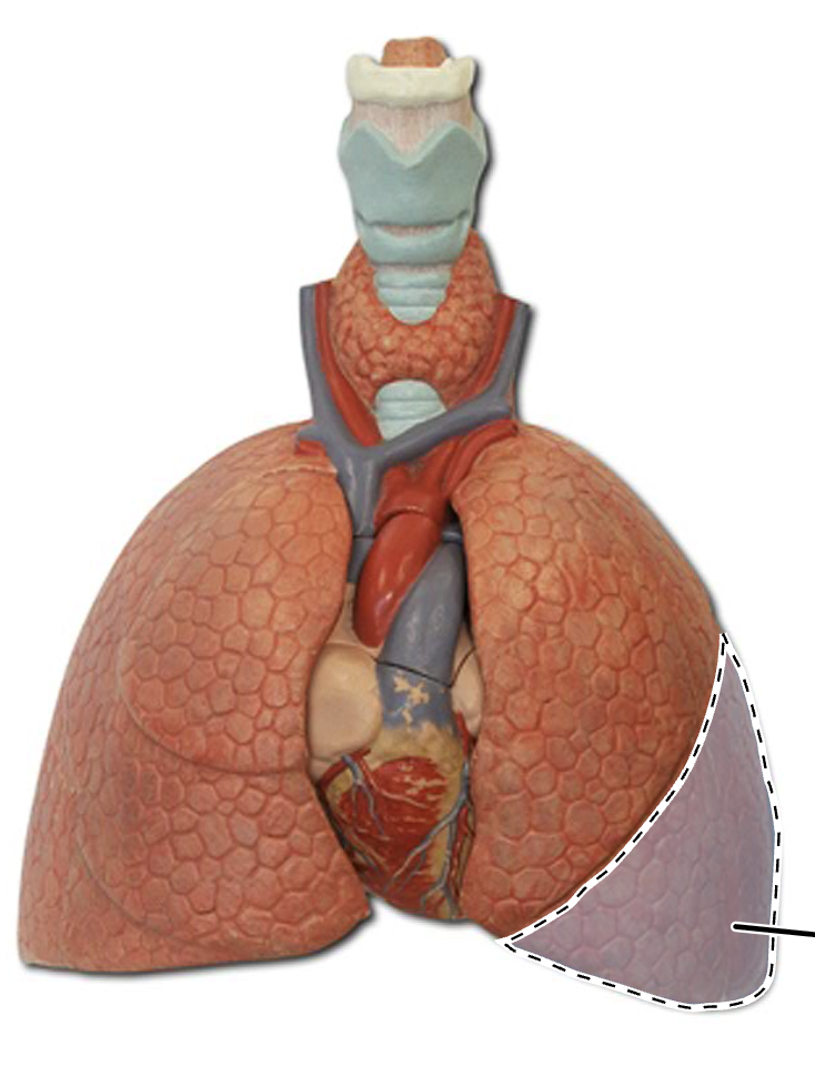
Inferior Lobe of the Left Lung
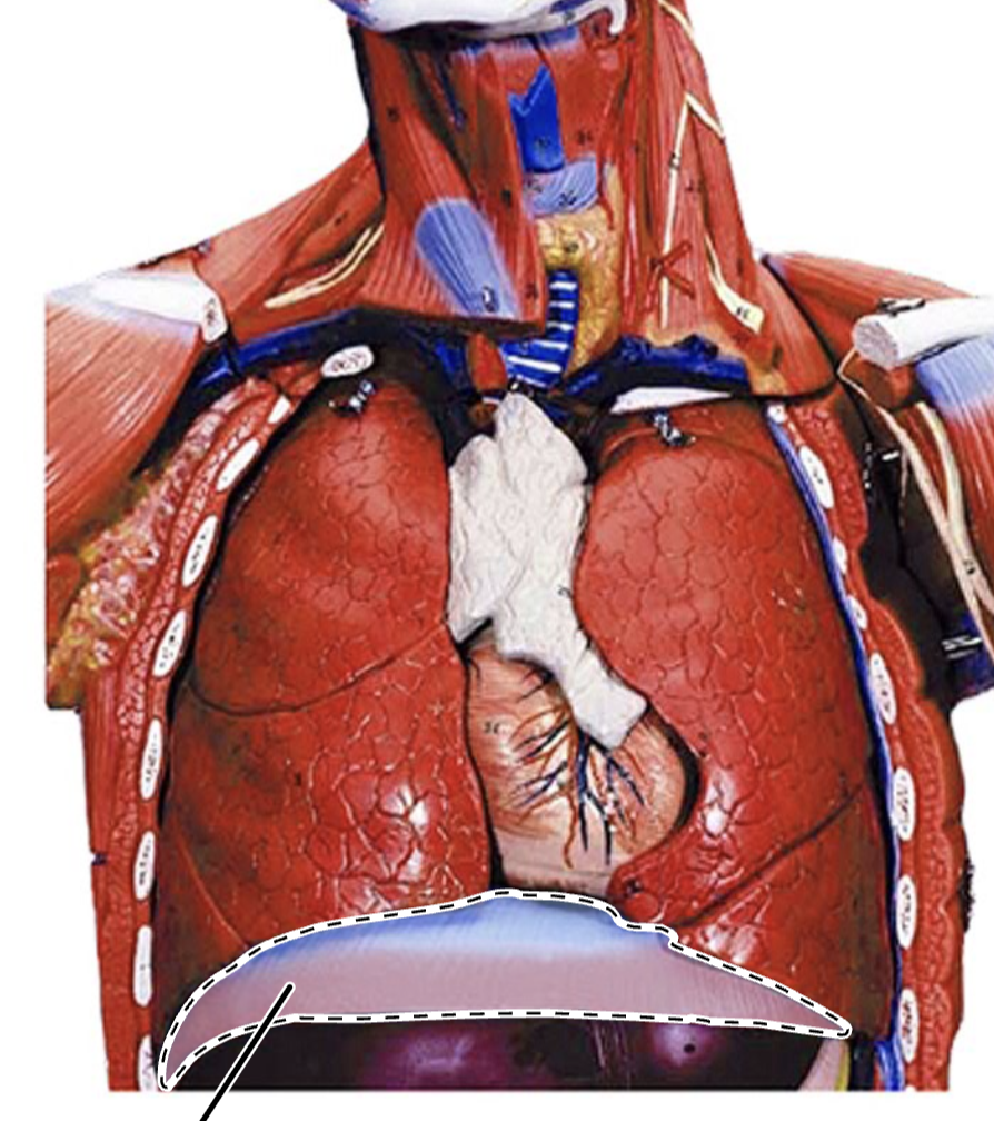
Diaphragm

3 (Lines Organs)
Visceral Pleura
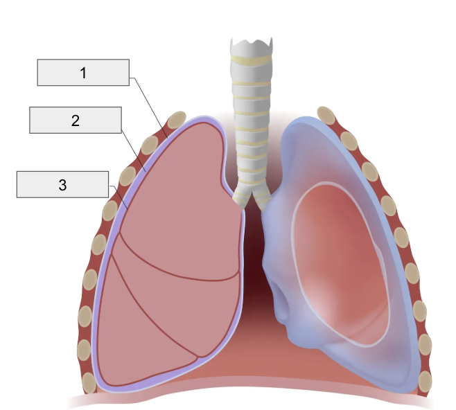
2 (Space in-between Pleura / Intrapleural Pressure is here)
Pleural Cavity

1 (Lines Cavity)
Parietal Pleura

4
Fungiform Papillae
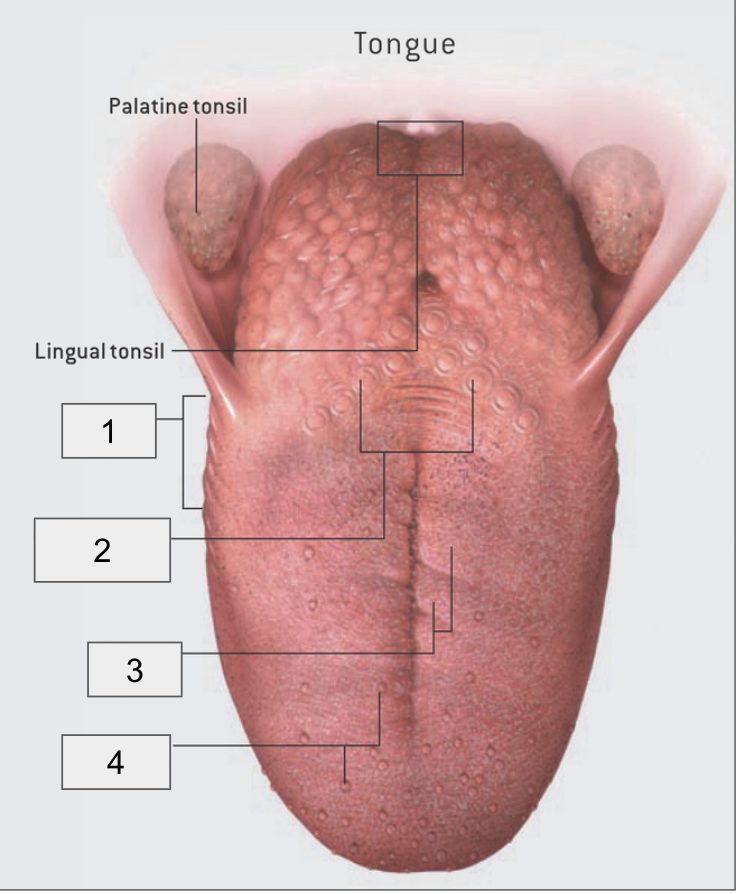
3
Filliform Papillae

2
Circumvallate Papillae

1
Foliate papillae

Incisors
Canine
Premolar
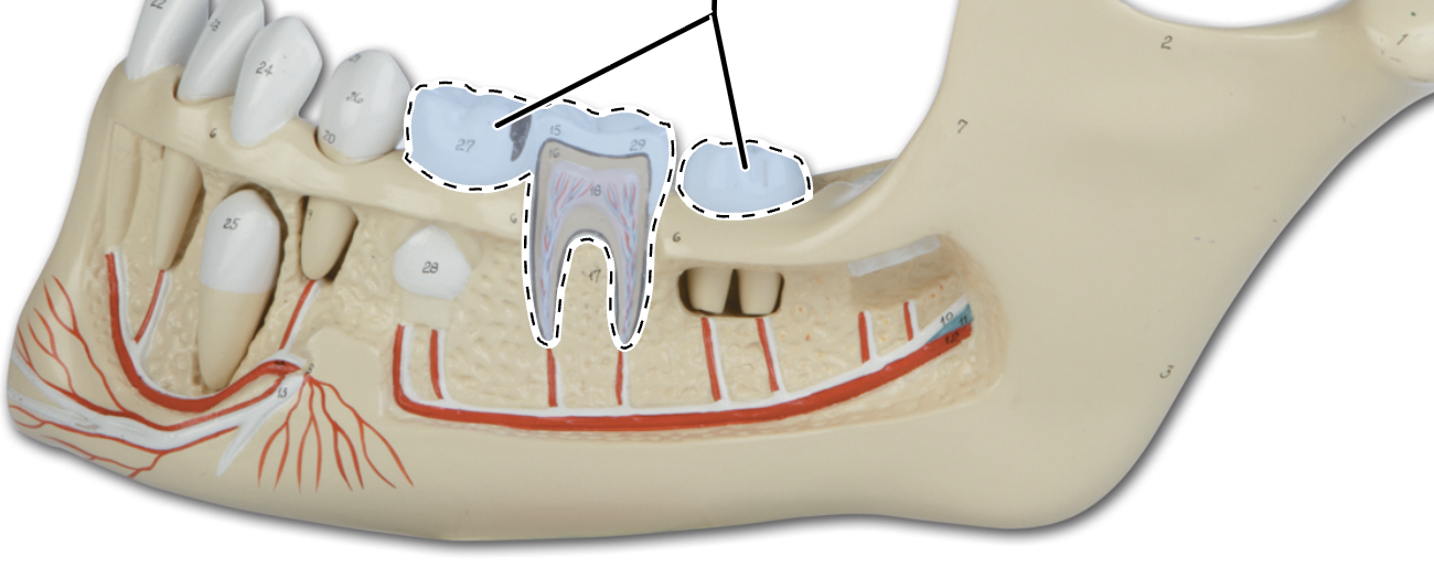
Molars
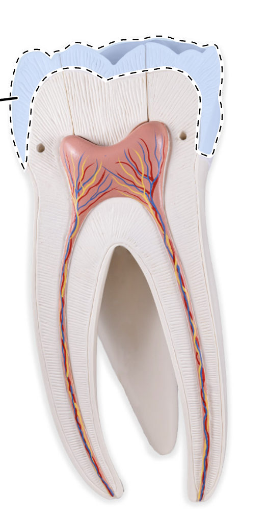
Enamel
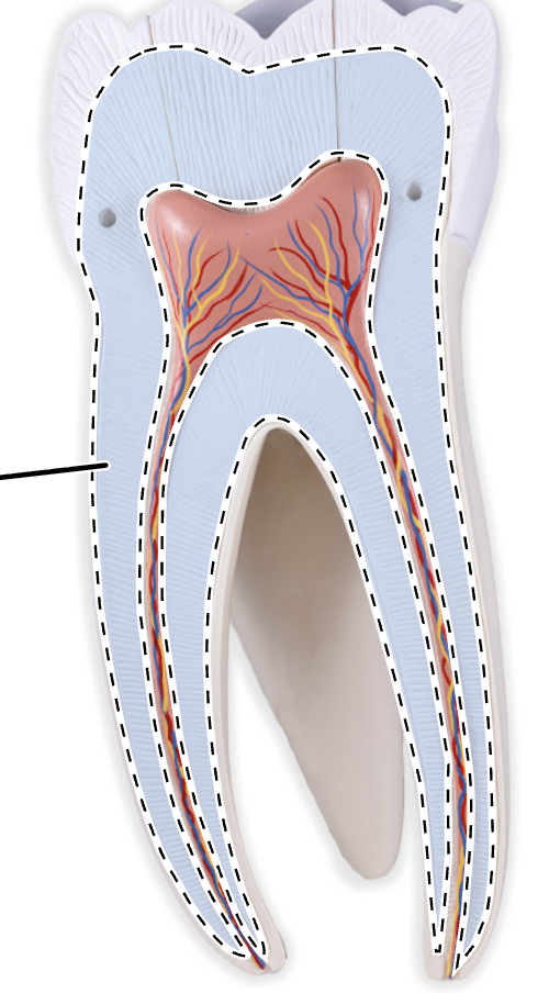
Dentin
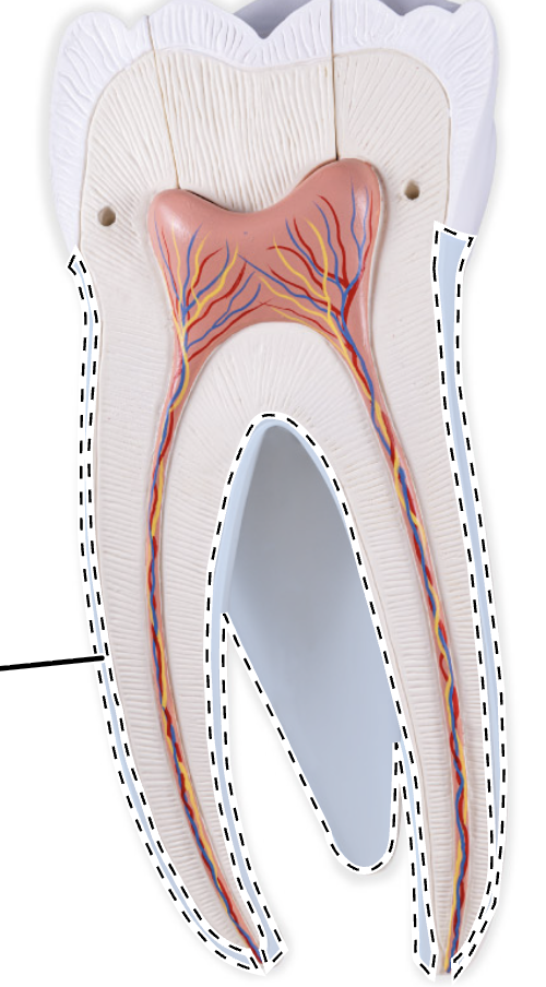
Cementum
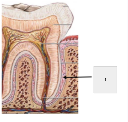
Periodontal Ligaments
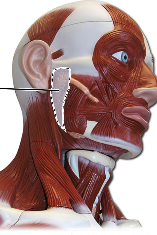
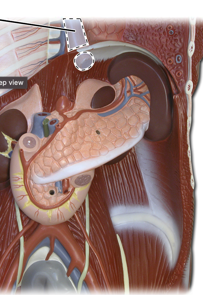
Parotid Gland
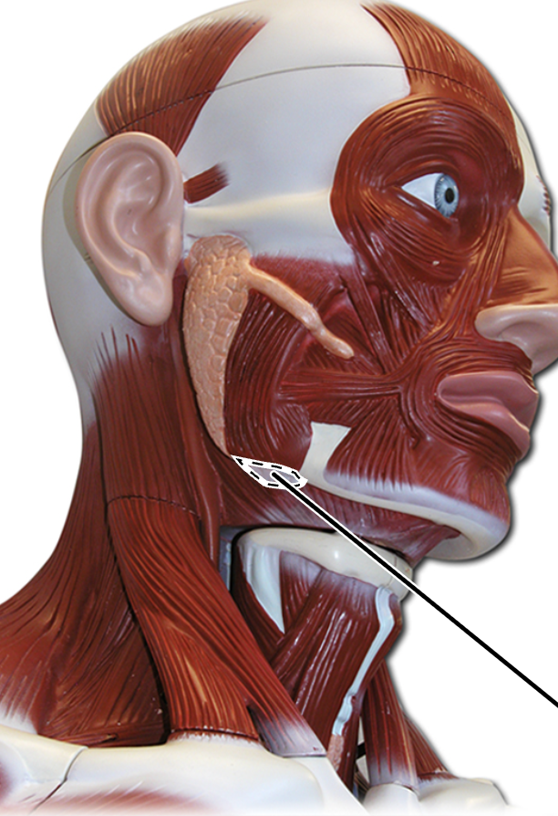
Submandibular Gland
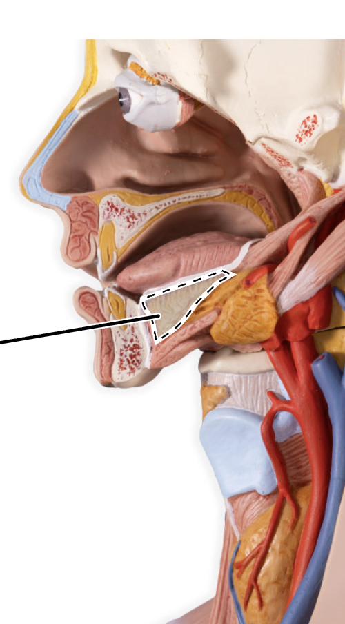
Sublingual Gland
Esophagus
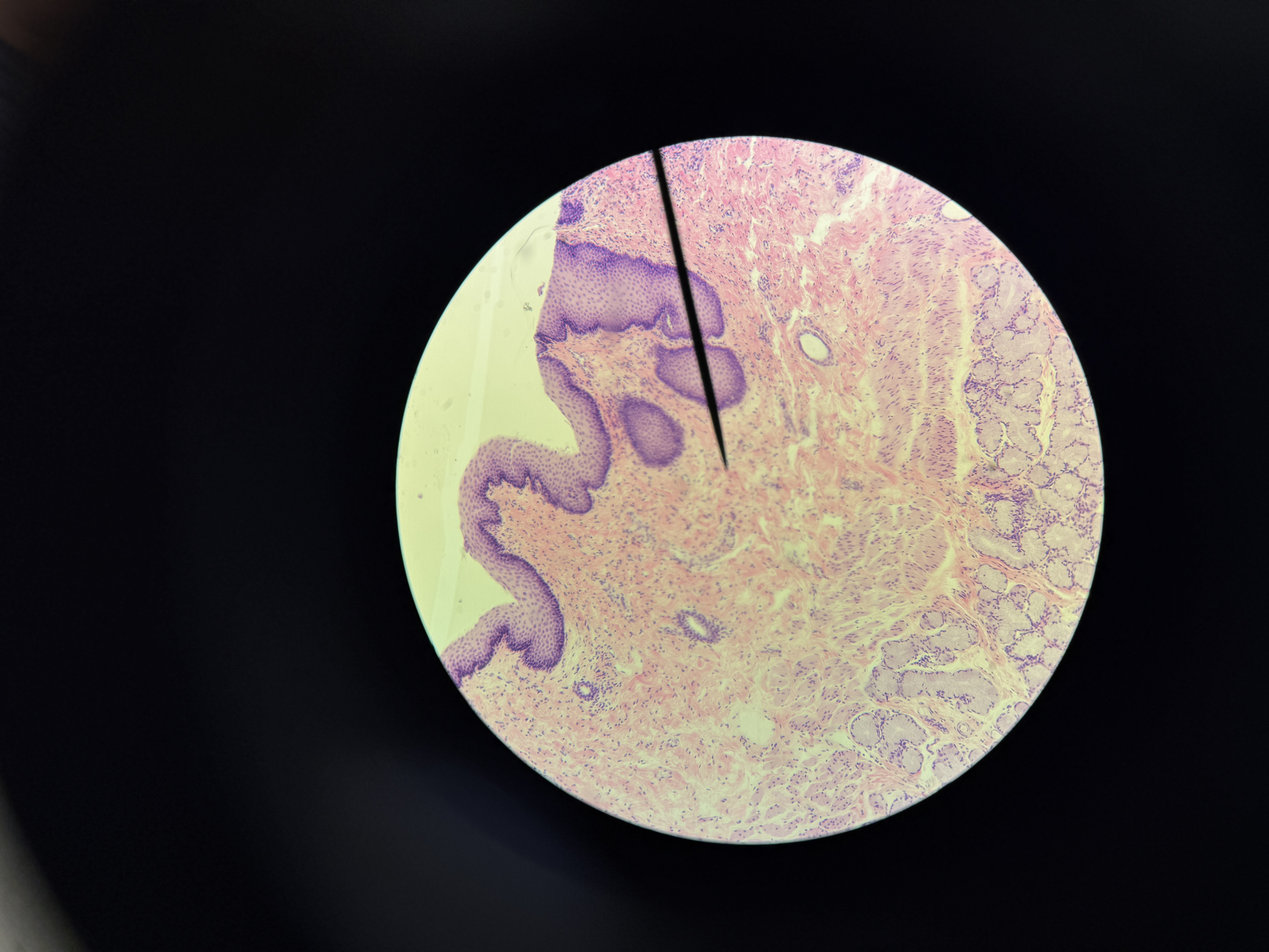
Esophagus (Histology)
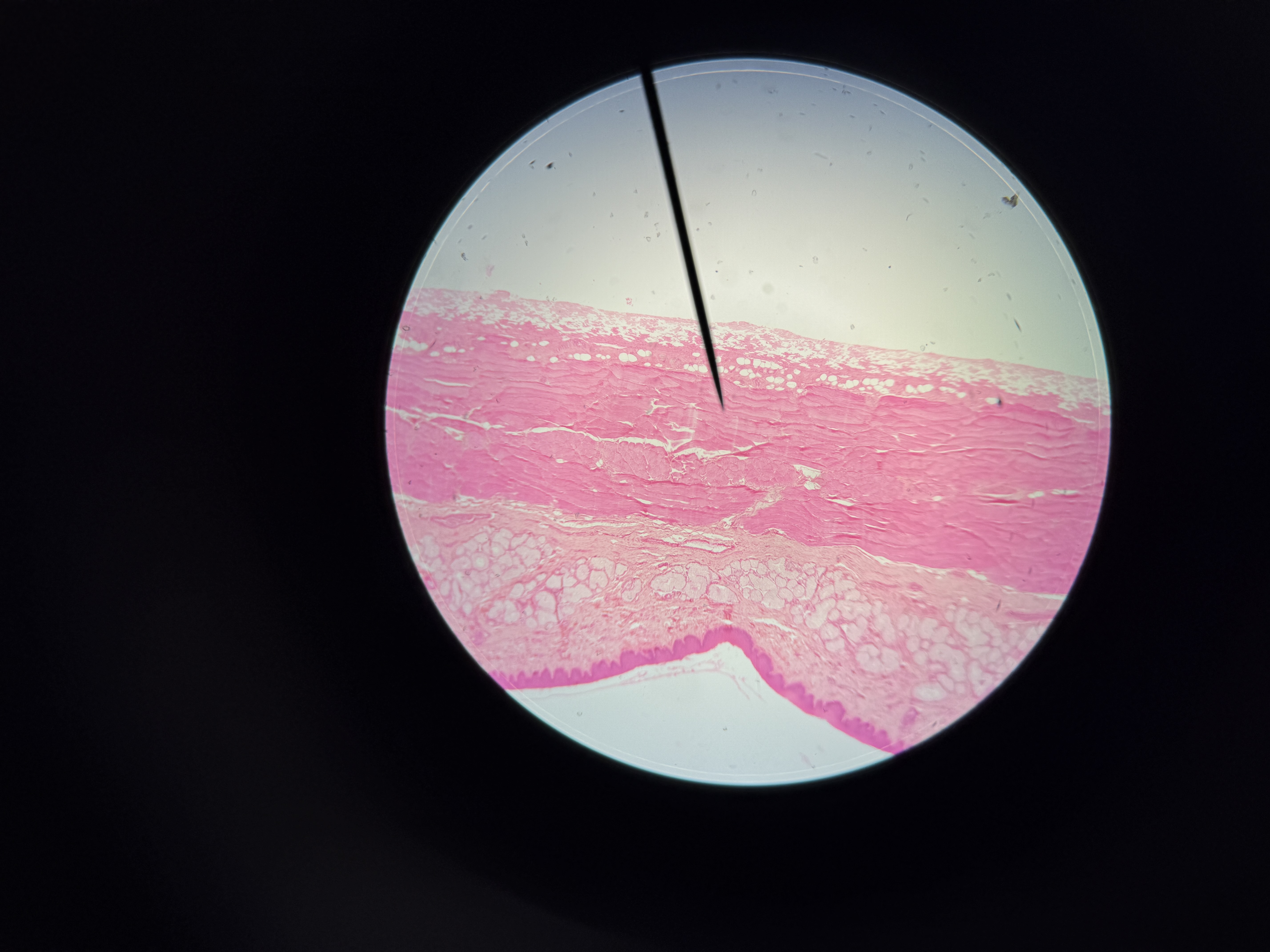
2
Esophagus (Histology 2)
Sphincter (opening) that leads into the stomach from the esophagus
Gastroesophageal sphincter

4
Stomach Cardia

1
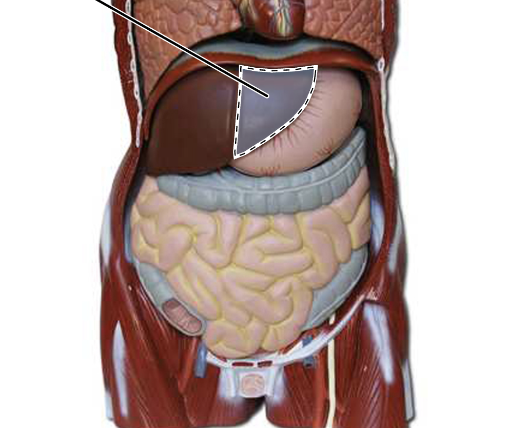
Stomach Fundus

2
Stomach Body

3
Pyloric Antrum
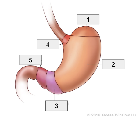
5
Pylorus
(Sphincter (Opening) from the stomach to the small intestine)
Pyloric Sphincter
Stomach Body Histology
2
Stomach Body Histology (2)
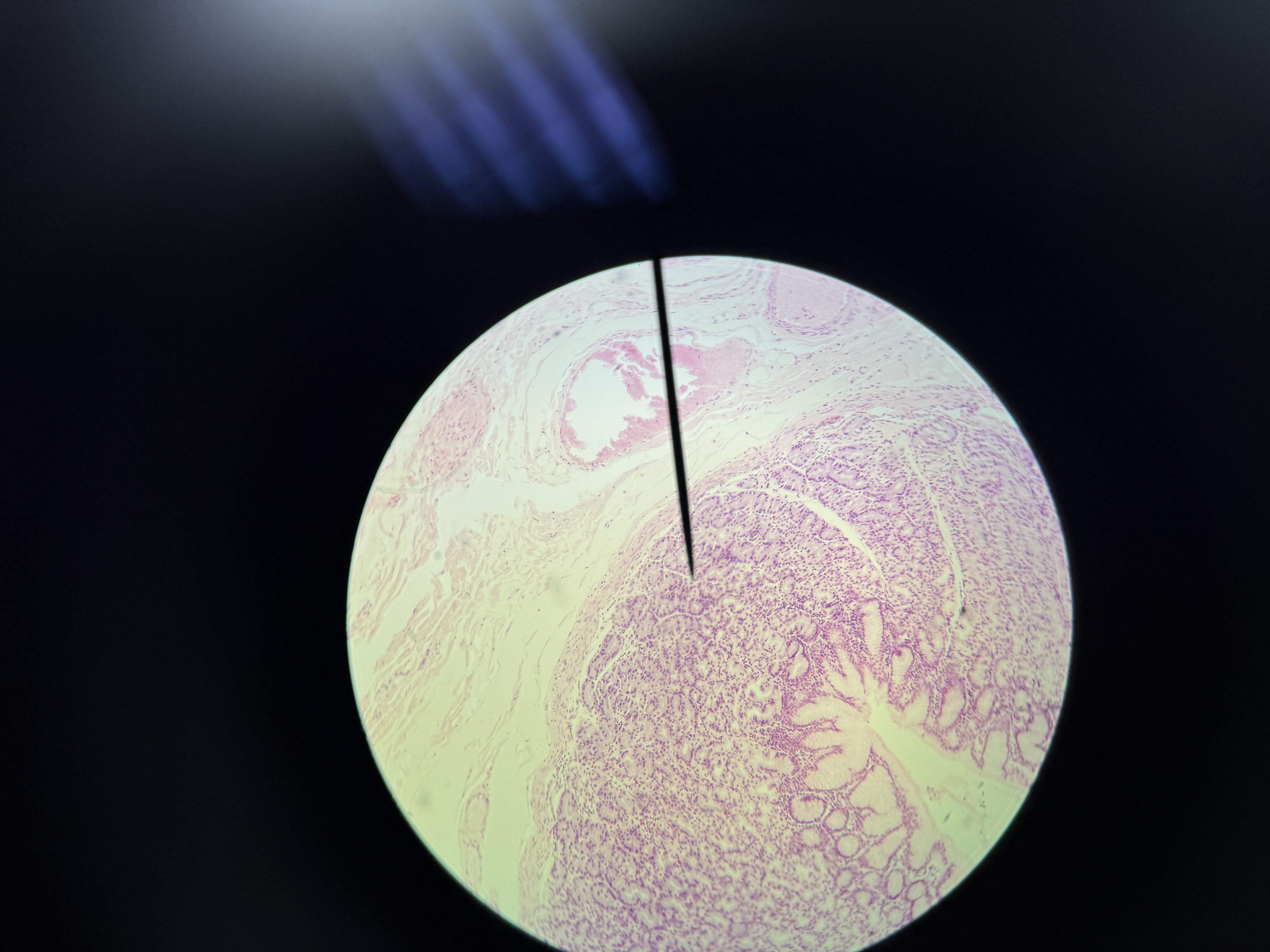
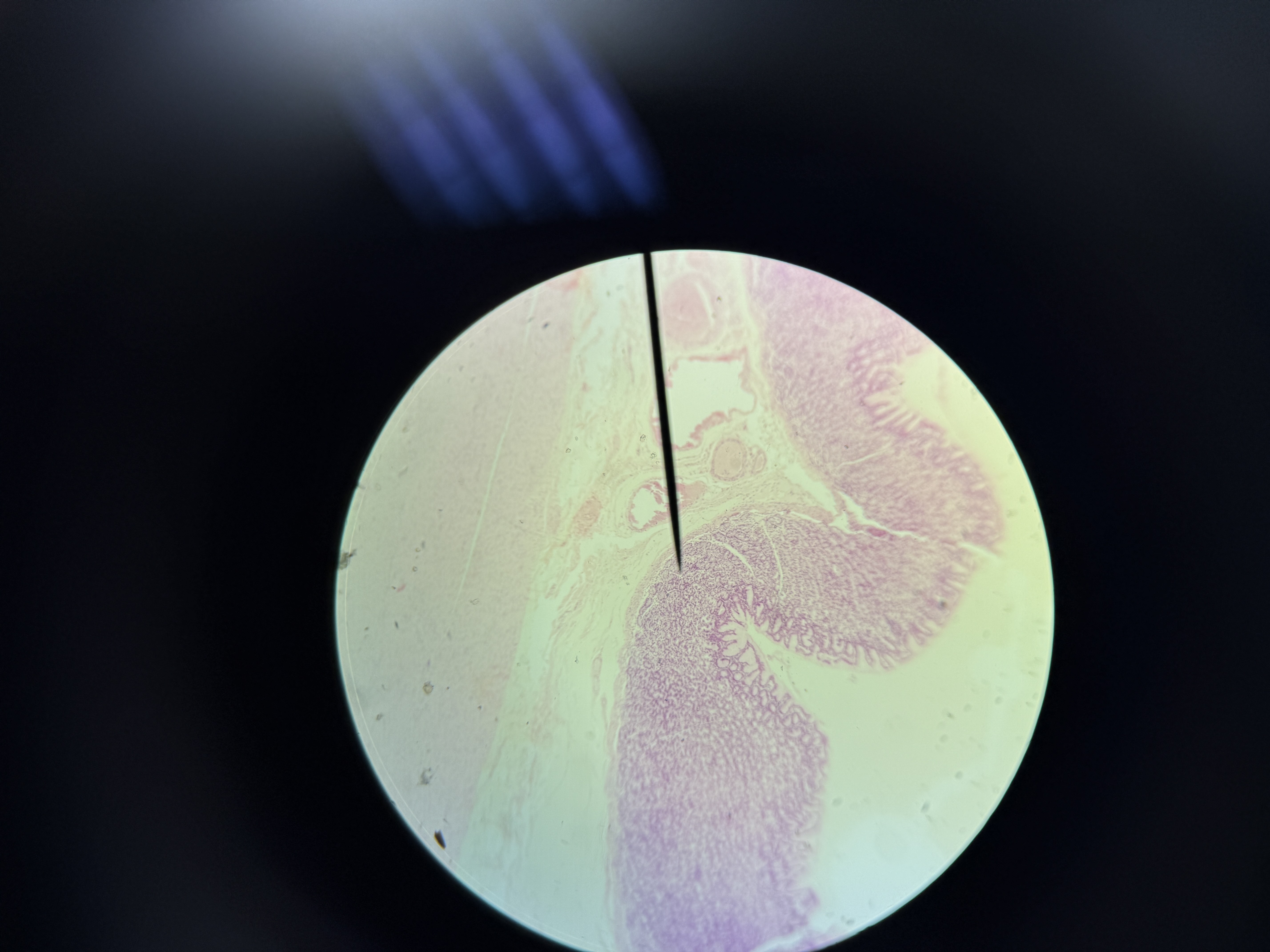
Small Intestine
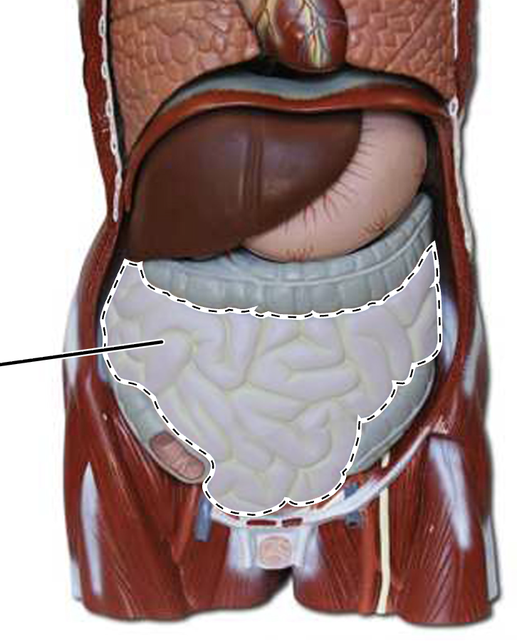
Small Intestine
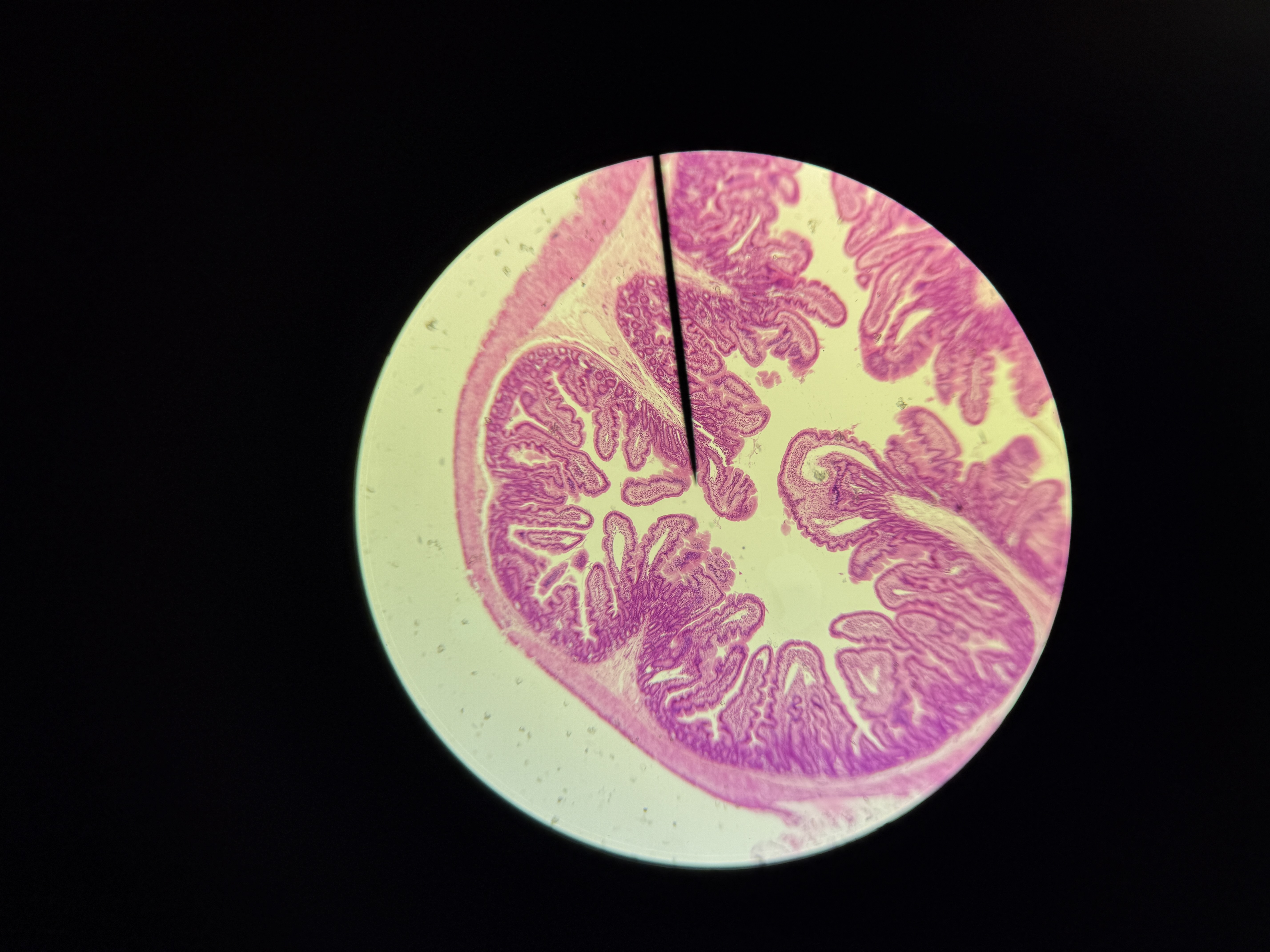
Duodenum
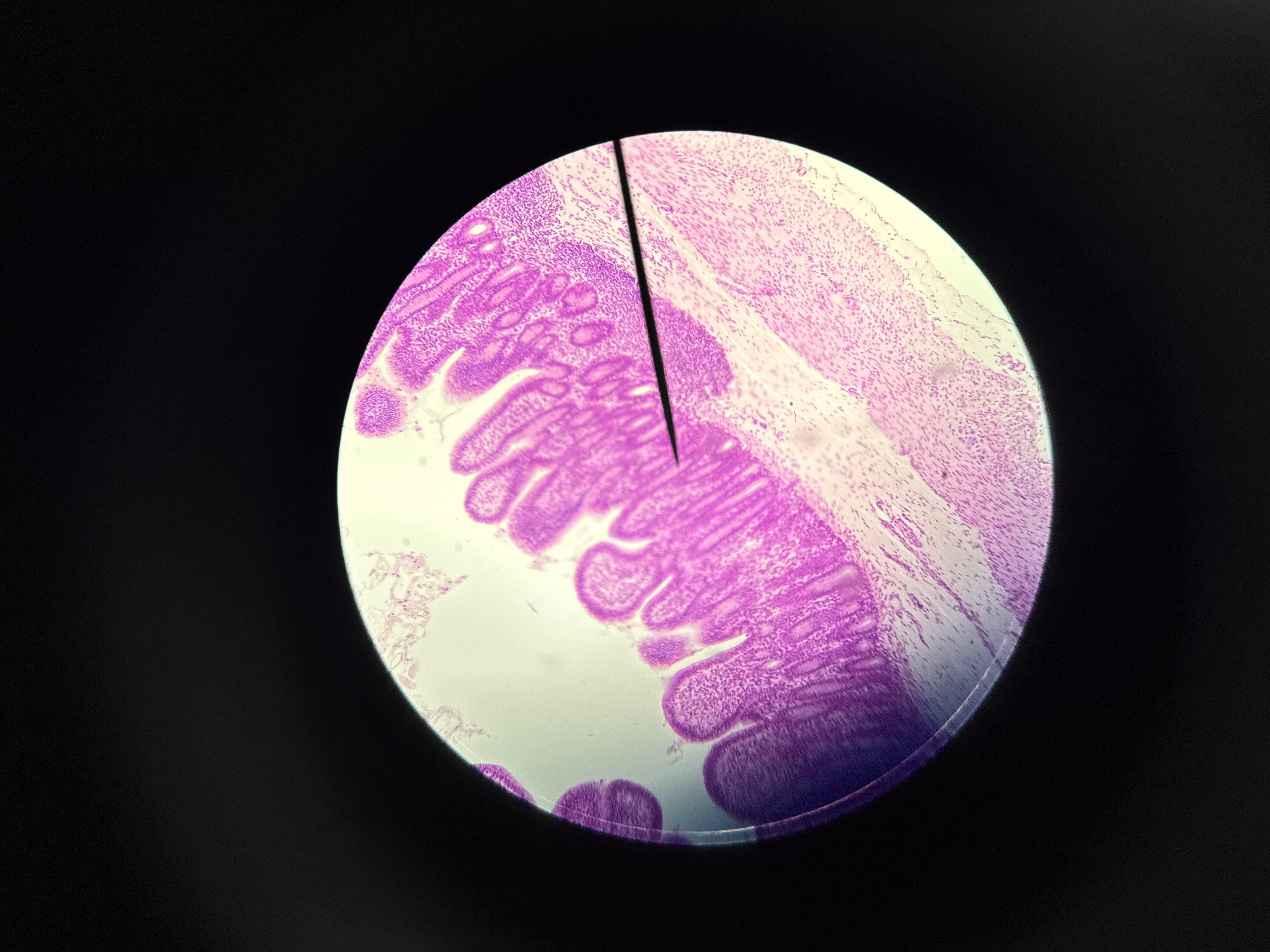
(Peyer Patches)
Ileum
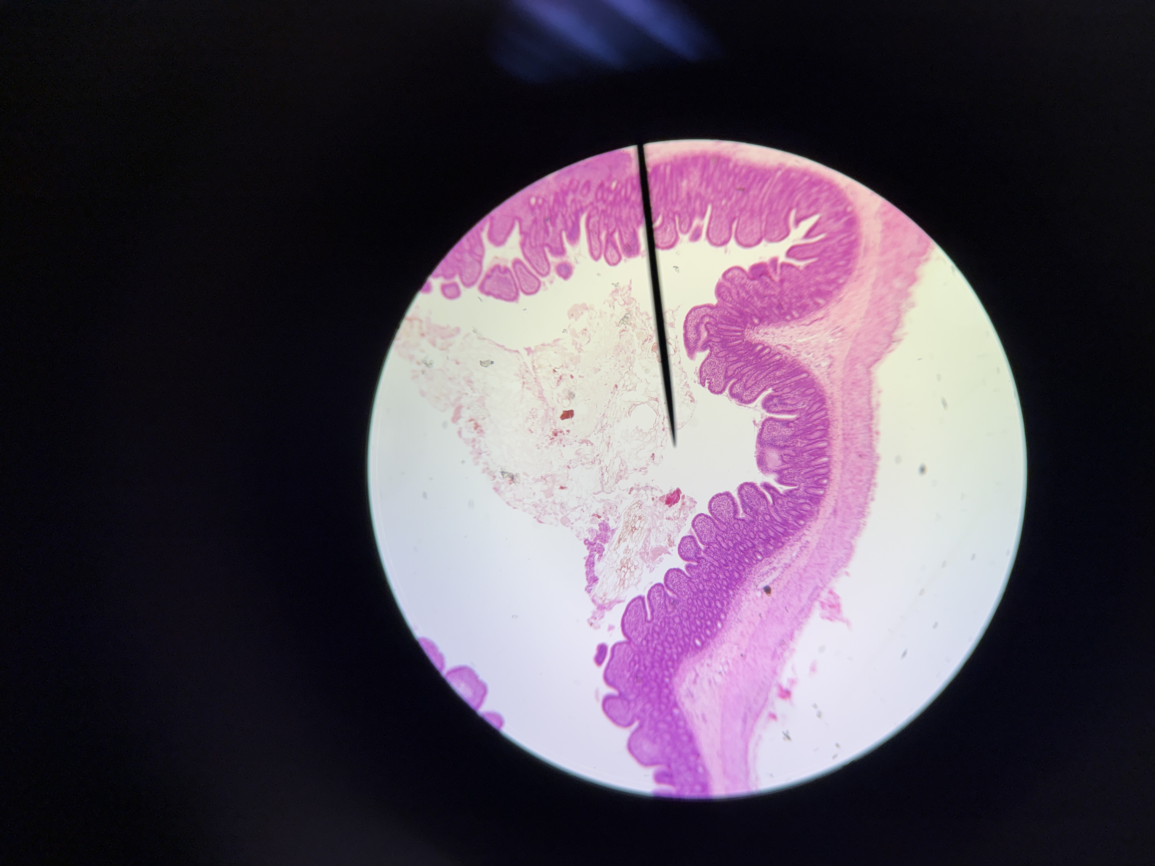
(more intestinal crypts)
Jejunum
Ileum
Jejunum
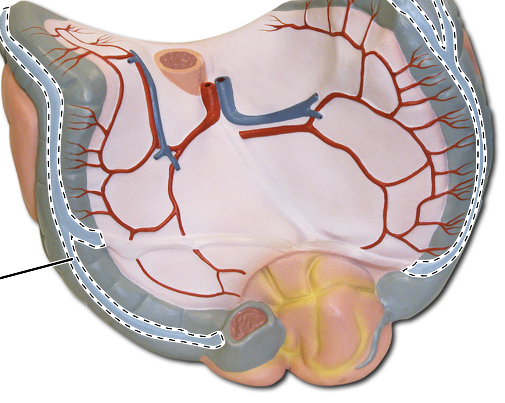
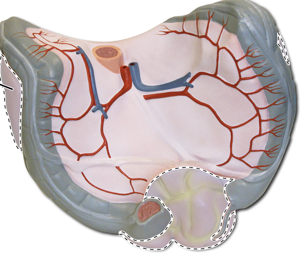
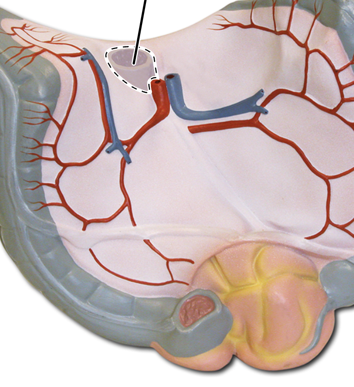
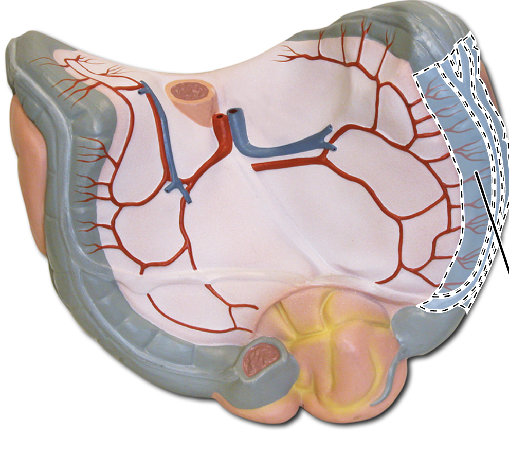
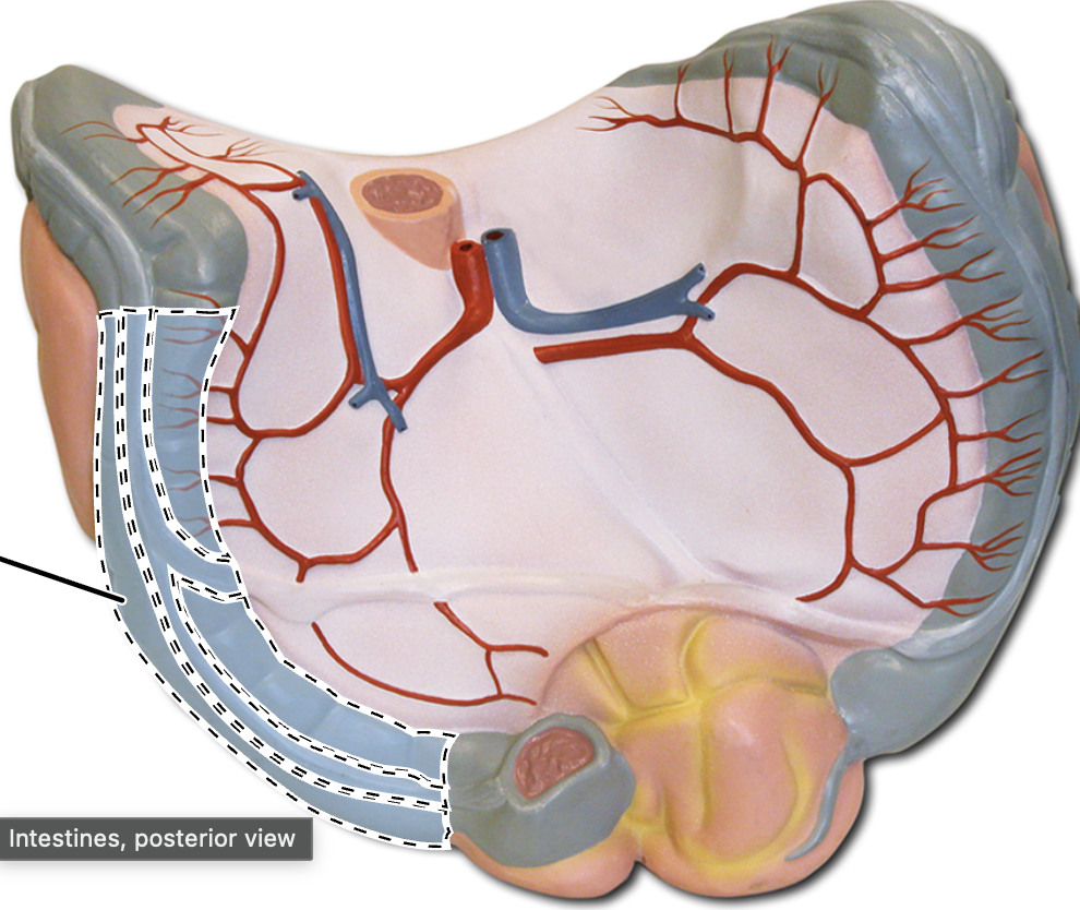
Taenia Coli
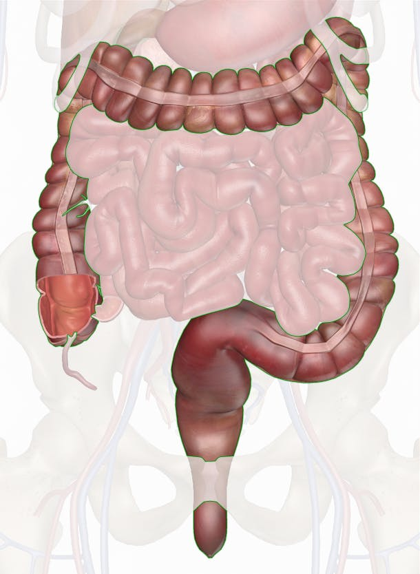
Large Intestine
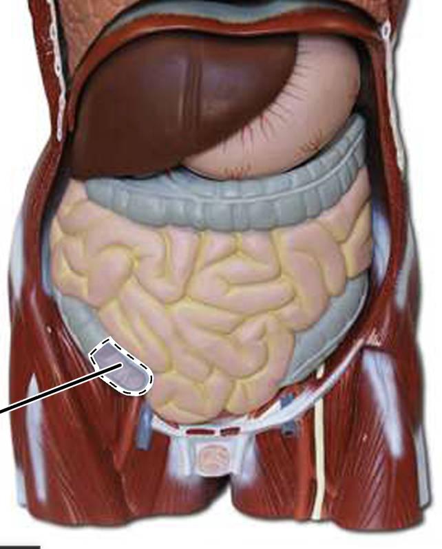
Cecum
Ascending Colon
Transverse Colon
Sigmoid Colon
Descending Colon
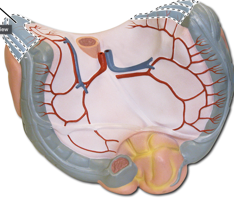
Haustra of Large Intestine
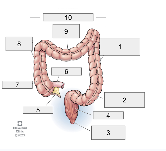
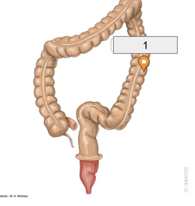
4
Rectum (Diagram)

3
Anal Canal (Diagram)

1
Descending Colon (Diagram)

2
Sigmoid Colon (Diagram)

6
Ileum (Diagram)

5
Appendix (Diagram)

7
Cecum (Diagram)

8
Ascending Colon (Diagram)

9
Transverse Colon (Diagram)

10
Large Intestine/Colon (Diagram)
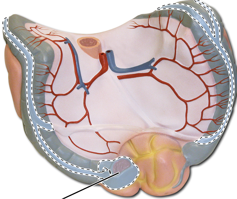
Epiploic appendages
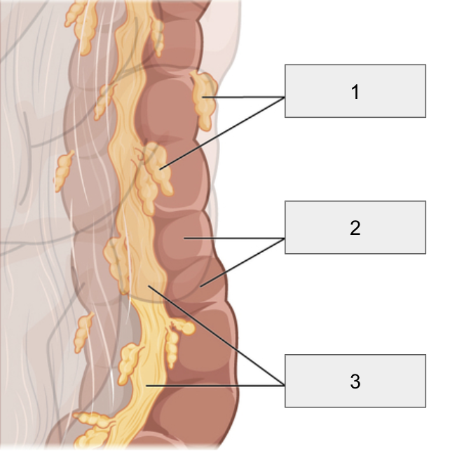
1
Epiploic Appendages

2
Haustra

3
Tenia Coli
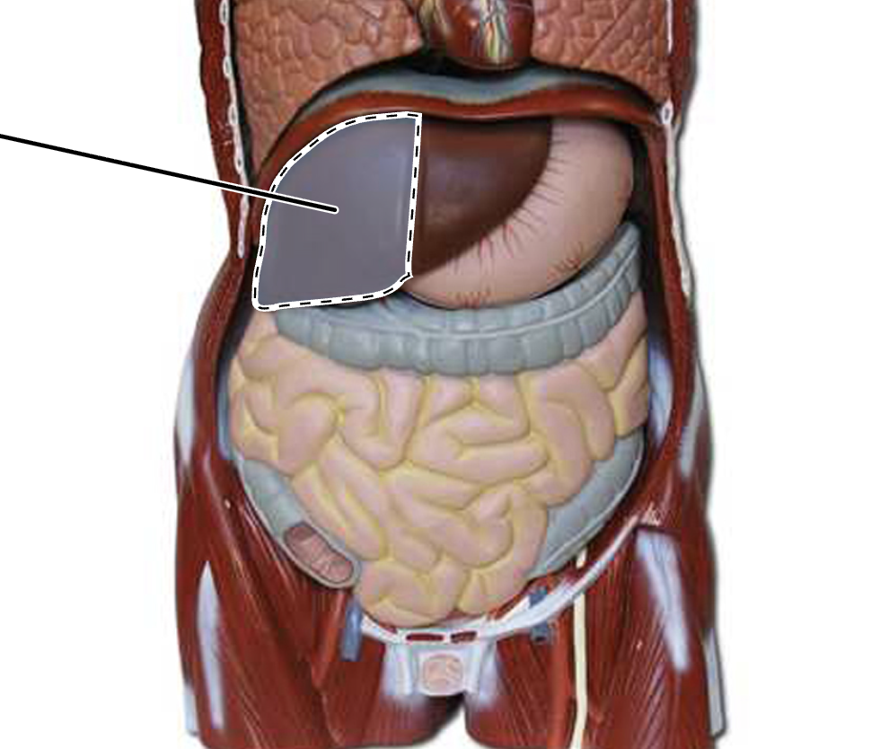
Right lobe of liver
Left Lobe of Liver
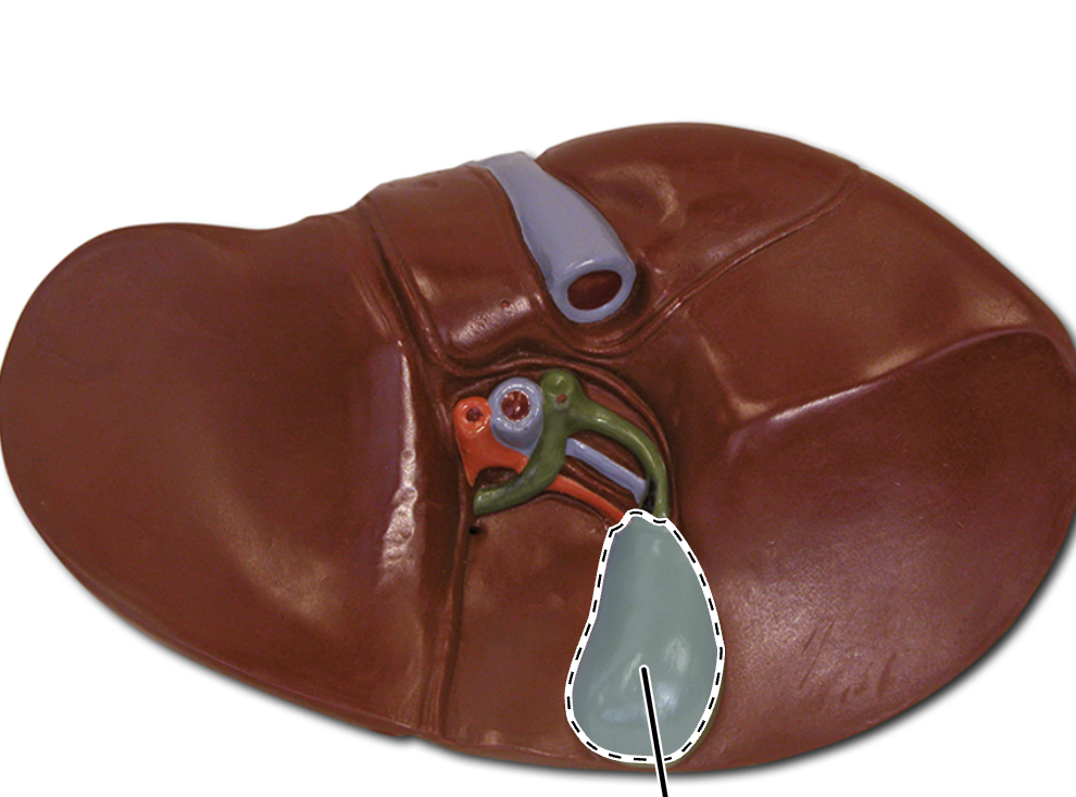
Gallbladder
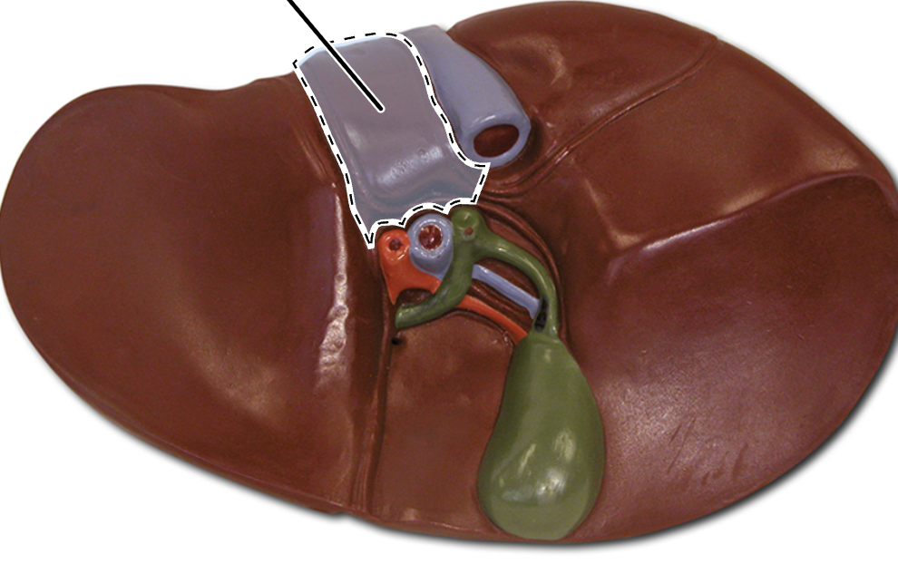
Caudate lobe of liver
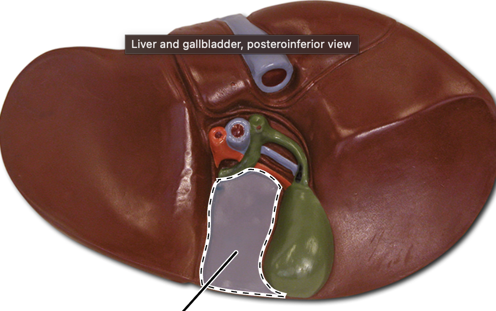
Quadrate Lobe of Liver