kidneys PART 3
1/33
There's no tags or description
Looks like no tags are added yet.
Name | Mastery | Learn | Test | Matching | Spaced | Call with Kai |
|---|
No analytics yet
Send a link to your students to track their progress
34 Terms
kidney failure
kidneys are unable to function effectively→ this prevents them from filtering blood
causes of kidney failure (2) [explain]
-kidney infections
→causes swelling of the kidney tissue,damaging the cells→ reduces their ability to filter blood
-high blood pressure
→damages the glomerulus→ so large molecules like proteins and blood cells can leak into the urine [proteins should remain in the blood, not enter Bowman’s capsule]
kidney failure can be detected by what [state defintion]
Glomerular Filtration Rate (GFR)→ a measure of the volume of blood that can be filtered by the kidneys every minute.
what does a low GFR and a high GFR show?
low GFR→ indicates kidney failure as kidneys are not filtering blood effectively
high GFR→ healthy kidney
what factors need to be considered when measuring GFR? explain (4)
age→ kidney function and GFR decline with age
gender→ men and women have different muscle mass
muscle body mass
exercise
effects of kidney failure (4)
-urea builds up in the blood
-high blood pressure
-decreased number of red blood cells→ reduce oxygen transport→ leads to tiredness
-cannot remove water from the blood→increased water levels in the blood→ fluid builds up in tissues
-increase levels of ions in the blood
waste products build up in the blood
-the damage kidneys cannot remove the waste products→ they builds up in the blood
-urea accumulates in the blood→ toxic to cells and can damage tissues, leading to vomiting and weight loss
electrolyte imbalance
-there’s less ultrafiltration so excess ions cannot be removed. Electrolytes build up in the blood→ affects the body’s osmotic balance
e.g. -Excess potassium can cause muscle weakness, abdominal cramps and fatigue. If very high, can lead to cardiac arrest
-imbalance of calcium and phosphate can lead to bone weakness
treatments for kidney failure
-renal dialysis
-kidney transplant
type of renal dialysis
haemodialysis
what does a dialysis machine contain?
-partially permeable membrane (separating blood and dialysis fluid)
-dialysis fluid
what does dialysis fluid contain?
glucose
-has the same glucose concentration as the blood
-there’s no concentration gradient→ glucose does not diffuse out of the blood.
urea
-no urea
-blood contains a high concentration of urea→ creates a steep concentration gradient
-Urea diffuses out of the blood into the dialysis fluid
ions/ salt
-normal ion concentration
-If blood ion levels are too low → ions diffuse into blood
If blood ion levels are too high → ions diffuse into dialysis fluid
-Movement only occurs where there's an imbalance, maintaining electrolyte balance
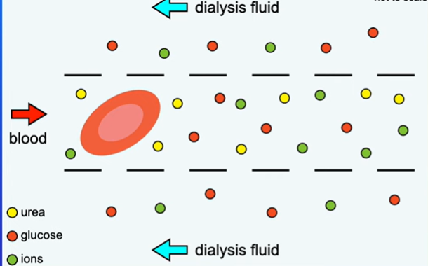
what remains in the blood and why?
glucose→ no concentration gradient→ required for respiration
proteins and blood cells→ too large to pass through membrane
how does dialysis fluid and blood flow in dialysis machine and why? name of this
the blood and dialysis fluid flow in opposite directions to maintain a steep concentration gradient [and increase rate of diffusion]
(counter-current flow)
how does haemodialysis work? (8)
-the patient’s blood is passed through a dialysis machine
-blood flows through tubes made of a partially permeable membrane surrounded by dialysis fluid
-Blood and dialysis fluid flow in opposite directions to maintain a steep concentration gradient
-as blood flows past the dialysis fluid, molecules pass through partially permeable membrane by diffusion
-The dialysis fluid has no urea so urea diffuses into the dialysis fluid down its concentration gradient
-the dialysis fluid has the same glucose concentration as blood, so glucose does not diffuse out of the blood.
-If there’s excess ions in the blood, they diffuse from the blood to the dialysis fluid. If there’s a low ion concentration in the blood, ions from the dialysis fluid flows into the blood
-Blood is filtered and the clean blood is returned to the patient’s body.
what blood vessel carries blood to the dialysis machine and returns to what blood vessel?
Blood is carried to the dialysis machine by an artery and returned to a vein. [like artery carrying blood to kidney]
why is the dialysis fluid constantly refreshed? (2)
-maintain concentration gradient between the blood and the dialysis fluid.
-prevents urea diffusing back into the blood
pros (4) and cons (3) of haemodialysis
pros
-Widely available in hospitals
-remove waste products from the blood
-keeps patient alive until a transplant is available
-monitored by trained professionals, reducing issues
cons
-requires regular sessions, which is time-consuming.
-patients need to manage their diet→ low protein and salt intake [low protein intake to reduce urea production. low salt intake to prevent build-up]
-risk of infection and blood clotting
how to reduce rejection of the kidney transplant (3)
-donor and recipient are matched for blood type/ antigens
-recipient takes immunosuppressant drugs
-use a close relative as donor as they have a closer tissue match
pros (3) and cons (4) of kidney transplant
pros
-no need for regular dialysis
-fewer dietary restrictions
-better quality of life, allowing a normal lifestyle
cons
-risk of immune rejection→ the immune system may recognise antigens on the donor organ as foreign and attack it
-shortage of donor kidneys
-Immunosuppressant drugs increase risk of infections
-surgery has risks
urine samples can be used to detect substances
what does the presence of glucose in urine indicate
what does the presence of blood in urine indicate
diabetes
kidney damage [red blood cells should remain in the blood and not enter Bowman’s capsule]
what hormone is present in the urine of pregnant women?
hCG
what do pregnancy test use
monoclonal antibodies that are specific to hCG
hCG is a protein. How does it enter Bowman’s capsule?
it’s small enough to enter the Bowman’s capsule
testing for pregancy (8)
-urine is applied to pregnancy test strip
-the test strip contains monoclonal antibodies attached to coloured beads
-the monoclonal antibodies are specific to hCG
-hCG binds to mobile monoclonal antibodies to form an hCG antibody complex
-urine carries hCG antibody complex up the strip to a window
-the window contains immobilised monoclonal antibodies that binds to hCG antibody complex
-this creates a coloured line, indicating pregnancy
-a second layer of immobilised antibodies bind to any remaining mobile antibodies with or without hCG→ this forms a second line to show that the test is working correctly. (Control line)
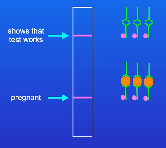
what happens if the woman is not pregnant? (3)
-there is no hCG in her urine
-The mobile monoclonal antibodies with colored beads does not bind to anything. only bind to the immobilised monoclonal antibodies in the second window
-only the control line will appear in the window
![<ul><li><p class="mb-4"><strong>Positive test</strong>: test line + control line</p></li><li><p class="mb-4"><strong>Negative test</strong>: only control line</p></li><li><p class="mb-4"><strong>Invalid test</strong>: No control line appears (test malfunction)</p></li></ul><p>[tip: a working test must show control line]</p>](https://knowt-user-attachments.s3.amazonaws.com/bb64882c-5bc5-4a24-970f-ff47c23ce65f.png)
Positive test: test line + control line
Negative test: only control line
Invalid test: No control line appears (test malfunction)
[tip: a working test must show control line]
examples of drugs that are tested
-anabolic steroids
-recreational drugs
anabolic steroids
drugs that build up muscle tissue→ increases strength
example of an anabolic steroid
testosterone
what’s used to test for anabolic steroids in urine (2
gas chromatography and mass spectrometry
testing for anabolic steroids
-gas chromatography vaporises urine sample and compounds are passed through a column
-Different compounds travel at different speeds, causing them to separate.
-mass spectrometry ionizes the separated compounds and identifies the compounds
-results compared with known substances
testing for drugs (4)
-urine is applied to test strip
-the test strip contains monoclonal antibodies specific to the drug
-the drug binds to the antibodies
-colour change indicates drug is present
-Chromatography and mass spectrometry used