Anatomy Chp 8
1/83
There's no tags or description
Looks like no tags are added yet.
Name | Mastery | Learn | Test | Matching | Spaced | Call with Kai |
|---|
No analytics yet
Send a link to your students to track their progress
84 Terms
joints
also called articulations; places where two or more bones meet
joint structure
determines function and range of motion
trade-off of joints
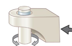
mobility vs. stability- more movement = less stability (high injury risk)
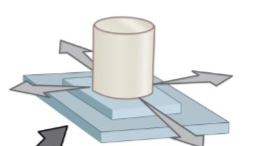
classification methods of joints
by range of motion (functional)
by structure (histological)
functional classification terms
synarthrosis, amphiarthrosis, diarthrosis
synarthrosis
immovable
amphiarthrosis
slightly moveable
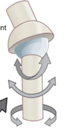
diarthrosis
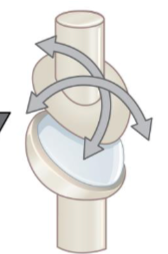
freely moveable (synovial joints)
synarthroses
bones held by dense connective tissue
little or no movement
function: strength, protection, force distribution
examples:
suture → skull bones
gomphosis → tooth in socket
synchondrosis → epiphyseal plate
synostosis → fused bones
amphiarthroses
bones connected by cartilage or ligaments
limited movement
examples
syndesmosis → ligament connects bones
symphysis → fibrous cartilage pad
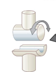
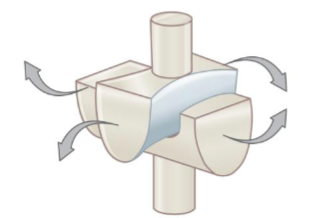
diarthroses
always have:
joint capsule
articular cartilage
joint cavity
synovial membrane
accessory structures
sensory nerves
blood vessels
synovial fluid functions:
lubrication
nutrient delivery
shock absorption
accessory structures
menisci
fat pads
ligaments
tendons
bursae
menisci
fibrous cartilage pads
fat pads
cushioning, space-filling
ligaments
capsular, extracapsular, intracapsular
tendons
stabilize joint via muscle tone
bursae
fluid filled sacs that reduce friction
tendon sheaths = elongated ____
synarthrosis
strongest joint
diarthrosis
weakest joint
increases stability
ligaments, joint shape, muscle tone
axes of motion
superior-inferior
medial-lateral
anterior-posterior
types of movement
angular motion, rotation, special movements
angular motion
flexion → decreases joint angle
extension → increases joint angle
hyperextension → beyond normal (injury risk)
abduction → away from midline
adduction → towards midline
circumduction → circular motion
rotation
medial (internal)
lateral (external)
pronation → palm faces posterior
supination → palm faces anterior
special movements
inversion/eversion (foot)
dorsiflexion/plantar flexion (foot)
lateral flexion (spine)
protraction/retraction (jaw/shoulders)
opposition/reposition (thumb)
temporomadibular joint (TMJ)
synovial hinge + gliding joint
fibrous cartilage (not hyaline)
articular disc divides joint into two cavities
high mobility, high dislocation risk
movements;
elevation
depression
protraction
retraction
side-to-side
intervertebral joints
intervertebral disc (symphysis)
two zygapophysial (facet) joints (synovial)
intervertebral disc structure
anulus fibrous → outer fibrous cartilage
nucleus pulposus → inner gelatinous core (shock absorber)
endplates → attach disc to vertebrae
aging effects of intervertebral joints
low water content, decreases height, high herniation risk
shoulder complex
only one attachment to axial skeleton → huge mobility
mobility > stability
stability mainly from muscles → rotator cuff
sternoclavicular joint
“master joint”
positions scapula
has articular disc
glenohumeral (shoulder) joint
ball-and-socket
shallow socket → easily dislocated
glenoid labrum deepens socket
elbow joint
hinge joint
actually 3 joints in one capsule
very stable due to:
bone shape
thick capsule
strong ligaments
radio-ulnar joints
allow pronation and supination
linked by interosseous membrane
wrist joint
radiocarpal joint → condylar
intercarpal joints → plane
doesn’t position hand much but affects tendon efficiency
hand joints
thumb carpometacarpal joint → saddle (opposition)
MCP joints → condylar
interphalangeal joints → hinge
hip joint
strongest synovial joint
ball-and-socket
acetabular labrum deepens socket
extremely stable → fractures more common than dislocations
knee joint
most complex hinge joint
two joints → tibiofemoral, patellofemoral
menisci crucial for stability
major ligaments → ACL/PCL, MCL/LCL
locking mechanism stabilizes knee in extension
ankle joint (talocrural joint)
hinge joint
dorsiflexion and plantar flexion
stability from ligaments and malleoli
foot joint
designed for support and flexibility
intertarsal, tarsometatarsal, MTP joints
aging joints
low cartilage
low synovial fluid
high stiffness and fracture risk
arthritis= inflammation of synovial joints
exercise helps slow degradation
joints
where two bones meet
fluid, cartilage, or fibrous tissue
tradeoff of flexibility and strength
synarthosis
no movement
types: fibrous, cartilaginous, bony fusion
fibrous synarthrosis
suture (sutural ligaments), gomphosis (periodontal ligaments)
cartilaginouse synarthoris
synchondrosis (ex: btwn ribs and sternum)
bony fusion synarthrosis
synostosis (ex: fusion of frontal bones)
amphiarthrosis
some movement
types: fibrous, cartilaginous
fibrous amphiarthrosis
syndesmosis
between tibia and fibula
between fibula and talus
cartilaginous amphiarthrosis
symphysis
pubic symphysis
intervertebral discs
diarthrosis (synovial)
free movment
key parts of diarthrosis
fibrous joint capsule
synovial membrane
articular cartilages
joint cavity containing synovial fluid
synovial fluid
lubricates articular cartilages and reduces friction
nourishes chondrocytes of articular cartilages
acts as shock absorber
accessory structures of a knee joint
bursa
fat pad
meniscus
tendons
blood vessels
nerves
ligaments
extracapsular
intracapsular
pivot joint
ex: btwn C1 and C2
hinge joint
ex: elbow
saddle joint
ex: btwn trapezium carpal bone and 1st metacarpal bone
ball and socket joint
ex: hip joint
condyloid joint
ex: btwn radius and carpal bones of wrist
plane joint
ex: btwn tarsal joints
abduction
away from longitudinal axis
frontal plane; lateral/medial
adduction
towards longitudinal axis
frontal plane; lateral/medial
flexion
decreases angle btwn bones; bend forward
anterior/posterior plane
extension
increases angle btwn bones'; bend backwards
anterior/posterior plane
hyperextension/flexion
movement beyond normal limits
types of angular motion
adduction/abduction
flexion/extension
rotation
supination/pronation (up/down)
eversion/inversion (moving feet lateral/medial)
dorsiflexion/plantar flexion (moving feet up/down)
posterior/anterior (ex: jaw)
inferior/superior (ex: jaw)
opposition/reposition
TMJ (temporomandibular joint)
2 synovial cavities in same place
very loose
allows for chewing
plane and hinge joint
intervertebral ligaments
ligamentum flavum
posterior longitudinal ligament
interspinous ligament
supraspinous ligament
anterior longitudinal ligament
intervertebral disc
vertebral endplate
anulus fibrosis
nucleus pulposus
zygapophysial joints
articular processes between vertebrae
herniated/bulging discs
damages anulus fibrosis causes herniation of nucleus pulposus
sternoclavicular joint
only joint for axial and upper appendicular
two synovial cavities
two plane joints
glenohumeral joint
ball and socket
greatest range of motion (high injury risk)
triaxial
elbow joint
3 joints within one capsule
2 hinge joints: humero-ulnar (strongest; trochlea and trochlear notch) and humeroradial (more flexible; capitulum and head of radius)
1 pivot joint: proximal radio-ulnar
proximal and distal radio-ulnar joints
pivot joints, allow rotation
proximal radio-ulnar joint
head of radius and radial notch of ulna
distal radio-ulnar joint
ulnar notch of radius and head of ulna
radio-ulnar ligament and interosseous membrane
stabilize distal joint, allows for supination/pronation
condylar joints
flexion/extension
adduction/abduction
circumduction
wrist joint
radiocarpal joint anf intercarpal joint
femur joint
ball and socket joint
fat pad absorbs shock
knee joint
works as hinge joint
ligaments, menisci, tendons, bursa and fat pad all stabilize joint
ligaments prevent hyperextension/flexion and allow for extensive adduction/abduction
cruciate ligaments allow locking/unlocking of joint and allow you to stand for long periods of time
ligaments that stabilize knee joint
anterior/posterior cruciate ligament
tibial/fibular collateral ligament
popliteal ligament
talocrural joint
hinge joint; talus, tibia, fibula)
limited dorsiflexion
limited plantar flexion
tibiotalar joint
hinge; main joint; bears body mass; supported by:
proximal tibiofibular joint: plane joint
distal tibiofibular joint: fibrous syndesmosis
fibulotalar joint: fibrous syndesmosis