Supporting Structures and Impressions
1/41
There's no tags or description
Looks like no tags are added yet.
Name | Mastery | Learn | Test | Matching | Spaced | Call with Kai |
|---|
No analytics yet
Send a link to your students to track their progress
42 Terms
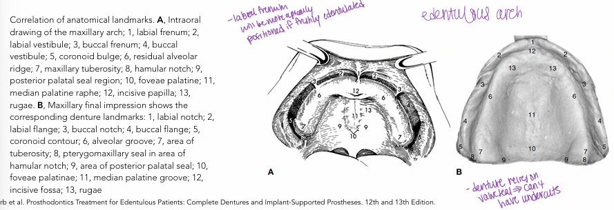
anatomy of supporting maxillary structures
residuala ridge
-the shape and size of the alveolar ridges change when the natural teeth are removed
-resorption following extraction of the teeth is rapid at first, but it continues at a reduced rate throughout life
-crest of the edentulous ridge is an important area of support; bone is subject to resorption, which limits its potential for support unlike the palate (resistant to resorption); ridge crest should be looked on as a secondary supporting area rather than a primary supporting area
incisive foramen
-located beneath incisive papilla
-lies nearer to the crest of the ridge as resorption progresses
-care should be taken that the denture base does not impinge on them
maxillary tuberosity
-enlargements are often fibrous but can be bony
-in case of excess tissue, it can prevent proper location of the occlusal plane and may interfere with the lower denture if it is not surgically removed
combination syndrome
-cascade of edentulism starting with losing teeth on the maxillary
-tend to lose bone in the anterior mandible last
-remaining tissue becomes flabby tissue and will require pre-prosth surgery
-use mucostatic impression (doesn’t pressurize region)
sharp, spiny processes
-frequently there are sharp spiny processes on the maxillary and palatine bones
-will need alveoloplasty
-tissue tend to be very thin on top of processes
torus palatinus
-hard bony enlargement that occurs in the midline of the roof of the mouth and is found in about 20% of the population
-covered by a thin layer of mucous membrane that is easily traumatized by the denture base unless a relief is provided
anatomy of peripheral or limiting structures
-the labial vestibule: runs from one buccal frenum to the other on the labial side of the ridge
-right and left buccal vestibule: extends from the buccal frenum to the hamular notch
-vibrating line: extends from one hamular notch to the other across the palate (have patient say “ah” in short bursts)
labial vestibule
-divided into a left and right labial vestibule by the labial frenum (fold of mucous membrane at the median line)
-labial notch in the labial flange of the denture must be just wide enough and just deep enough to allow the frenum to pass through it without manipulation of the lip
-orbicularis oris forms the outer surface of the labial vestibule
-buccal frenum forms the dividing line between the labial and buccal vestibules
-levator anguli oris attaches beneath the frenum and consequently affects the position of the frenum
-orbicularis oris pulls the frenum forward, buccinator pulls it backward
buccal vestibule
-lies opposite the tuberosity and extends from the buccal frenum to the hamular notch
-size varies with the contraction of the buccinator muscle, position of mandible, and amount of bone lost from maxilla
-size and shape of the distal end of the buccal flange of the denture must be adjusted to the ramus and the coronoid process of the mandible and the masseter muscle
-extent of the buccal vestibule can be deceiving because the coronoid process obscures it when the mouth is opened wide
-mucous membrane of hamular notch consists of thick submucosa made up of loose areolar tissue
-tissue can be safely displaced by the posterior palatal border of the denture to help achieve a posterior palatal seal
vibrating line
-imaginary line drawn across the palate that marks the beginning of motion in the soft palate when an individual says “ah”
-not the same as the junction of hard and soft palate
-not a well-defined line- should be described as an area rather than a line
-distal end of the denture should extend at least to the vibrating line
-in most instances it should end 1-2mm posterior to the vibrating line
-extends from one hamular notch to the other
-at the midline, it usually passes about 2mm in front of the fovea palatinae
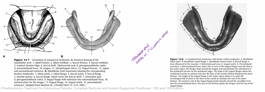
anatomy of supporting mandibular structures
support for mandibular denture
-comes from the body of the mandible
-peripheral seal is provided by the form of the denture’s border as determined by the macroscopic and microscopic anatomy of the limiting structures
-presence of the tongue and its individual size, form, and activity complicate the impression procedure and pt’s ability to manage the denture
-retention of a mandibular denture is constantly threatened by tongue movements
-average available denture-bearing area for an edentulous mandible is 14cm² (edentulous maxilla is 24cm²)
residual ridge
-mucous membrane of the crest of the lower residual ridge capable of providing good soft tissue support for the denture when securely attached to the underlying bone
-crest of the residual ridge may not be favorable as the primary stress-bearing area for a lower denture because underlying bone is often cancellous
buccal shelf
-area between the mandibular buccal frenum and the anterior edge of the masseter muscle
-bound medially by the crest of the residual ridge, laterally by the external oblique ridge, and distally by the retromolar pad
-bone of the buccal shelf covered by a layer of cortical bone and the shelf lies at right angles to the vertical occlusal forces
mylohyoid ridge
-soft tissue usually hides the sharpness of the mylohyoid ridge
-shape and inclination of the ridge vary greatly among edentulous patients
-anteriorly, mylohyoid ridge with its attached mylohyoid muscle lies close to the inferior border of the mandible
-posteriorly, often lies flush with the superior surface of the residual ridge after resorption
mental foramen
-as resorption takes place, mental foramina will come to lie closer to the crest of the residual ridge
-mental nerves and blood vessels may be compressed by the denture base unless relief is provided
genial tubercles
-usually lie away from the crest of the ridge
-with resorption, genial tubercles become increasingly prominent
-not a problem until at level of basal bone
torus mandibularis
-bony prominence usually found bilaterally and lingually near the first and second premolars midway between the soft tissues of the floor of the mouth and the crest of the alveolar process
-covered by extremely thin layer of mucous membrane
-often needs to be removed surgically because it can be difficult to provide relief within the denture for the torus without breaking the border seal
labial vestibule
-runs from labial frenum to buccal frenum
-length and thickness varies with the amount of tissue that has been lost
-extent of denture flange in this area often limited because of the muscles that are inserted close to the crest of the ridge
-mentalis muscle = particularly active muscle in this region
-depth of the flange will be determined by the turn of the mucolabial fold, which is the line of flexure of the mucous membrane as it passes from the mandible to the lip
-when pt’s mouth opens wide, the orbicularis oris muscle becomes stretched which narrows the sulcus → would displace the mandibular denture if the flange was unnecessarily thick
-mandibular dentures and impressions will always be narrowest in the anterior labial region
buccal vestibule
-buccal flange (starts immediately posterior to the buccal frenum) swings wide into the cheek and is nearly at right angles to the biting force
-extent of the buccal vestibule influenced by the buccinator muscle, which extends from the modiolus anteriorly to the pterygomandibular raphe posteriorly and has its lower fibers attached to the buccal shelf and the external oblique ridge
-external oblique ridge does not govern the extension of the buccal flange because the resistance encountered varies widely
-buccal flange may extend to the external oblique ridge, up onto it, or over it depending on the location of the mucobuccal fold
-denture should cover completely the buccal shelf (despite the fact that it will rest directly on the fibers of the buccinator muscle)
-distobuccal border must converge rapidly to avoid displacement by the contracting masseter muscle, whose anterior fibers run outside and behind the buccinator muscle in this region
distal extension
-limited by the ramus of the mandible, buccinator muscle fibers that cross from the buccal to the lingual side as they attach to the pterygomandibular raphe and the superior constrictor muscle, and the sharpness of the lateral bony boundaries of the retromolar fossa (formed by a continuation of the internal and external oblique ridges ascending the ramus)
retromolar pad
-triangular soft pad of tissue at the distal end of the lower ridge
-mucosa is composed of a thin, non-keratinized epithelium
-submucosa contains glandular tissue, fibers of the buccinator and superior constrictor muscles, pterygomandibular raphe, terminal part of the tendon of the temporalis muscle, and loose alveolar tissue
-denture base should extend approximately 1/2-2/3 over the retromolar pad
mylohyoid muscle
-forms floor of mouth
-arises from the whole length of the mylohyoid ridge
-ridge is sharp and distinct in the molar region and becomes almost indiscernible anteriorly
-medially, fibers join those from the mylohyoid muscle of the opposite side
-posteriorly, fibers continue to hyoid base
-extension of the lingual flange under the mylohyoid ridge cannot be tolerated in function because it will interfere with the action of the mylohyoid muscle when it contracts & this will displace the denture
-in posterior region, lingual flange can go beyond the mylohyoid muscle’s attachment to the mandible because the mucolingual fold is not in this area
-impression may depart from the stress-bearing area of the lingual surface of the ridge, moving away from the body of the mandible to be suspended under the tongue in soft tissue on both sides of the mouth, thereby reaching the mucolingual fold of soft tissue for a boarder seal
retromylohyoid fossa
-lingual flange moves into this fossa, ceasing to be influenced by the action of the mylohyoid muscle and can move back toward the body of the mandible producing the typical S curve of the lingual flange
sublingual gland region
-in the premolar region, sublingual gland rests above the mylohyoid muscle
-when floor of the mouth is raised, gland comes quite close to the crest of the ridge and reduces the vertical space available for the extension of the flange in the anterior part of the mouth
anatomy of peripheral or limiting structures- anterior region
-extends from lingual frenum back to where the mylohyoid ridge curves above the level of the sulcus
-premylohyoid fossa can be palpated here and premylohyoid eminence can be seen on impressions
-lingual border of the impression in this anterior region should extend down to make contact with the mucous membrane floor of the mouth when the tip of the tongue touches the upper incisors
-lingual flange will be shorter anteriorly than posteriorly
-at premylohyoid fossa, flange becomes larger as it extends below the level of the mylohyoid ridge
anatomy of peripheral or limiting structures- middle region
-extends from premylohyoid fossa to the distal end of the mylohyoid ridge, curving medially from the body of the mandible
-curvature caused by the prominence of the mylohyoid ridge and the action of the mylohyoid muscle
-when the middle of the lingual flange is made to slope toward the tongue, it can extend below the level of the mylohyoid ridge
-tongue rests on top of the flange and aids in stabilizing the lower denture on the residual ridge
-seal of lower denture maintained during these movements because the lingual flange remains in contact with the mucolingual fold in the alveololingual sulcus
-in this area, flange rests not on mucous membrane in contact with bone but on soft tissue; when mylohyoid muscle is relaxed, there is space between the flange and the floor of the mouth, but contact is reestablished when the floor of the mouth is raised
anatomy of peripheral or limiting structures- posterior region of peripheral
-flange passes into the retromylohyoid fossa
-as it does, it no longer is influenced by the action of the mylohyoid muscle & the flange can turn laterally toward the ramus to fill the fossa and complete the typical S form of the correctly shaped lingual flange
objectives of an impression
-provide support, retention, and stability for the denture
impression requirements
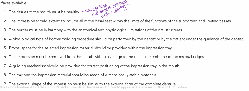
mucostatic v. mucodisplacing impression technique
-mucostatic: intended to record the shape of the tissues with a minimum of displacement
-mucodisplacing: intended to displace the border tissues to a predetermined extent
most important part of the impression-making process
-tray
-properly formed tray enables the dentist to carry the impression material to the mouth and control it without distorting the soft tissue that surrounds it
-large: distort the tissues around the borders of the impression and will pull the soft tissues under the impression away from the bone, distorting the dimensions of the sulcus in the process
-small: border tissue will collapse inward onto the residual ridge, which will distort the accurate recording of the border extension of the denture and prevent the proper support of the lips by the denture flange
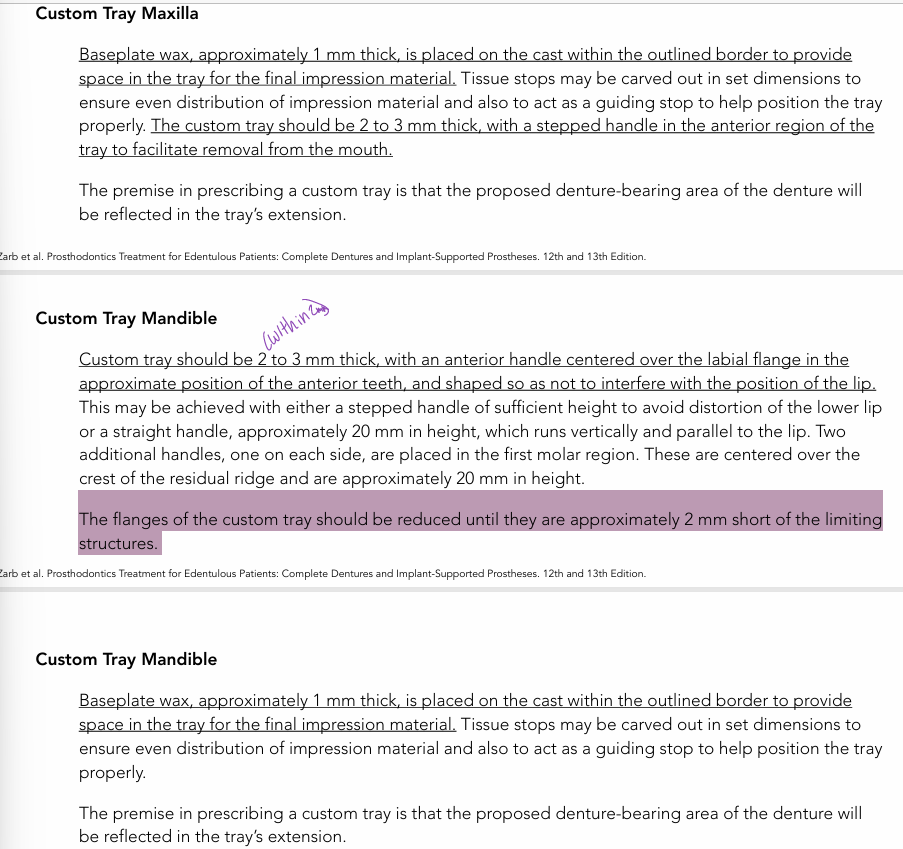
custom trays
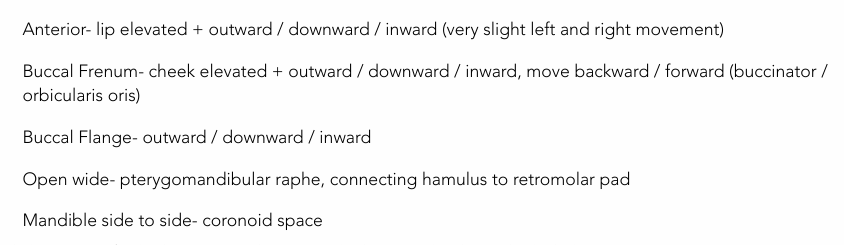
border molding maxilla
maxillary impression process
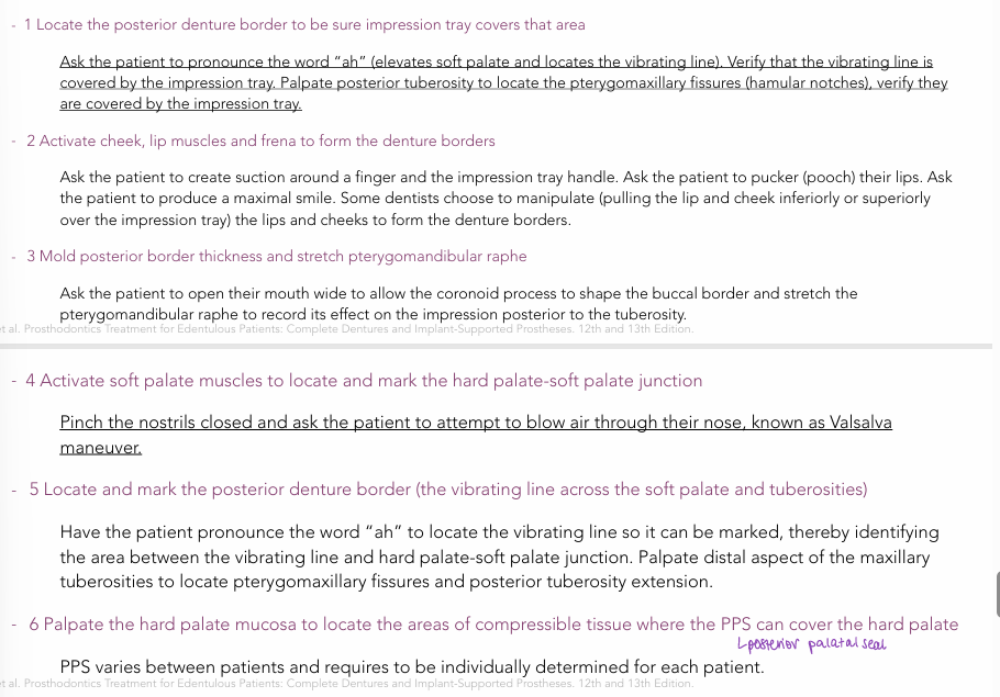
making final impression- maxilla
-align labial frenum
-push tray apically applying pressure near M1 without displacing anterior (in area of labial frenum)
-keep pushing down until hamular notches “lock in”
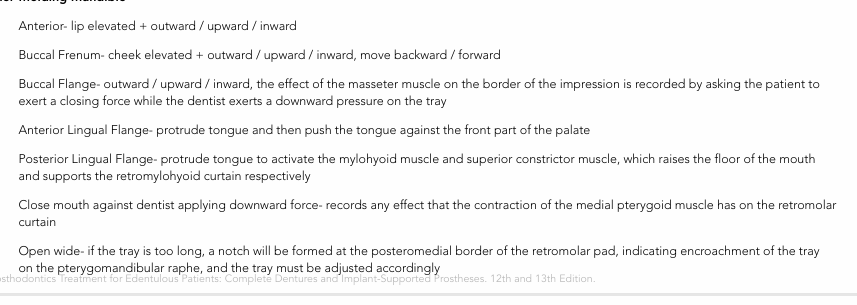
mandible border molding
mandible impression process
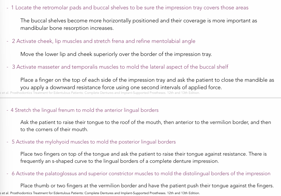
making final impression- mandible
-tray is rotated into the mouth in the horizontal plane with the anterior handle until it is over the residual ridge
-at this time, the patient is asked to raise the tongue slightly and the tray is moved downward towards its final position
-dentist’s index fingers of each hand are placed on top of the posterior handles and (with alternating gentle pressure) the tray is seated until the buccal flanges come into contact with the mucosa covering the buccal shelf
areas requiring relief in impression
-secondary stress-bearing areas
-palatal torus
-median palatine raphe
-mandibular tori
-retromylohyoid ridge
-undercuts of sharp bony prominence on ridges
maxillary and mandibular stress-bearing areas
-maxillary: primary firm tuberosities, primary hard palate on either side of palatal raphe, secondary alveolar ridge, secondary rugae
-mandibular: primary buccal shelves, primary retromolar pads, secondary alveolar ridge