Lab Practical
1/74
There's no tags or description
Looks like no tags are added yet.
Name | Mastery | Learn | Test | Matching | Spaced | Call with Kai |
|---|
No analytics yet
Send a link to your students to track their progress
75 Terms
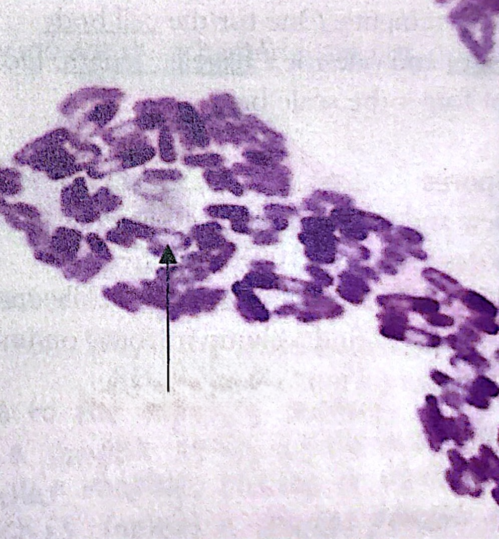
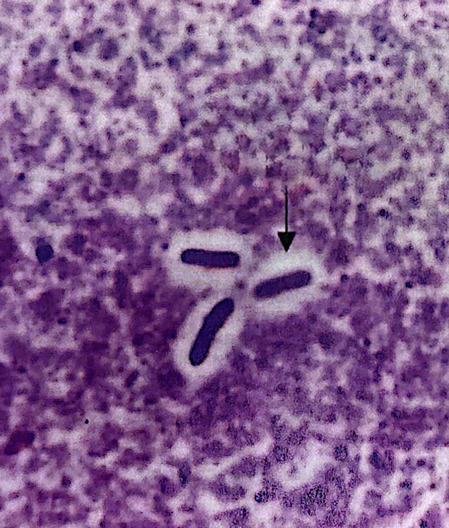
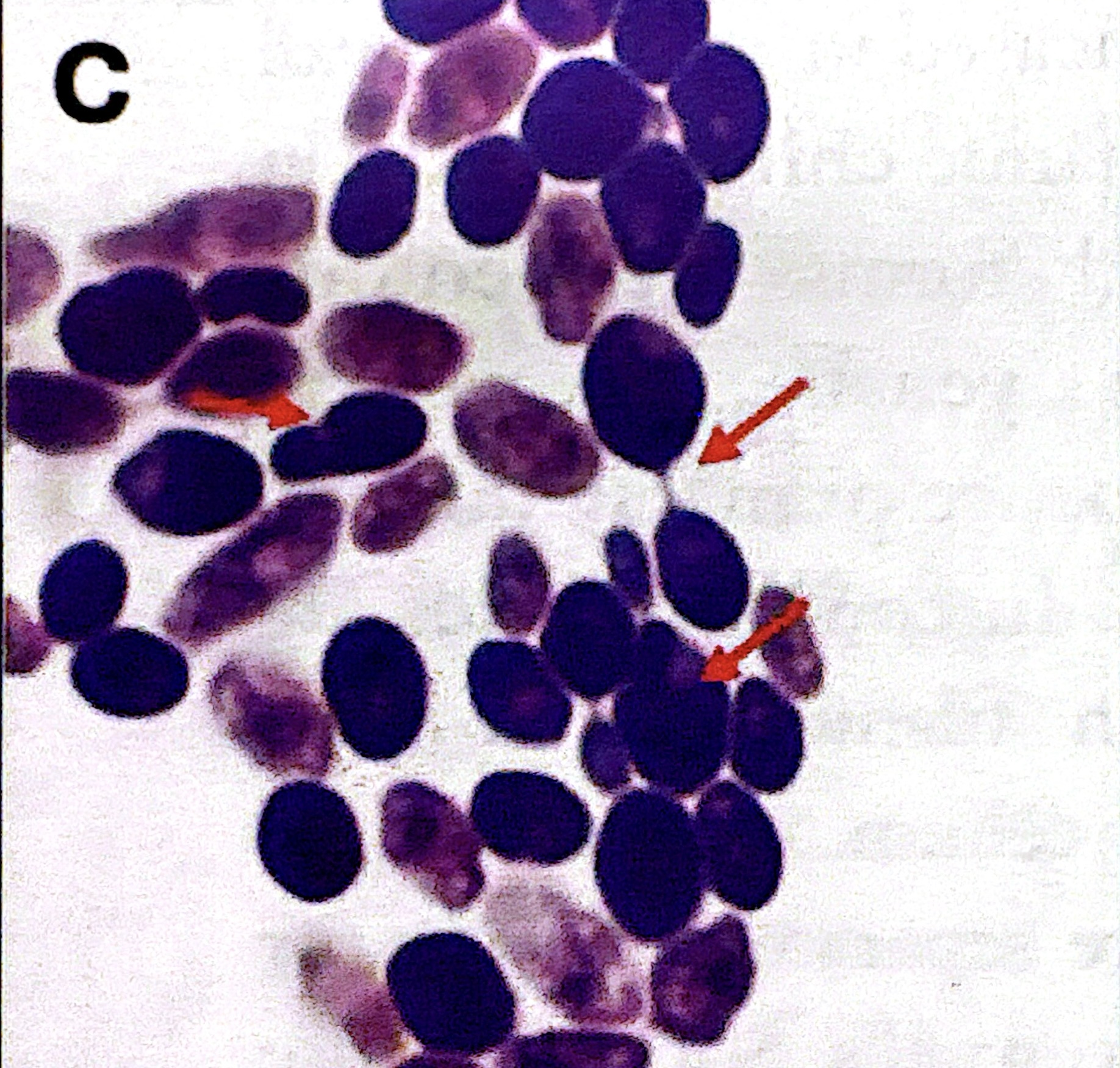
Gram positive
Purple gram stain
Gram Negative
Pink gram stain
Cocci
sphere cell morphology
Bacilli
rod cell morphology
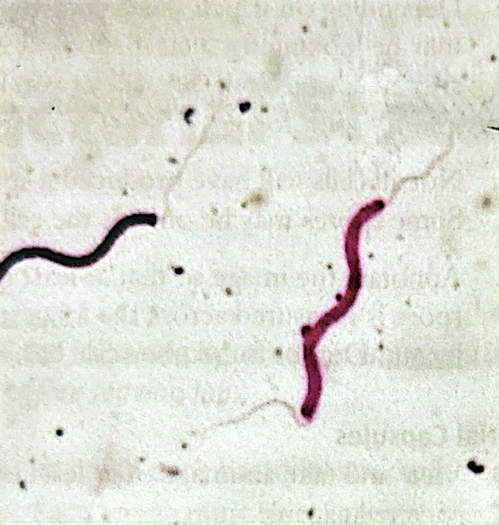
Spirilla
squiggle cell morphology
Vibrios
boomerang cell morphology
Corkscrew
spiral cell morphology
Endospore
a dormant extremely heat-resistant dehydrated structure
Resistant to chemicals and thus difficult to stain, as they are dehydrated and water-soluble dyes won't penetrate it
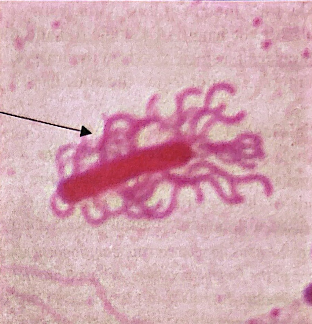
Capsule
Distinct, firm, and sticky polysaccharide layer
easiest to find towards the edge of the sample where the stain is the thickest
Flagella
specialized structures/appendages that many bacteria use for movement
Monotrichous
Single flagellus
Amphitrichous
has flagella at both ends

Lophotrichous
tuft of flagella at one of both ends
Peritrichous
has flagella surrounding the cell
Non-Motile
MTM has red precipitate with a small area beyond the stab line
Motile
MTM has red precipitate in a widespread area beyond the stab line
Motile
MTM has cloudiness spreading from the stab line
(# of colonies * total dilution factor)/ volume of culture plated (mL)
CFU/mL formula
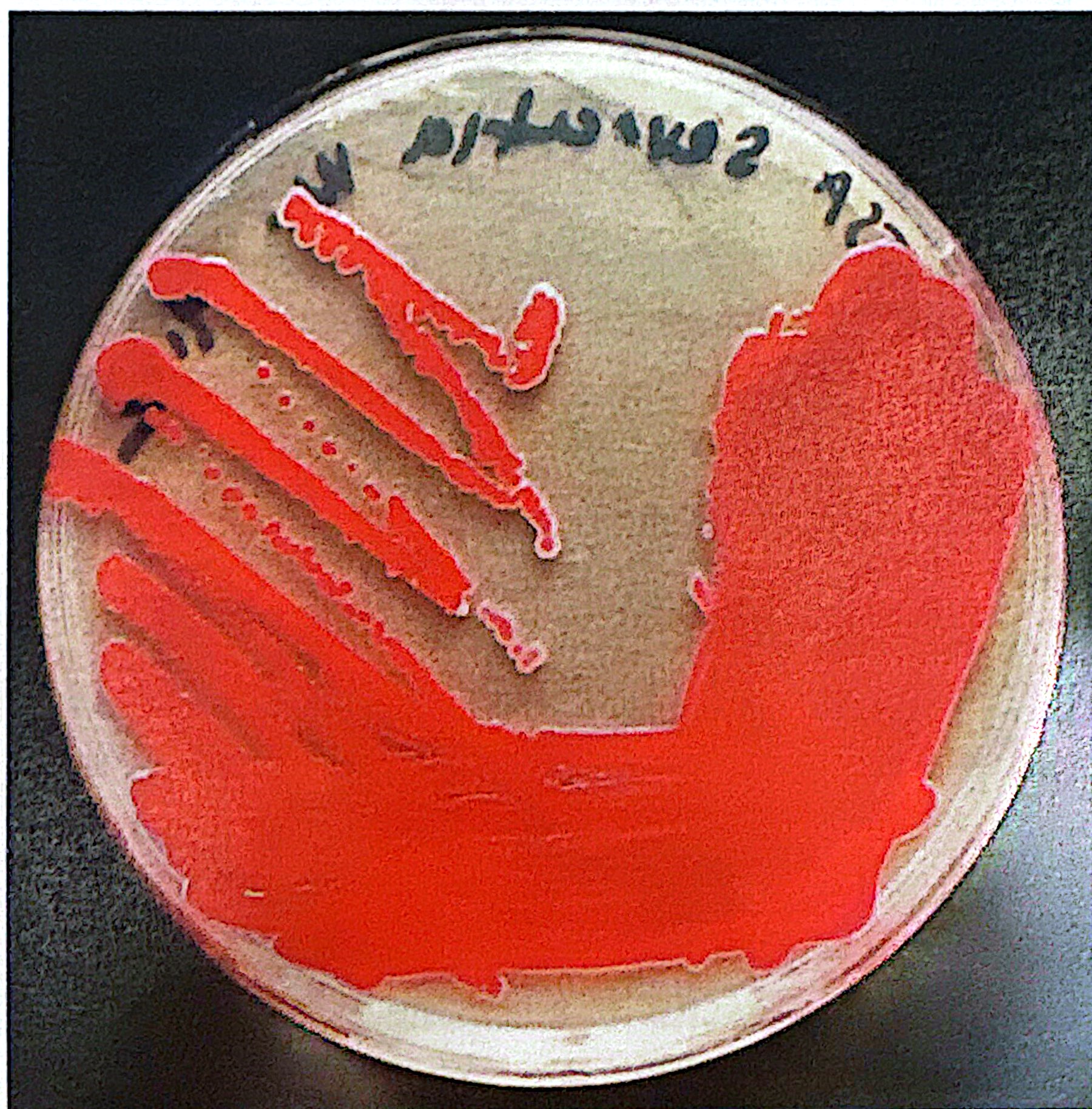
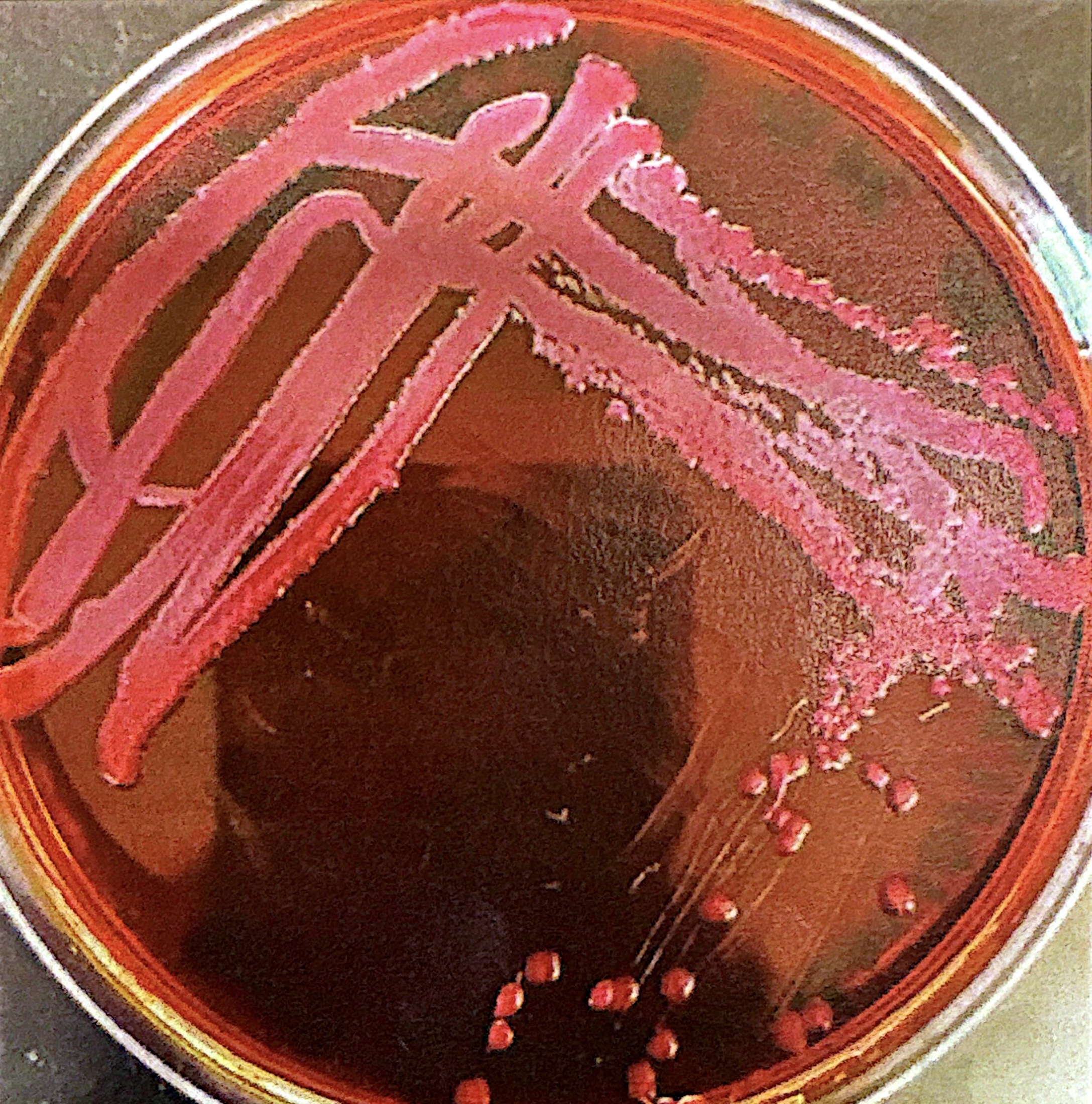
Prodigiosin, Serratia marcescens
Red pigment on solid media
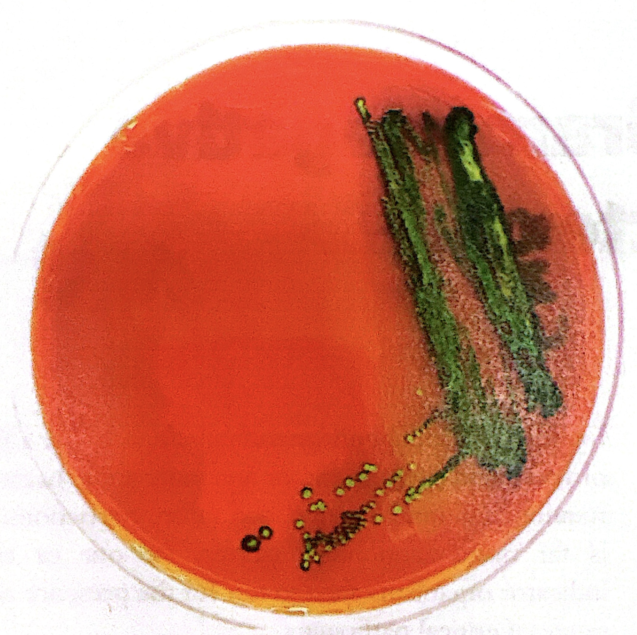
Pyocyanin, P. aeruginosa, water soluble
Blue-green pigment. Red in pH < 5.
Water Soluble
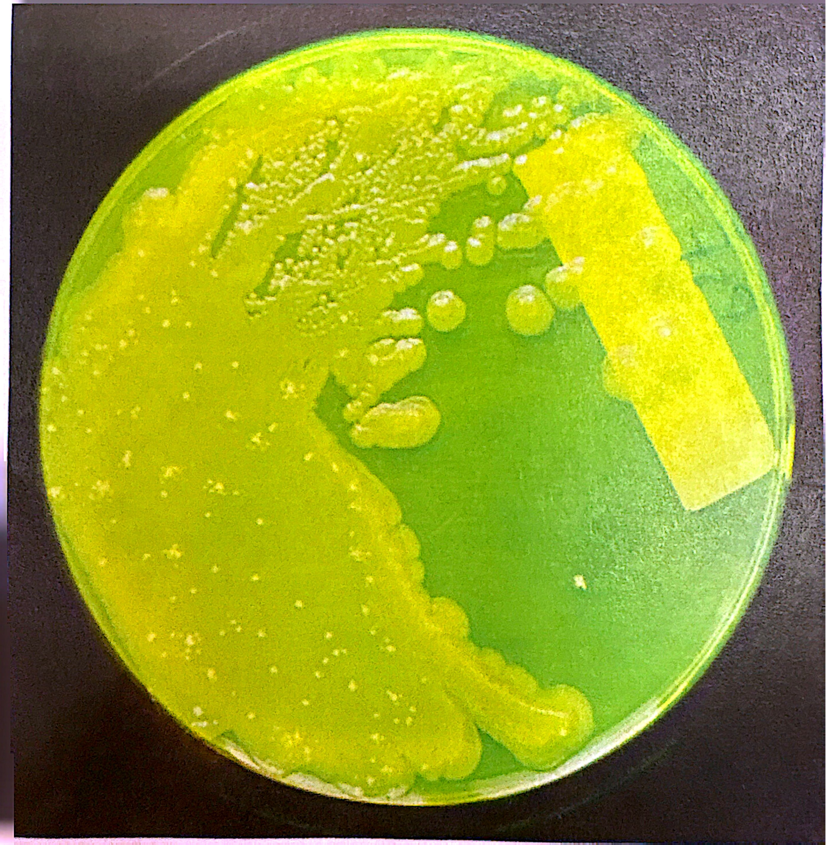
The pigment has spread into the water-based agar. Can be blue, green, or green-yellow pigments
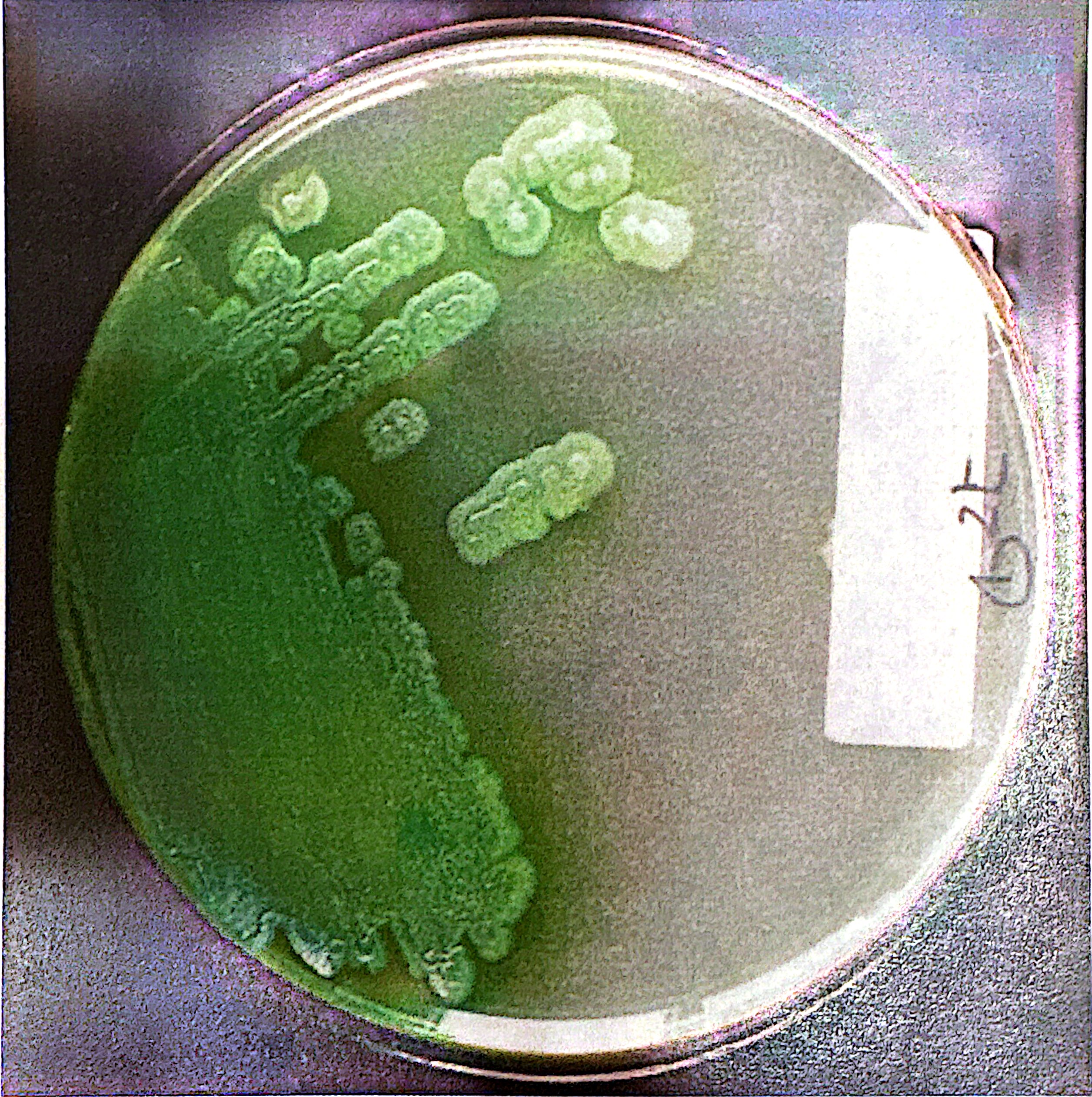
Pyoverdine/Fluorescein, Pseudomonas aeruginosa and P. fluoresceus, water soluble
Yellow-green pigment
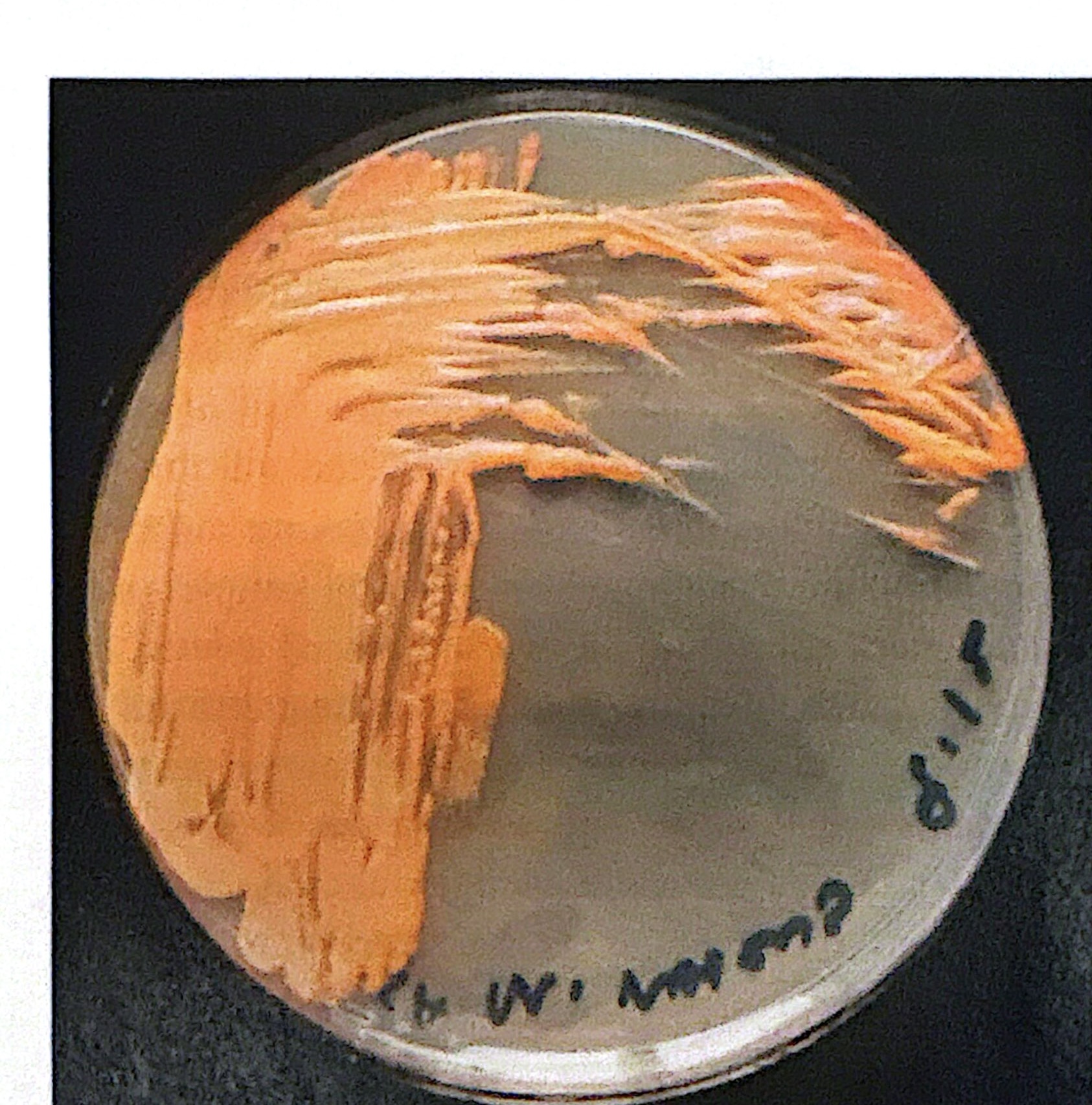
Canthaxantin/Cartenoid, Micrococcus roseus
Red-orange pigment. specific example produces pink colonies
Violacein, Chromobacterium violaceum
Purple pigment
Beta hemolysis
zone of clearing around the colony in SBA. Staph aureus and group A and B strep
Gamma hemolysis
no zone of clearing around colonies in SBA
Alpha hemolysis
produced by strep when the bacteria produce substances that oxidize the hemoglobin in red blood cells, turning the agar to a greenish brown.
Possessed by S. pneumoniae
staphylococci
If it grows on MSA, it is
Mannitol fermenter
yellow growth on MSA.
Non-Mannitol fermenter
No growth on MSA. red
Group A Strep
No growth in presence of bacitracin
Group B strep
Growth in presence of bacitracin
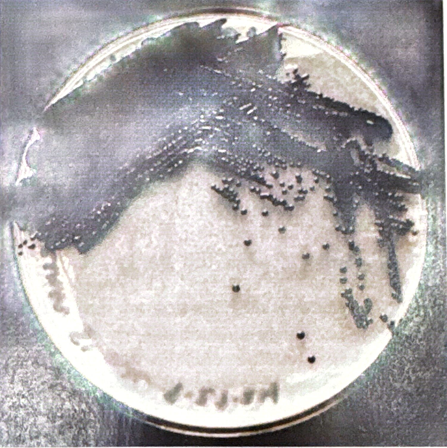
Gram negative, non-lactose fermenter
colorless or light lavender colonies on EMB
Gram negative, lactose fermenter
dark/dark purple colonies on EMB
Gram positive
No growth on EMB
E. Coli, lactose fermenter
green metallic sheened colonies on EMB
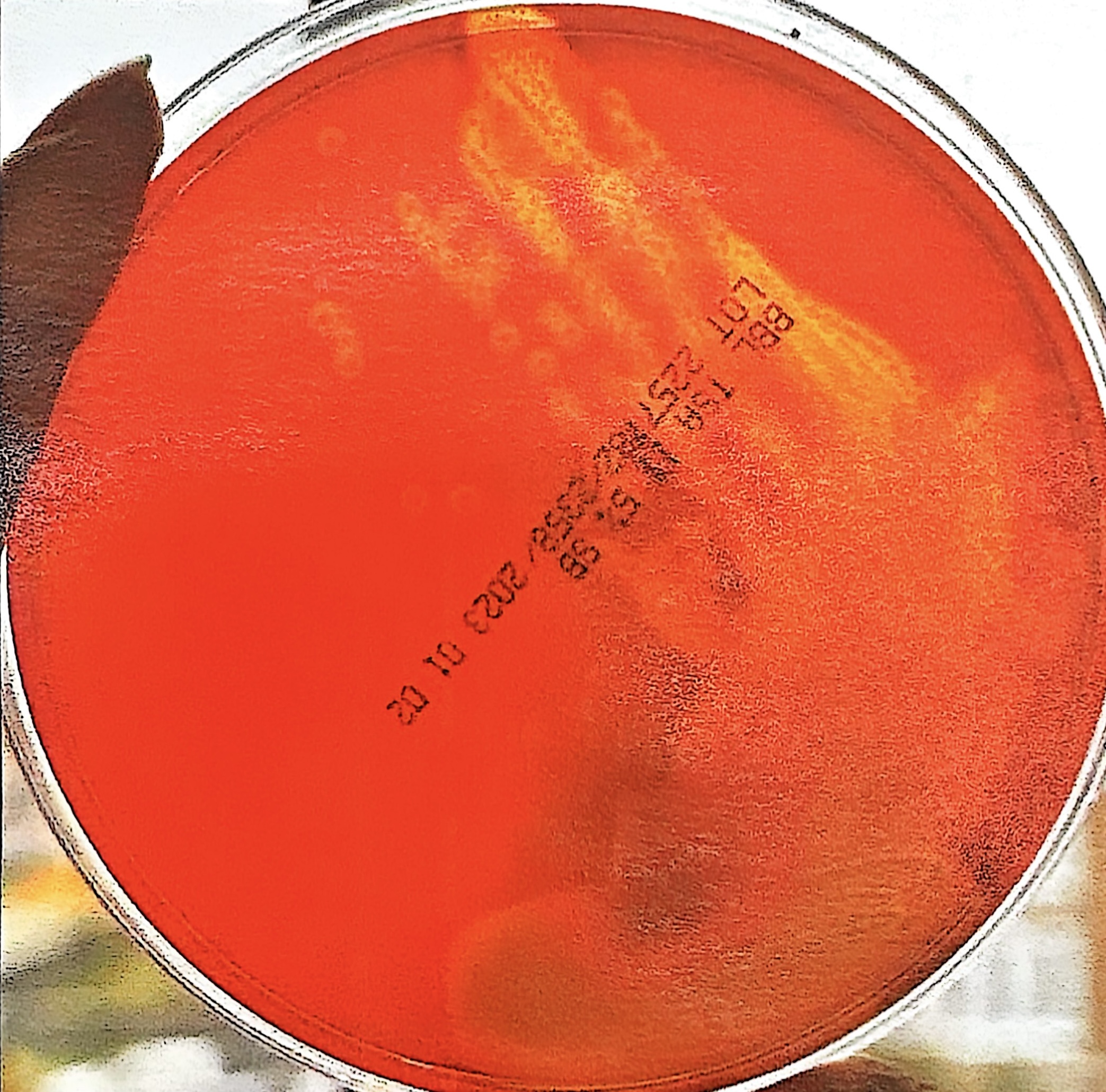
gram negative, lactose fermenter
red agar around colonies on MAC
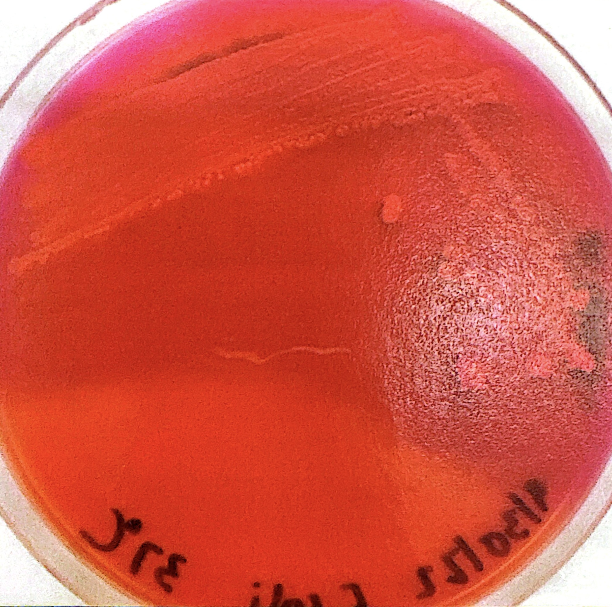
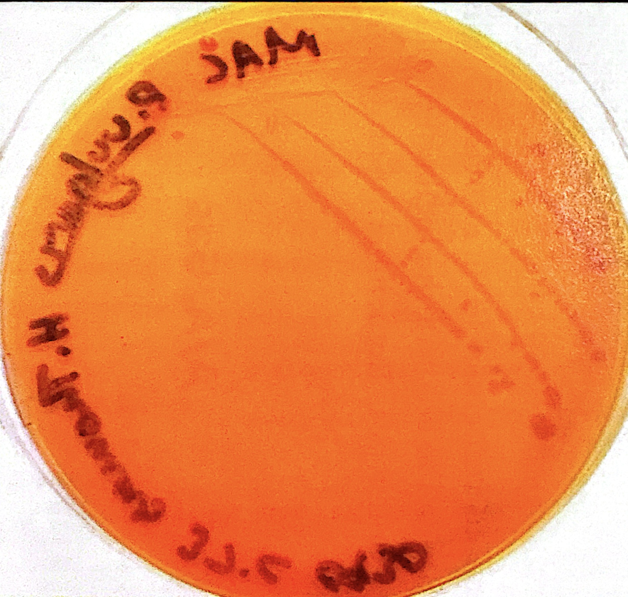
gram negative, non-lactose fermenter
orange MAC agar
Citrate only carbon source
bright blue Simmon’s Citrate Agar
Citrate not only carbon source
green Simmon’s Citrate Agar
positive
pink Urea Broth
negative
yellow or peach colored urea broth
Glucose only fermenter
Red Slant Yellow Butt, TSIA
Glucose and Sucrose and/or Lactose fermenter
Yellow Slant Yellow Butt, TSIA
gas
TSIA agar lifted
hydrogen sulfate
Black precipitate butt, TSIA
EMB
positive lactose fermentors produce acid, which results in methylene blue + eosin Y dye complexes that form dark/dark purple colonies
E. Coli
Ferments lactose/produces acid quickly on EMB. The acid reacts with the media dyes and produces dark colonies with green metallic sheen.
MAC
Neutral red indicator reddens with agar around colonies in presence of acid from lactose fermentors. Non-lactose fermenters turn agar more orange.
Simmon’s Citrate Agar
bromothymol blue green at neutral pH but becomes blue as pH increases due to citrase metabolizing citrate, which forms alkaline product sodium carbonate.
Urea Broth
differentiates presence of Urease, which hydrolyzes urea into ammonia and carbon dioxide. phenol red indicator turns pink as ammonia increases the pH.
Phenol red
in TSIA slants and MSA. turns yellow when pH decreases (acid from bacteria fermenting carbohydrates). Positive is pink in urea brother as pH increases with ammonium production
Red, Yellow
Slant/Butt TSIA color that indicates an organism can only ferment glucose.
As the small concentration of glucose is fermented, organisms moves on to break down peptones aerobically releasing alkaline byproducts and turning the slant red
Anaerobic conditions in butt means glucose is not oxidizes, so as glucose fermentation acid builds up it turns butt yellow
Yellow, Yellow
Slant/Butt TSIA color that indicates an organism can ferment glucose and either lactose or sucrose or both.
Slant is yellow because lactose and sucrose are at 10x the concentration of glucose, meaning more production of acid.
After 24 hours the sugars may deplete and bacteria moves to peptones in the slant which produces basic ammonia turning slant the other color.
gas
bacteria that use thiosulfate as a terminal electron acceptor produce hydrogen gas which lifts agar up out of the tube. CO2 may also be produced through fermentation that may be visible as agar bubbles/cracks
Hydrogen Sulfate
Produced through bacteria’s reduction of thiosulfate. Combines with iron that produces a black precipitate visible in a TSIA butt. Requires an acidic environment, so it’s production assumes it’s covering a yellow/acidic butt.
positive
Yellow carbon fermentation test
negative
Red carbon fermentation test
gas
bubble in the inverted tube (Durham tube) in carbon fermentation test
Aspergillus. V, M, P, C
reproduces with asexual conidiophores that produce conidia.
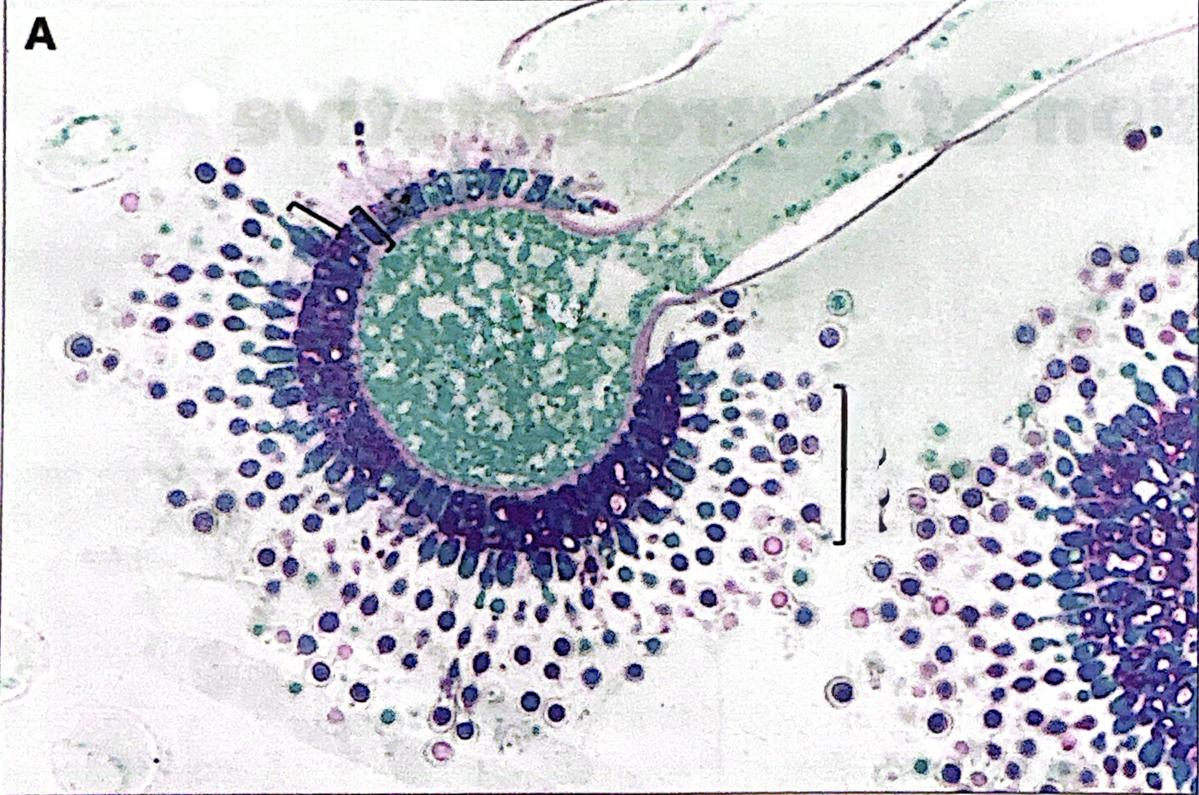
Vesicle, V
Bulbous head containing conidia-producing cells in Aspergillus. Either uniseriate (one) or biseriate (two) layers of conidia-producing cells
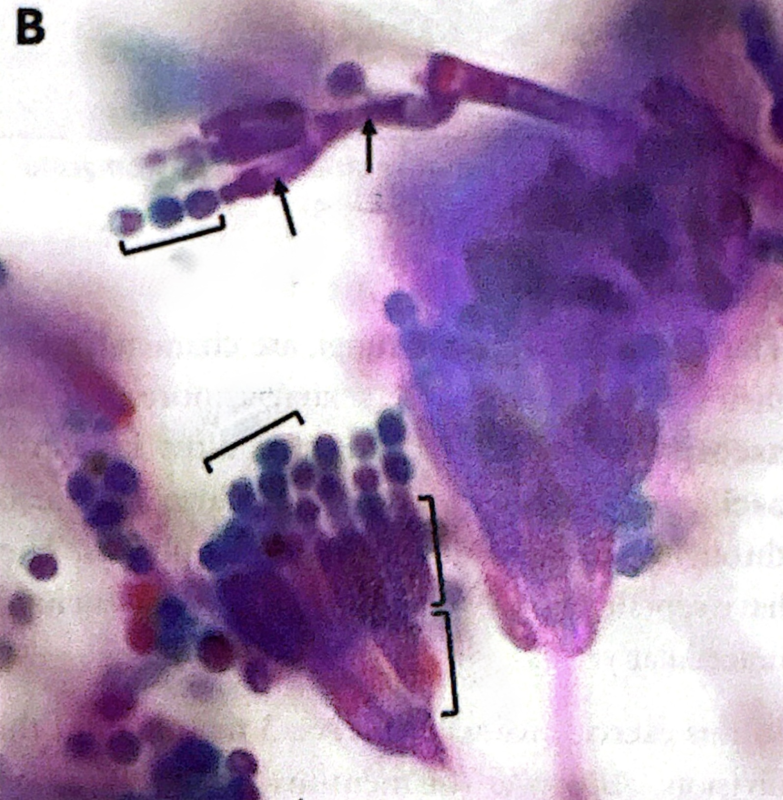
Phialides
flask-shaped cells that arise from metulae. Structure in Aspergillus and Penicillium
Metulae, M
where phialides arise from, surround the vesicle. Structure in Aspergillus and Penicillium
Conidia, C
non-motile spores. Circular and structure in Aspergillus and Penicillium
Pencillum. M, P, C
blue-green molds that undergo asexual reproduction through branched conidiophores
Saccharomyces cerevisiae
budding yeast. Oval shaped and reproduce through asexual budding (“budding” yeast)
Rhizopus stolonifera. S, C, Sp, Sm
Reproduces asexually with sporangiospore contained within sporangia formation
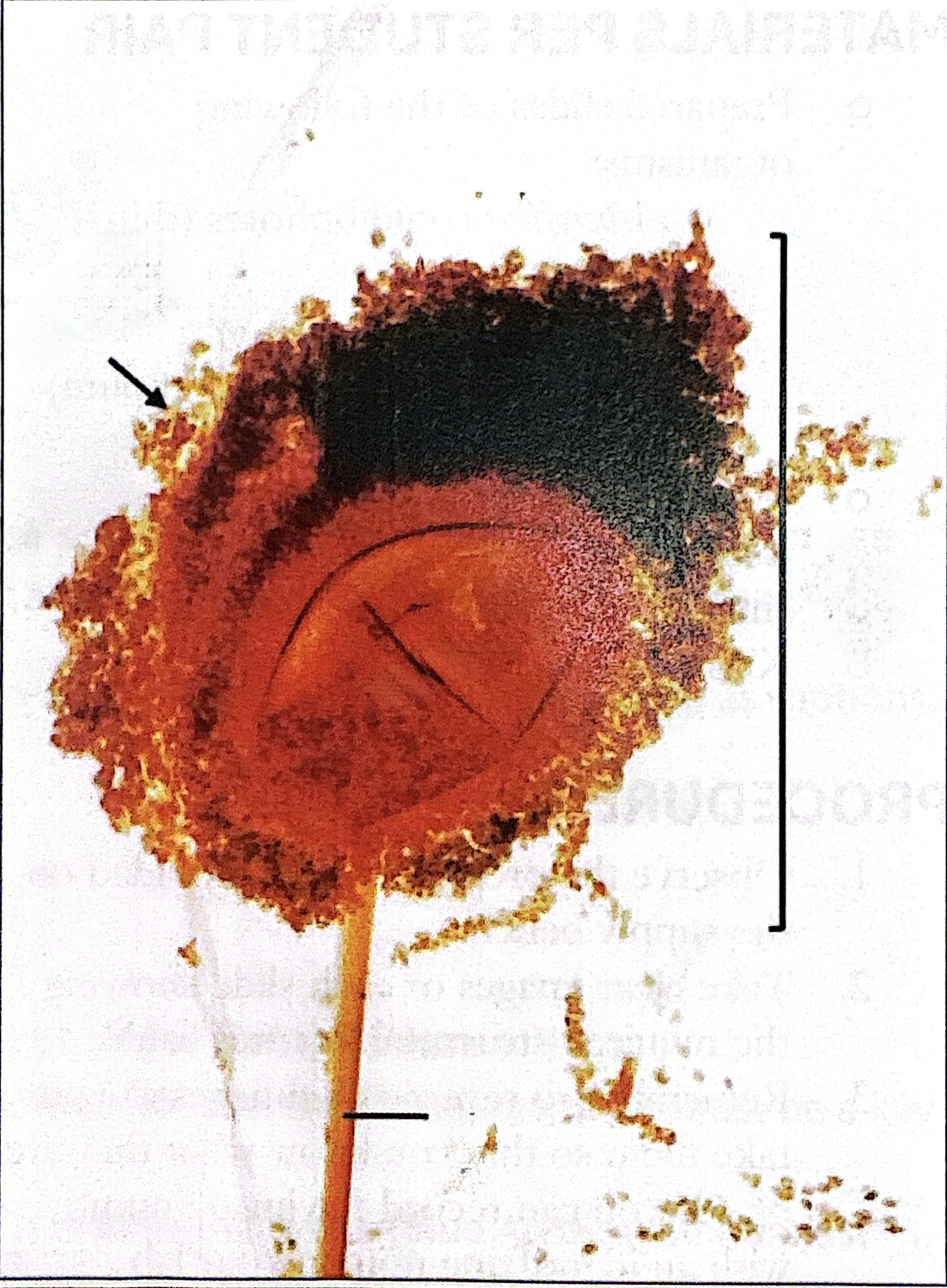
Sporangiophore. S
aerial hyphae that emerge near the rhizoids.(the branched line) in Rhizopus.
Sporangium. Sm
contains the columella in Rhizopus.
Sporangiospore, Sp
surround the columella and capped by sporangium in Rhizopus
Columella, C
dome-shaped structure within each sporangium which supports spore development in Rhizopus
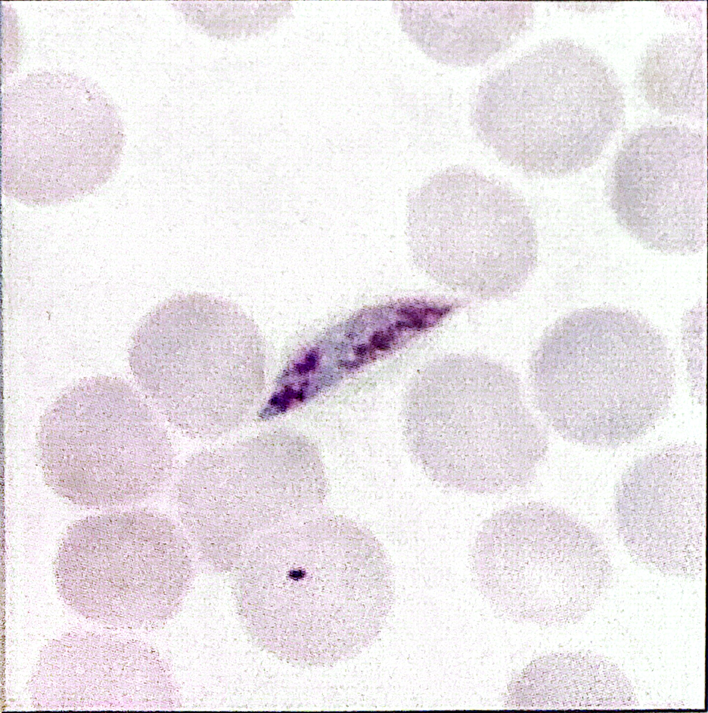
Ring stage
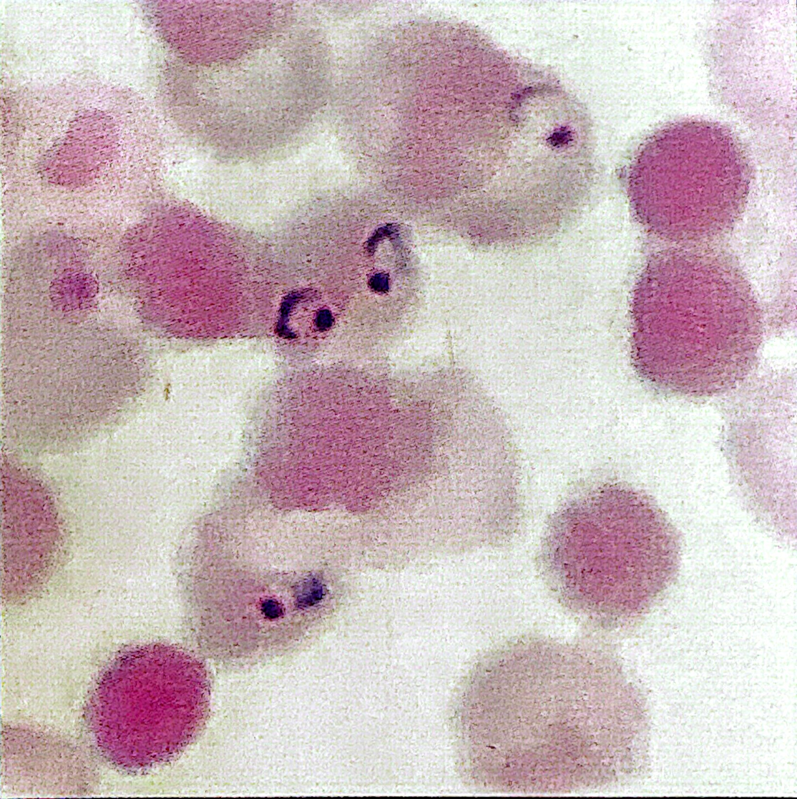
Mature Trophozoite
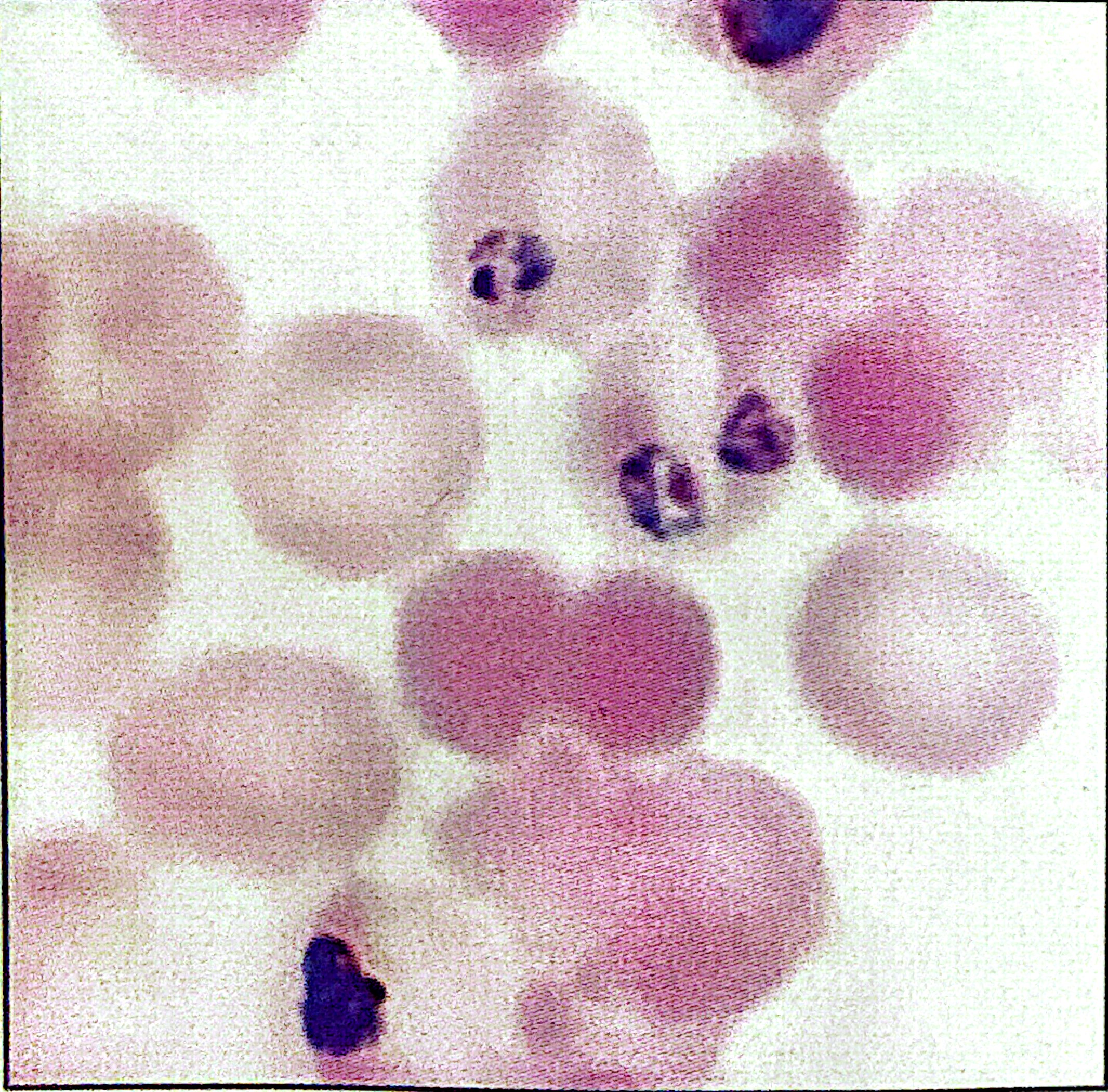
Schizont

Gametocyte