Vascular Malformations and Retinal Telangiectasia
1/32
There's no tags or description
Looks like no tags are added yet.
Name | Mastery | Learn | Test | Matching | Spaced | Call with Kai |
|---|
No analytics yet
Send a link to your students to track their progress
33 Terms
Retinal telangiectasia
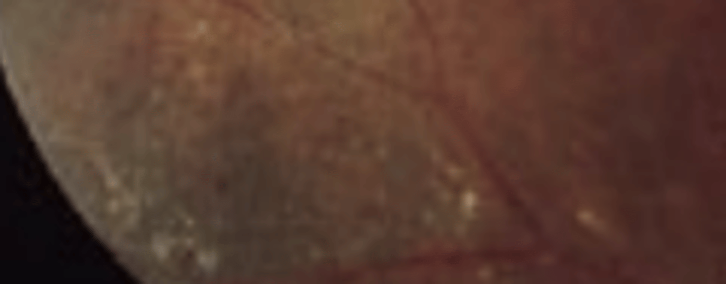
vascular abnormalities of the retina (can occur anywhere on the body) characterized by a loss of endothelial cells and pericytes resulting in the thickening of the capillary wall and formation of multiple aneurysms. Compromise of the blood retinal barrier results in exudation into the intraretinal or subretinal space.
smaller vessels
Vascular dilation and tortuosity seen in retinal telangiectasias is distinguished from other vascular pathologies such as diabetic retinopathy as it occurs in...
Secondary
retinal telangiectasia occurring secondary to systemic conditions such as BRVO or diabetic retinopathy**. Other conditions include irradiation, sickle cell disease, ROP, and eales.
hyperfluorescence, leakage
Retinal telangiectasias will present on fluorescein angiography with early ____ and late _____
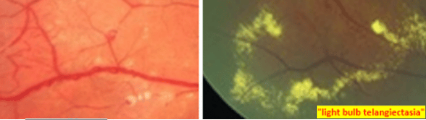
Idiopathic macular telangiectasia, MacTel)
a heterogenous group of conditions characterized by telangiectatic alterations of the juxtafoveolar capillary network in one or both eyes differing in appearance, pathogenesis, and management. Are thought to be a groupd of primary neurovascular degenerative disease which lead to a loss fo photoreceptors.
temporal
Changes to vessels seen in macular telangiectasias occurs ____ to the fovea
Group I (Aneurysmal)
congenital group of IJFTs that are unilateral with easily visible telangiectasias occurring primarily in males 40yo and causing visual loss due to macular edema. Degree of vision loss is variable.
Group IA
subgroup of group I IJFTs that affects more of the mid-peripheral fundus. Is proposed to be a possible sub-form of Coat's disease.
Group IB
subgroup of group I IJFTs that causes focal juxtafoveal telangiectasis confined to 2 clock hours. Has much better visual acuity outcomes.
CME, Laser photocoagulation, intravitreal steroids and anti-VEGF
The treatment of group I IJFTs is an attempt to prevent vision loss caused by ____. _____ is the mainstay treatment, but _____ may be employed as well.
Group II (Perifoveal)
group of IJFTs that are bilateral with more subtle telangiectasias occurring primarily in middle aged (>50 yo) patients and causing vision loss due to retinal atrophy. CNVM is common. Is associated with systemic disease (DM, HTN, CVD).
tamoxifen, crystalline deposits, cystic spaces, temporal
Both macular telangiectasia type II and ____ toxicity will present with ____ and _____ in the macular area. Determine if it is macular telangiectasia by the presence of telangiectatic vessels. Additionally, changes due to telangiectasia will occur ____ to the macula
Group IIA
most common acquired subgroup of group II IJFTs having gold crystalline deposits adjacent to retinal atrophy occurring temporal to the fovea. Risk of CNVM and must be determine proliferative or non-proliferative. Treatment for proliferative is primarily anti-VEGF therapy. No treatment for non-proliferative.
Group IIB
subgroup of group II IJFTs that is similar to group IIA, but without refractile deposits and pigmented plaques.
Group III (Occlusive)
group of IJFTs that are very rare. Is characterized by progression destruction of the perifoveal capillary network usually in association with medical or neurological disease.
Group IIIA
subgroup of group III IJFTs that is the most severe form usually associated with systemic disease such as polycythemia, multiple myeloma, and chronic lymphatic leukemia. Presents in patients 60 yo as progressive loss of central vision due to occlusion of parafoveal capillaries. Prognosis is poor with no treatment.
Group IIIB
subgroup of group III IJFTs that is the same as group IIIA, but with neurological disease. Presents with deep tendon reflex hyperactivity and other CNS symptoms.
Coat's disease
a rare idiopathic unilateral retinal telangiectasia most commonly developing in young males (earlier presentation worse prognosis). Occurs with periods of exacerbations, but overall is progressive with each episode. Is a loss of blood retinal barrier that commonly results in exudation and subsequent retinal detachment. Neovascularization is also common. Patient presents with unilateral vision loss, strabismus, and leukocoria. Refer to retina for neovascularization, macular edema, retinal detachment, and neovascular glaucoma.
Leber's miliary aneurysms
a mild variant of Coat's disease, having minimal exudation.
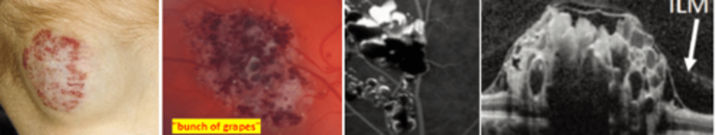
Cavernous hemangioma
rare benign tumors of the inner retina or surface of the ONH which are congenital (autosomal dominant) and unilateral. Is associated with systemic/CNS disorders such as cranial nerve palsies and epilepsy. Breaks in the ILM can result in epiretinal membrane formation. Hemorrhage may rarely occur.
vitreous hemorrhage and epiretinal membrane, MRI/CT
Complications of cavernous hemangioma are rare but include ____(2). Patient should also have a ____ imaging and a consult to evaluation the central nervous system.
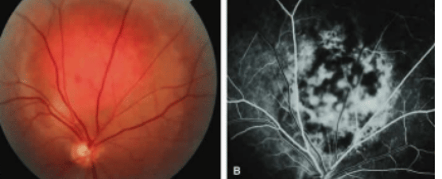
Capillary hemangioblastoma (retinal hemangioma)
rare vascular tumors of the retina or ONH seen within 2 decades of life. Are sight threatening as these vessels tend to leak. Is associated with Von Hippel Lindau syndrome especially when multiple lesions are present. Risk of macular edema and retinal detachment (exudates) as well as neovascularization (ischemia). Refer for systemic work-up and treatment of retinal complications.
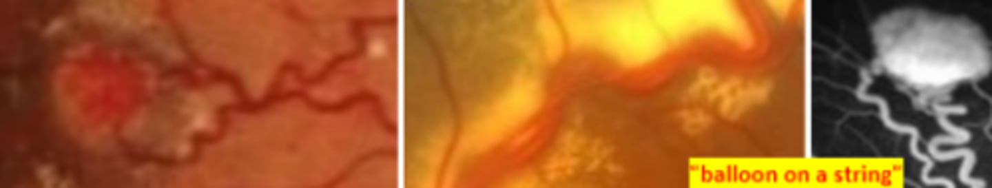
steal phenomenon
The ____ explains that capillary hemangioblastomas cause retinal ischemia as they rely on blood supply from the retina.
Von Hipple Lindau disease
phacomatosis occurring due to an autosomal dominant mutation in chromosome 3. Involves capillary hemangioma formation in the central nervous system and is associated with renal and pancreatic carcinoma. Ocular and genetic testing indicated.
chromosome 3
von hippel lindau disease is associated with a mutation of which chromosome?
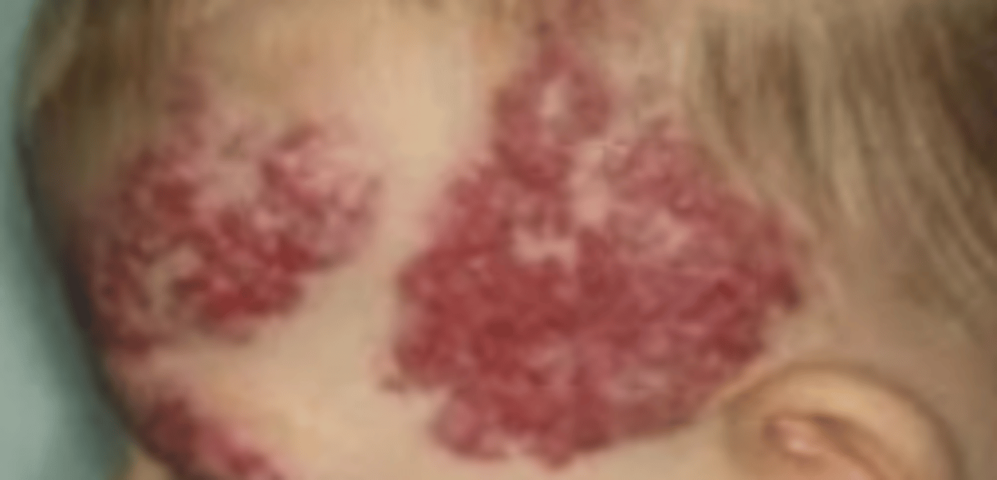
Racemose hemangioma
a congenital advanced type of arteriovenous malformation resulting in a direct communication between arteries and veins with no capillary beds occurring unilaterally. Can be independent or occur alongside Wyburn Mason syndrome. Vessel occlusion may occur due to compromise of blood flow. Effect on vision is variable. Refer for systemic and neuro consult.
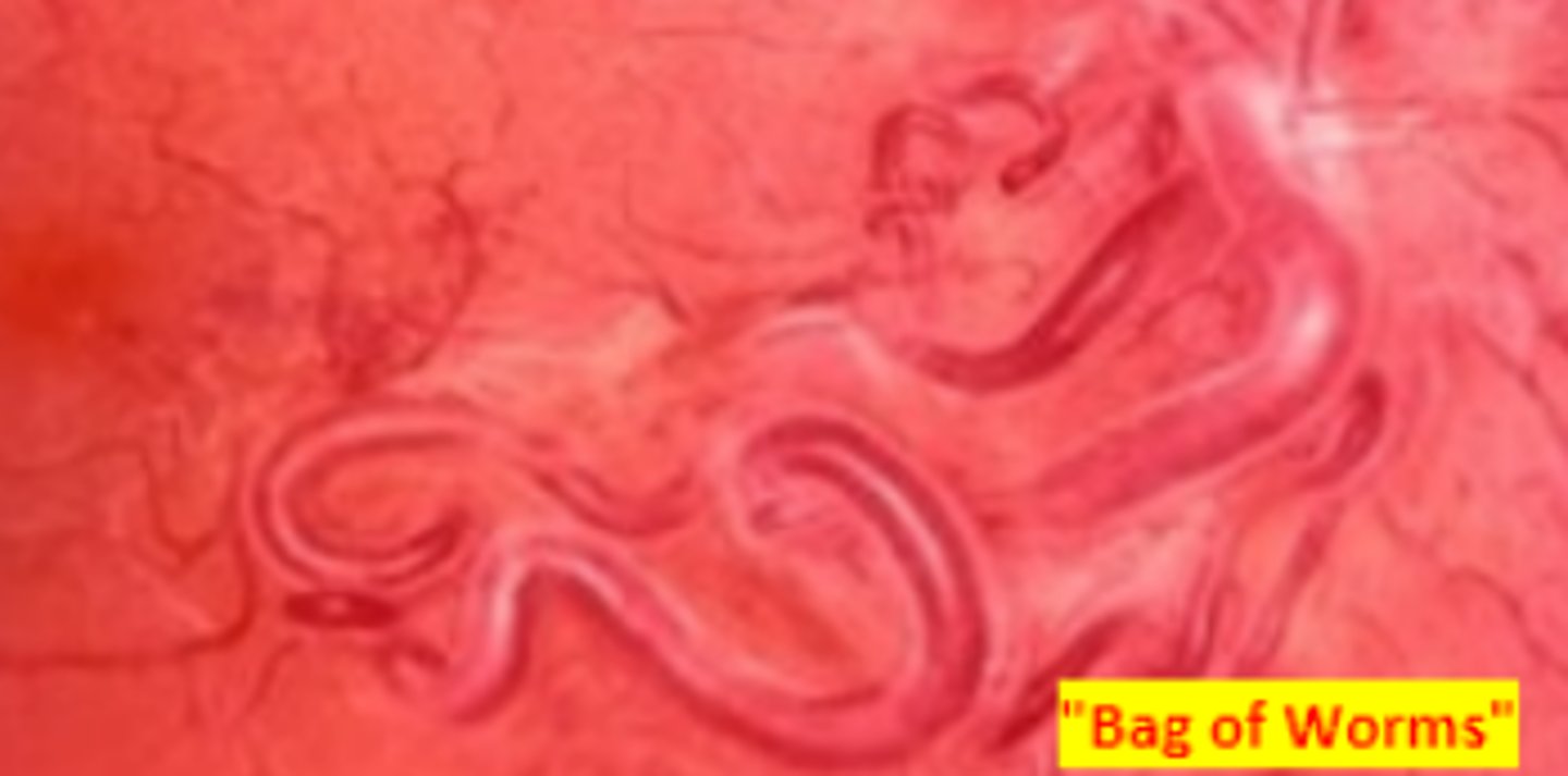
Wyburn Mason Syndrome (Bonnet Dechaume Blanc syndrome)
congenital phacomatosis resulting in AV malformation (racemose hemangioma) in the brain and ipsilateral lesions of the retina/ONH. Skin lesions are highly variable in presentation.
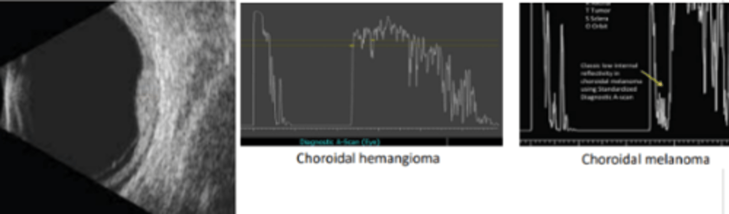
Choroidal hemangioma
a benign vascular tumor of the choroid. No treatment unless there is macular involvement or extensive fluid leakage. Laser therapy and irradiation is available.
Solitary
choroidal hemangioma that has no systemic association and appears as an ovoid mass in the 2nd-4th decade. Hypermetropia may occur due to elevation of the retina. Asymptomatic if not involving the visual axis.
Diffuse
congenital choroidal hemangioma that is associated with Sturge Weber syndrome.
Sturge Weber Syndrome
phacomatoses characterized by cutaneous nevus flammeus (diffuse choroidal hemangioma) involving the first and second branch of the trigeminal nerve. Ipsilateral choroidal and meningeal (possible seizures) hemangiomas are also present at birth. Is associated with ipsilateral secondary glaucoma.
Amelanotic choroidal melanoma, choroidal metastasis, RPE detachment
three differential diagnosis for choroidal hemangioma.
acoustically solid, high internal reflectivity
Compared to an amelanotic choroidal melanoma, a choroidal hemangioma will appear ____ on B scan and will have _____ on A scan