Biology-Unit 1-AOS 1
 Call Kai
 Learn
Learn Practice Test
 Spaced Repetition
 Match
Match Flashcards
 Knowt Play
1/104

Earn XP
Description and Tags
Name | Mastery | Learn | Test | Matching | Spaced | Call with Kai |
|---|
No analytics yet
Send a link to your students to track their progress
105 Terms
Characteristics of living things
Movement (self-generated), Respiraton(release energy from nutrients through aerobic respiration, Sensitivity (sense and react to stimili), Growth (increase and develop overtime) , Reproduction (producing offspring), Equilibrum (maintaining homeostasis through enviroment changes), Excretion ( Removing wastes)
Cell Theory
1. All living things are made up of cells 2.Cells are the smallest/basic units of life 3.All cells come from pre-exisiting cells
Prokaryotic cells
A group of single celled organisms with no nucleus and a circular group of DNA. (Bacteria and Archea)
Eukayotic cells
A group of single/multi-celled organisms with a nucleus and linear strands of DNA (Animals, Plants)
Charcteristics of Prokaryotic cells
-No nucleus -Circular DNA -Plamatids -No membrane-bound organelles -Unicelluar only -replicate by Binary fission
Charcteristics of Eukaryotic cells
-Have a nucleus -DNA is linear chromosones -Membrane-bound organelles -Unicelluar or multicelluar -replicate by mitosis (somatic cells) Meiosis (gametes)
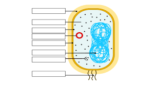
Fill in the prokaryotic cell
Plasmid DNA, Cytosol, Circular DNA, Riobosome, Plasma membrane
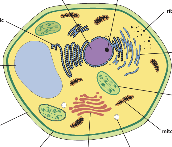
Fill in the eukaryotic/Plantcell
Vacuole
-Mitochondrion
-Nucleus(linear DNA)
-Cytosol
-Golgi appartus
-Vesticle
-Smooth endoplastic reticulum
-Rough endoplastic reticulum
-lysosome
-Nucleous
-Cell wall
-Chloroplast
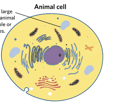
Fill in Animal Cell
-Small/broken upVacuoles
-Mitochondrion
-Nucleus
-Vesticle
-Smooth endoplastic reticulum
-Rough endoplastic reticulun
-Ribosome
-Golgi Appartus
What are not membrane bound organelles?
Ribosomes, Cell Wal, Cytoskeleton
Differences between Plant/Animal cells
Plant cells have a Cell wall
Plants cells have chloroplasts
Plants cells have a large central Vacuole
Organelle
Cellular structure that has a role to keep a cell working
Cytosol
Fluid that surrounds organelles inside a cell
Cytoplasm
includes the cytosol and organelles inside (not Nucleus)
Nucleus
DMO that contains cell’s DNA inside a nucleous that makes ribosomes
Ribosomes
Made of ribosomal DNA (rRNA), makes proteins (floats in cytoplasm or attaches to rought ER)
Rough endoplastic reticlum (RER)
coated in ribosomes, so it can make or modify proteins
Smooth endoplastic reticlum (SER)
Produces Lipids and no ribosomes
Golgi apparatus
Packaging, sorting and modification of proteins for use in a cell
Lysosome
Contains digestive enzymes that break down cell waste
Mitochondrion
Site of aerobic celluar respiration which produces ATP for celluar process, OWN DNA AND RIBOSOMES
Chloroplast
Does photosynthesis in plants and has their own DNA and ribosomes
Vacuole
Used for water/solute storage (maintans plant structure)
Plasma membrane
Selectively permable membrane barrier that controls what enters and leaves the shell
Cell wall
Provides structure to plants, bacterial and fungal cells
Vesicles
Carries substances in or out of cell
Cytoskeleton
Keep’s cell’s shape and transporting vesicles out of cel
Photo Synthesis (opposite of Celluar respiration)
Process of converting light energy, carbon dioxide and water into glucose and oxygen
Celluar respiration (opposite of Photo synthesis)
Converts glucose into ATP
Why do large cells struggle with nutrient uptake and waste removal?
Low surface area to volume ratio, so diffusion across the membrane is less efficient
Why are small cells better at nutrient uptake and waste removal?
High surface area to volume ratio, allowing faster diffusion.
What is the Plasma membrane?
Phospholipid bilayer which separates the intracelluar enviroment from the extracelluar enviroment (semipermable, embedded with proteins, carbohydrates and cholestral
Phospholipid
type of lipid that makes up the cell membrane
-Phosphate head
-two fatty acid tails
Phosphate head
Hydrophillic part of phopholipid that is polar and faces watery inside and outside of cell.
Fatty acid tails
hydrophobic part of phospholipid that is non-poalr
Amphillic
Molecules that are hydrophillic and hydrophobic
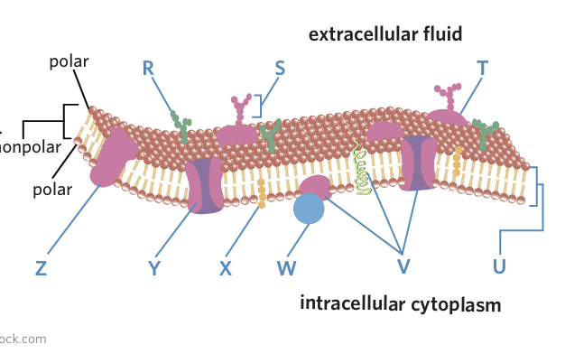
Fill in
glycolipid • cholesterol • glycoprotein • carbohydrate • integral protein • protein channel • peripheral protein • phospholipid bilayer • transmembrane protein
Phospholipid bilayer
Two layers of phospholipids that make up the cell membrane
Function of protein in Plasma membrane
form channels and help with transport (embedded in Plasma membrane)
Intregral protein
Permanently part of membrane
Transmembrane proteins
Go all the way through the bilayer
Peripheral proteins
Temporaily attached to membrane
Carbohydrates
in chains, outside of cells to proteins and helps with recognition, signalling and adhesion
Cholesterol
A lipid that helpes maintain membrane fluidity
Passive transport
The movement of molecules through a semipermable membrane and down the concentration gradient .
Concentration gradient
The difference in solute concentration between two adjacent atoms
Diffusion
Movement of molecules from high concentration to low concentration ‘down their concentration gradient’
Explain diffusion
particles are always moving which overtime leads to an even dispersion of particles in the area
What types of molecules can freely diffuse across the plasma membrane?
-small, uncharged and hydrophobic molecules (O2 and C02)
-Non polar
What is facilitated diffusion?
type of passive transport where moleucles move through a phospholipid bilayer with the aid of a membrane protein
What goes through facilitated diffusion?
-Large molecules, polar molecules and charged molecules (ions)
-as well as non-pol and small because it is FASTER
What can molecules use for facilitated diffusion?
Protein channels and carrier proteins
-(specific to certain molecules)
-
Hypertonic solutions
The solution has a higher solute concentration than the cell. Water flows out of the cell, causing it to shrivel or shrink.
Hypotonic
The solution has a lower solute concentration than the cell. Water rushes into the cell, causing it to swell and potentially burst.
Isotonic
The solution and cell have equal solute concentrations. Water moves in and out equally, meaning the cell retains its normal size and shape.
tonicity
a measure of the relative concentration of solutes on either side of a semipermeable membrane, described as hypertonic, hypotonic, or isotonic
Conformational change
Carrier proteins adapt for a specific molecule
-returns to og shape after transport
eg Glucose carrier only glucose
Osmosis
Diffusion of water across a semi-permable membrane from low solute concentration to high solute concentration hypotonic>hypertonic, with the cocentration gradient
Membrane is seletively permable so
many solutes like sugar cannot cross easily
-water can cross easily
-instead of sugar moving water moves
Example of osmosis
-more sugar inside the cell
-less sugar outside the cell
Inside= high solute concentration
outside= Low solute concentration.
SO water moves from low solute to high solute(dilutes the sugar until concentrations become equal)
Active transport
Movement of substances across a membrane against their concentration gradient
-requires ATP
-Uses protein pumps
What is ATP
Adenosine triphosphate
define Protein-meditated transport
type of active transport which involves using membrane proteins to move molecules across a membrane, against their concentration gradient
requires
-energy (ATP)
-membrane proteins (protein pump and carrier proteins)
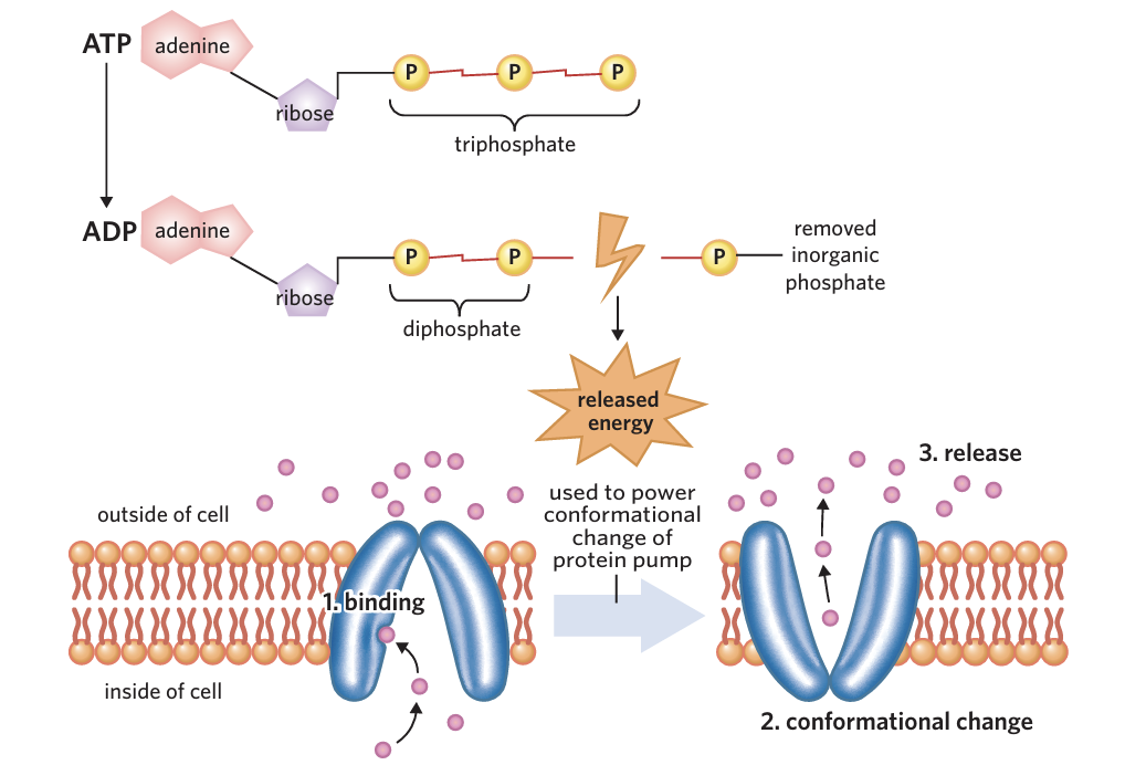
Steps of protein-meditated active transport
1.Binding- Target molecule binds to protein pump
Conformational change-ATP is broken down
ATP> ADP+P and energy is realeased so conformational change can occur in protein pumo
Release- Target molecule is pushed through the protein and released on the other side of plasma mebrane
Visualise the protein-meditated process
Bulk Transport (large molecules)
Type of active transport that uses vesicles to move large molecules or groups of molecules across a membrane (cytosis)
-Exocytosis
-Endocytosis
Exocytosis (opposite of endocytosis)
-Moves large substances out of cells
Vesicle with secretory products is transported to the plasma membrane (vesicular transport)
Fusion- Vesicle membrane fuses with cell membrane
Release- Secretory products are released from the vesicle, outside the cell
*Adds phospholipids to membrane, then releases it’s contents outside the cell
*plasma membrane increases slightly
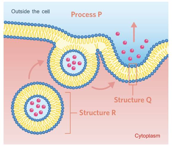
What is happening
-Moves large substances out of cells
Vesicle with secretory products is transported to the plasma membrane (vesicular transport)
Fusion- Vesicle membrane fuses with cell membrane
Release- Secretory products are released from the vesicle, outside the cell
*Adds phospholipids to membrane, then releases it’s contents outside the cell
*plasma membrane increases slightly
Visualise endocytosis
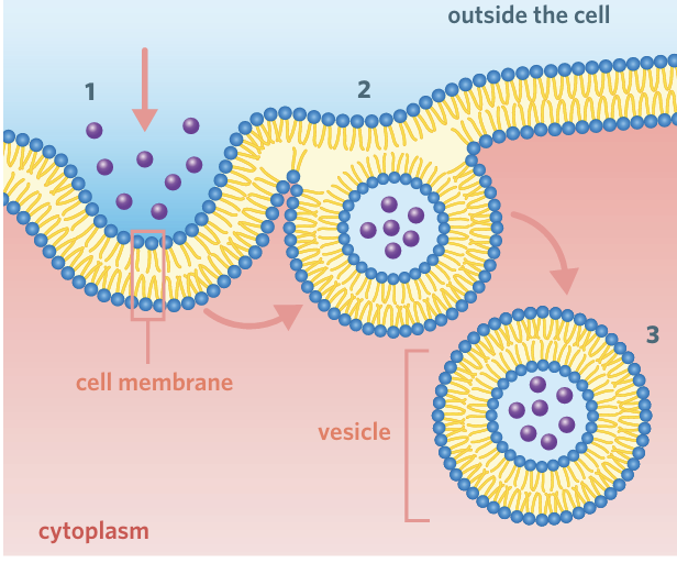
Endocytosis(bulk transport) FTB fold the bubble
moves large substances into of cell
1.Fold- plasma membrane folds inwards that fills with target molecules and extracelluar fluid
2.Trap- Target molecules become enclosed as the membrane keeps folding
3.Bud- Vesicles pinches off membrane
*If taken in large amounts cell could shrink
What is the purpose of cell replication?
Growth and development- organisms increase their number of cells.( Cell size stays the same)
Maintainence and repair-Old/damaged cells are replaced, ensuring function of organism
Reproduction-Single-celled organisms reproduce by making identical copies
Plasmid
circular group of DNA that is separate from chromosome
Cytokineis
Division of the cytoplasm and formation of two genetically identically daughter cells
Septum
A dividing wall formed during binary fi
Steps of binary fisson DECS
Orginal cell
DNA replicated
Elongation
Cytokinesis and formation of septum
Two genetically identical daughter cells
Exponetial growth
1 bacerium
20 minutes> 2
40 minutes> 4
60 minutes> 8
What is the eukaryotic cell cycle
process where a cell grows, replicates it’s DNA, and divides into two idential daughter cells
Interphase-Celluar growth and duplication of chromosomes
Mitosis-Separation of sister chromatids and the formation of two new nuclei
Cytokineis- Division of the cytoplasm and formation of two daughter cells
Queiesnt
dormant cells which can re-enter the cell cycle
Terminally differentiated
Cells that are fully specialised and can no longer enter the cell cycle
Chromatid
one half of a double-standed chromosome
Centromere
Structure which holds sister chromatids together
Chromosome
Structure composed of DNA
Interphase
G1 phase
Cells grows
-proteins are synthesised
-organelles replicate
G0 phase
-cells that not required/terminally differntiated
S phase (synthesis)
-DNA replicated
-each chromosone forms
-idential sister chromatids
G2 Phase
-Cell continues growing
-proteins produced for mitosis
-prepares for division
Somatic
body cells except reproductive cells
Mitosis
Division of the nucleus producing two identical nuclei
Phases of mitosis Pass Me A Taco
Prophase
-Chromatin condeses into chromosones
-Centrioles migrate to poles of cell
-Spindle fibres form
-Nuclear membrane breaks down
Metaphase
-Chromosomes line up at the equator
-spindle fibres attach to centromeres
Anaphase
-Sister chromatids separate to opposte poles
-Centrosomes spilt
Telophase
-Chromosomes decondense
-Two nuclear membrane form
Two nuclei are produced
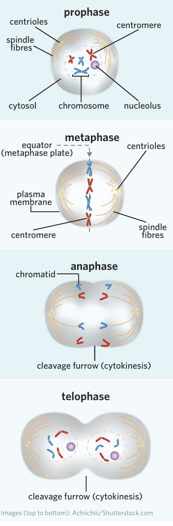
Cleavage furrow
an indentation of the plasma membrane during cytokinesis
Cytokinesis-Animal cell
Cleavage furrow forms
Cell pinches in two
Cytokinesis-Plant cell
-Cell plate forms
-Develops into a new cell wall
Cell cycle check points
G1 checkpoint: checks for growth, DNA damage and nutriends
G2 checkpoint: DNA replicated correctly and resources
Metaphase checkpoint: checks that chromosomes are lined up.
Apoptosis
Controlled and regualted death of cells used to remove
-Damaged cells
-Diseased cells
-Unnesscary cells
1.Mitochondrial pathway (intrisic)
2.Death receptor Pathway (extrinsic)
Steps of Mitochondrial pathway (intrisic)
*Triggred by internal cell damage
1.Mitochondrial detect damage
2.Cytochrome C is released
3.Capase enzymes are activated
4.Apoptosis begins
2.Death receptor Pathway (extrinsic)
*Trigged by external signals
1.Death signalling molecules bind to death receptors on the cell membrane
2.Capase enzymes are activated
3.Apoptosis begins
Stages of Apoptosis DSBBP don’t squish big big pickles
*after Capases are activated
1.Digestion of cell’s contents-capases break down proteins and organelles
2.Cell shrinks
3.Membrane blebbing-Plasma membrane buldges
4.Apoptosis bodies form-Cell break into small vesicles
5.Phagocytes enguld them by phagocytosis
Necrosis
-Unregulated death
-Cell damage, causes cell to burst and inflammation
Phagocytosis
After apoptosis, phagocytes engulf and digest the free-floating apoptotic bodies by phagocytosis.
phagocyte
a cell of the immune system responsible for engulfing and destroying harmful microorganisms and foreign material
Tumour formation
If the cell cycle is disrupted or insufficient, damaged cells can replicate exponentially, leading to the development of tumours and cancers.
Maligant tumours
-invades nearby tissue
-Can spread through blood
-Spread called metastasis