SAM.HI4: Non-Malignant Splenic Dz
 Call Kai
 Learn
Learn Practice Test
 Spaced Repetition
 Match
Match Flashcards
 Knowt Play
 Podcast
1/72

There's no tags or description
Looks like no tags are added yet.
Name | Mastery | Learn | Test | Matching | Spaced | Call with Kai |
|---|
No analytics yet
Send a link to your students to track their progress
73 Terms
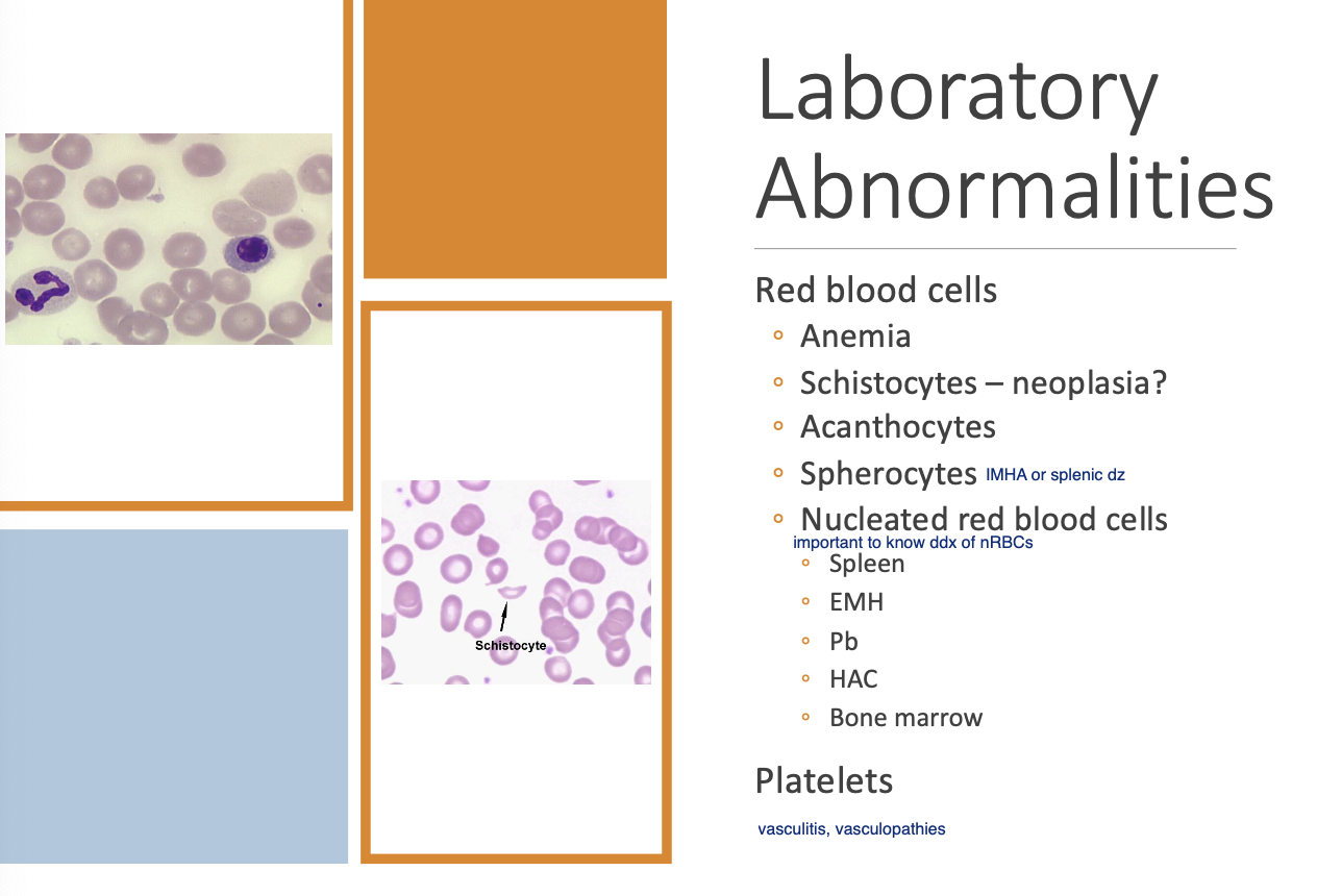
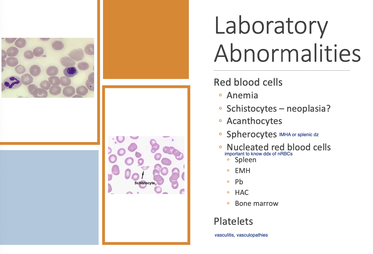
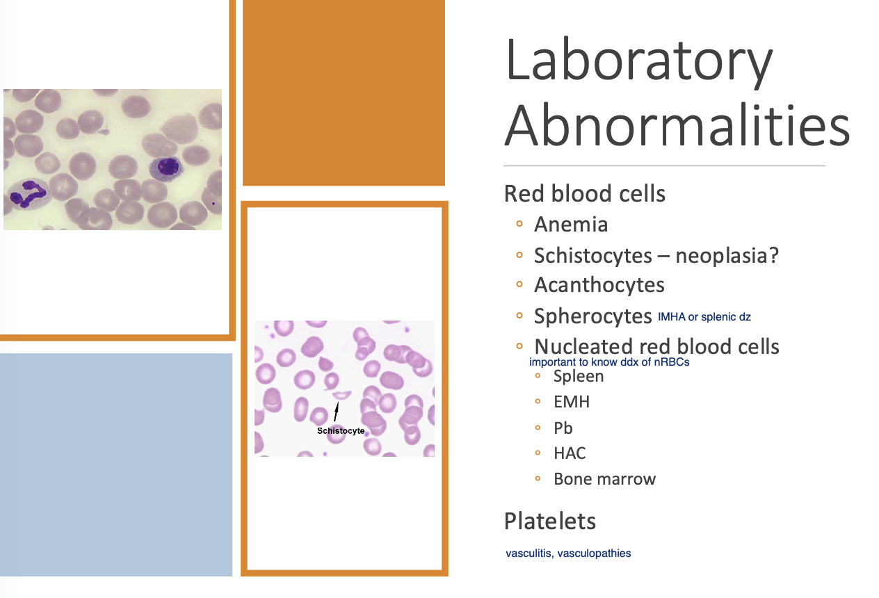
Changes in RBCs seen w/ splenic dz (5)
Anemia.
Schistocytes.
Acanthocytes.
Spherocytes
nRBCs.
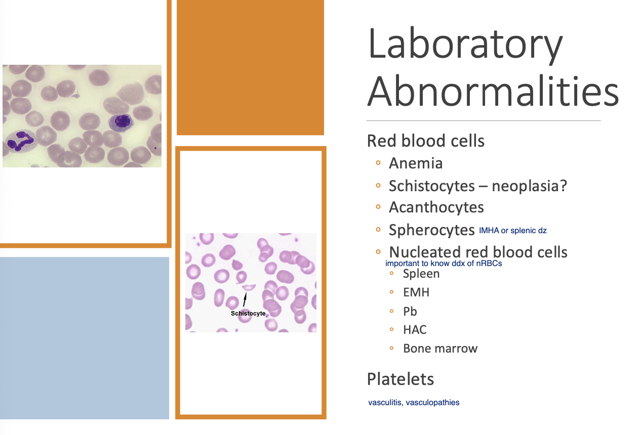
Schistocytes are highly suggestive of
Neoplastic splenic dz
nRBC can be seen w/
benign conditions such as EMH (spleen is not able to prevent release of immature cells when stimulated)
Leukoerythroblastic response
Increased nRBC and immature WBCs that can be seen w/ splenic dz.
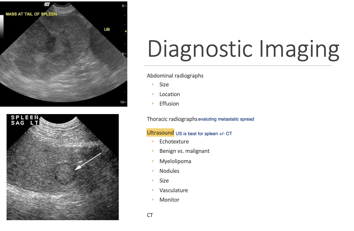
Rads in Dx of splenic dz
size and position of the spleen.
evidence of effusion.
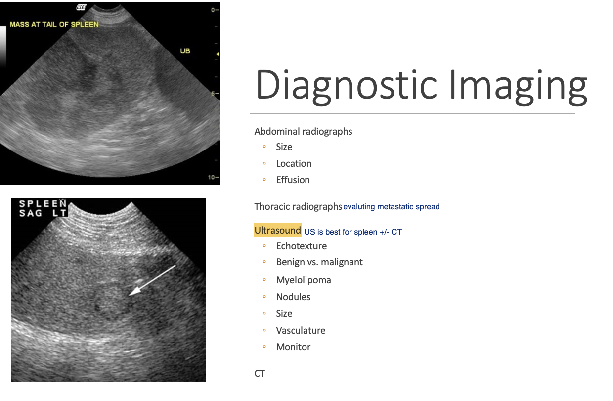
Generalized or focal splenomegaly on rads
may result in a mass effect in the mid-abdomen w/ displacement of other abdominal organs.
Rads w/ splenic torsion or hemorrhage (2)
decreased detail +/- effusion
torsion - abnormal placement of the spleen.
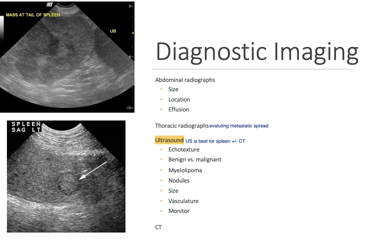
FNA is useful in the dx of dzs that cause
diffuse splenomegaly and some focal lesions
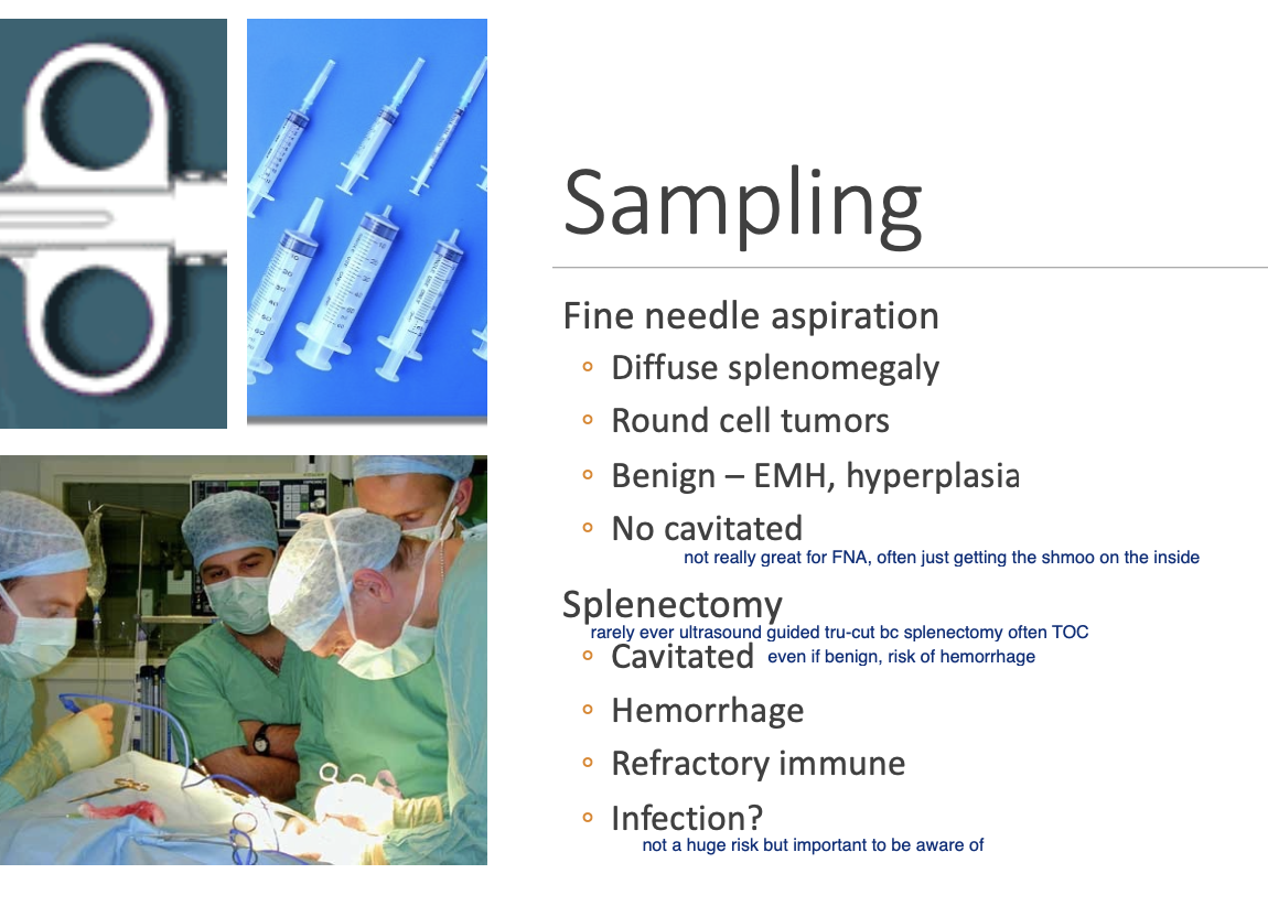
Which lesions have low yield w/ FNA
cavitated masses - not reco due to hemorrhage risk
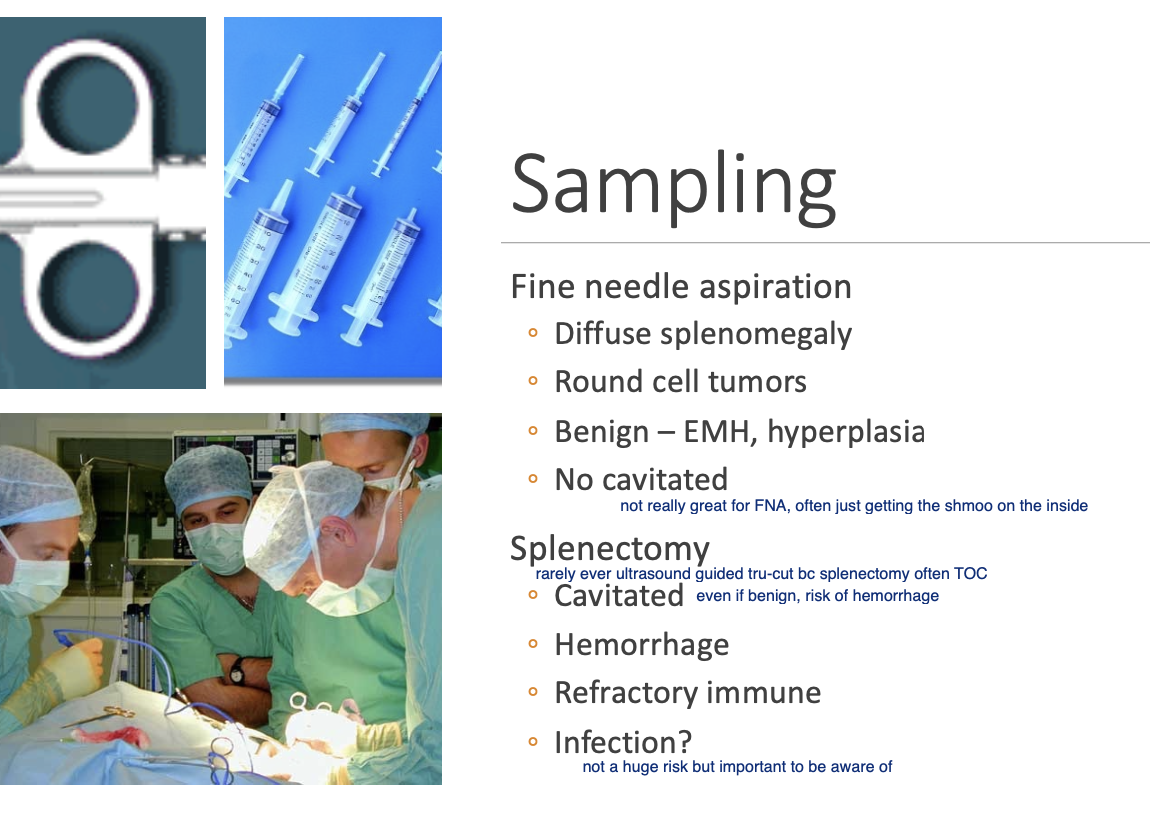
Which method is often performed for cavitated lesions/hemorrhage
Splenectomy > splenic biopsy
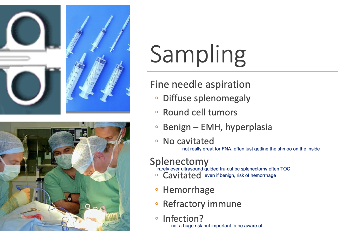
Removal of the spleen may predispose animals to developing certain infections (3)
Mycoplasma spp.
E. canis.
Babesia canis.
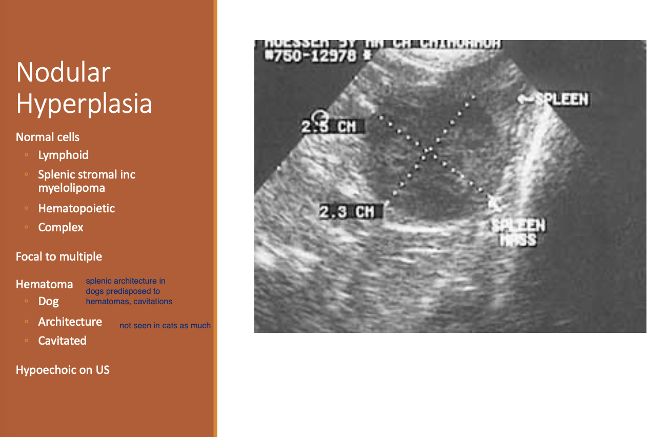
Nodular Hyperplasia occurs as a result of
proliferation of normal cells of the spleen.
4 types of Nodular Hyperplasia.
Lymphoid.
Splenic stromal.
Hematopoietic.
Complex.
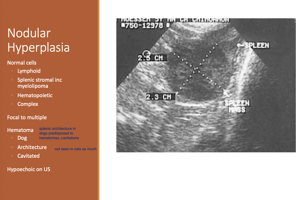
Which splenic pathology is a form of MH
Splenic myelolipoma.
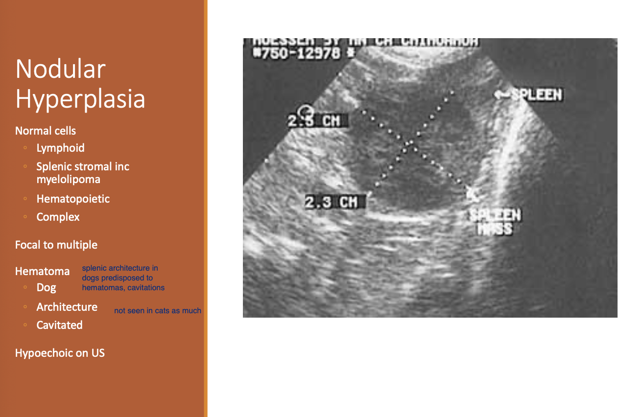
Lesions of nodular hyplerplasia
focal, but there may be multiple.
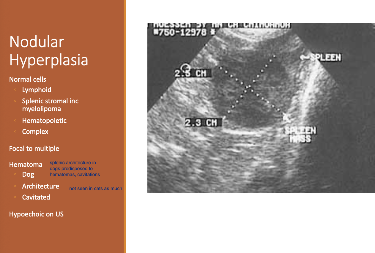
Nodular hypoplasia on US
hypoechoic
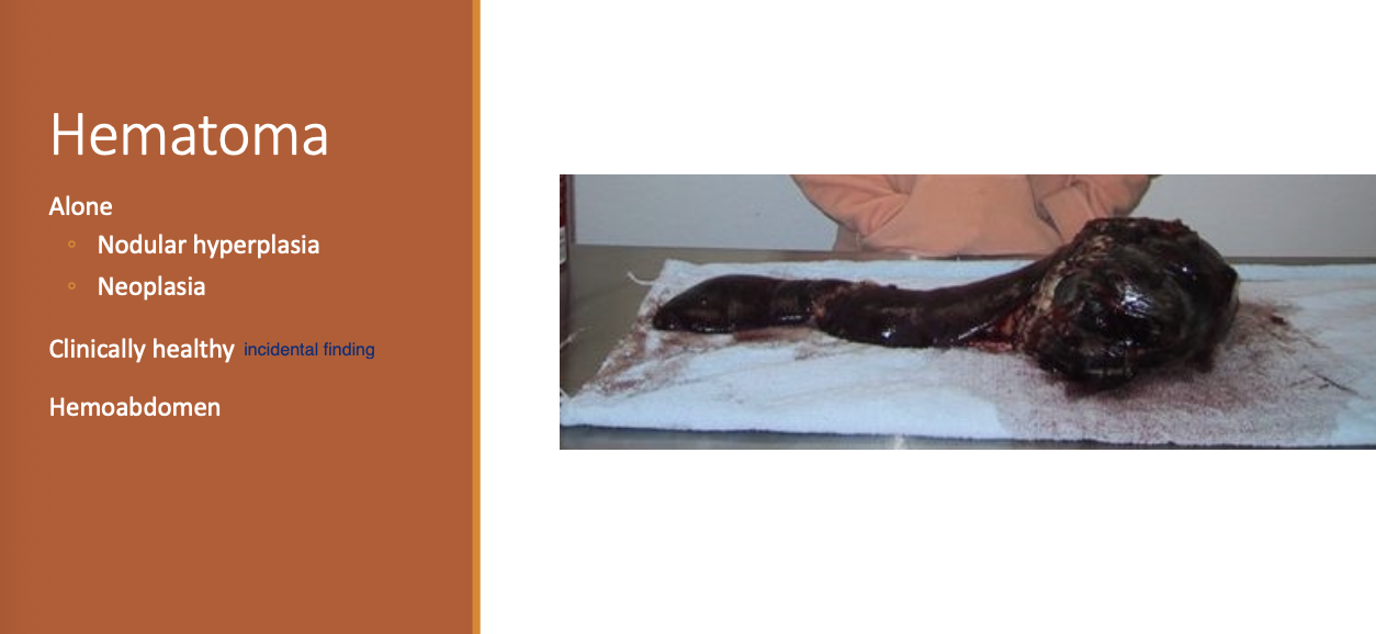
How can nodular hyperplasia lead to hematoma in the dog?
distortion of the splenic vasculature by hyperplasia
Why is it suspected that cats are less likely to have splenic hematomas?
different anatomy of the spleen and orientation of the vasculature.
Splenic hematomas are more common in
large breeds - GSD and standard poodles.
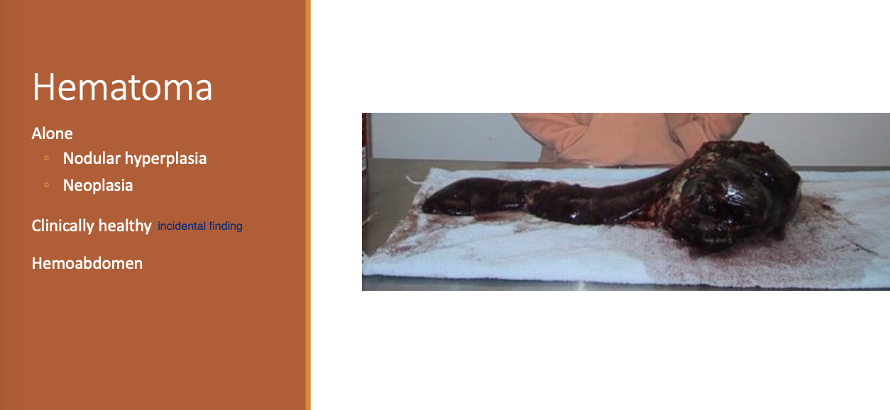
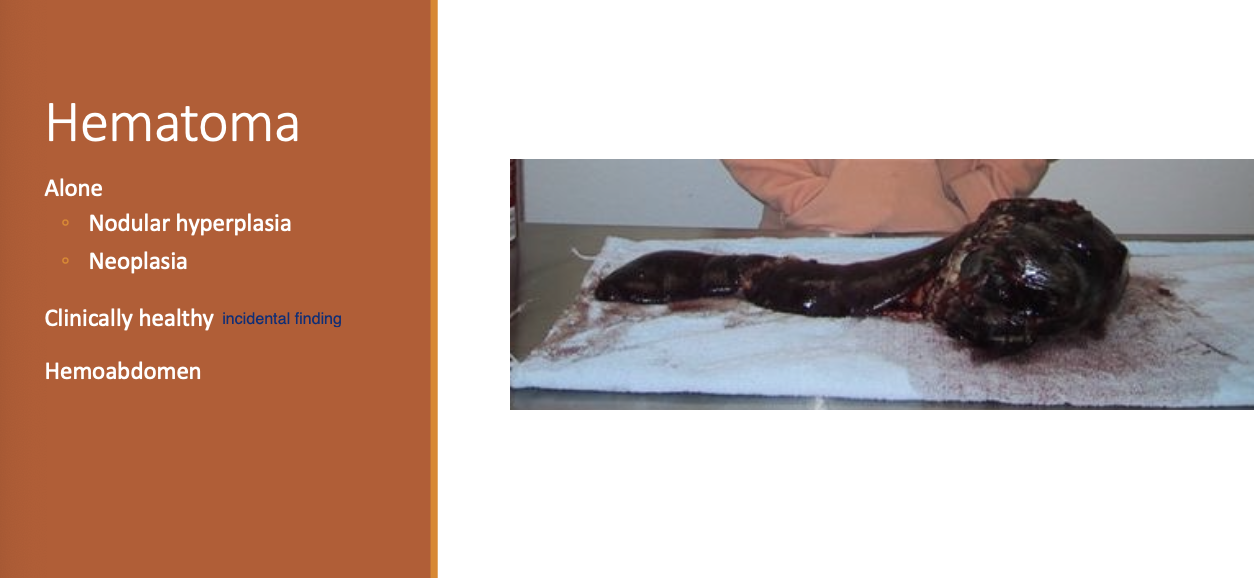
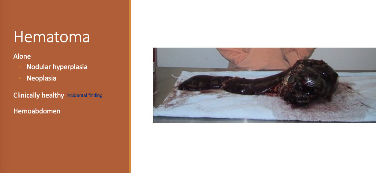
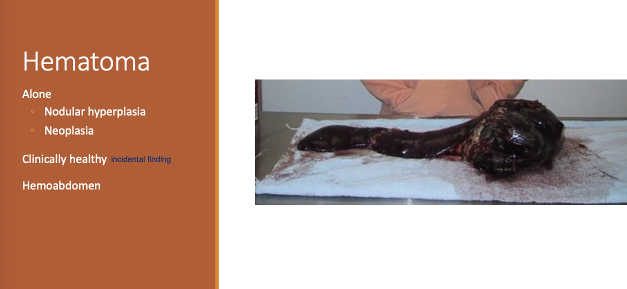
Hematomas may occur
alone or be associated w/ MH, neoplasia, trauma, or idiopathic.
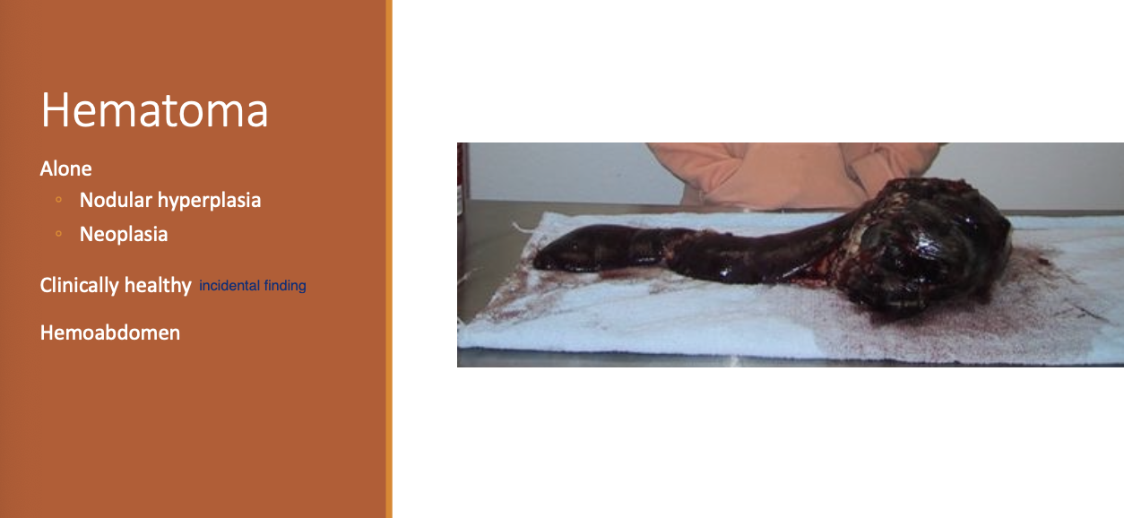
Clinical manifestation of dogs w/ hematomas
healthy
hemoabdomen
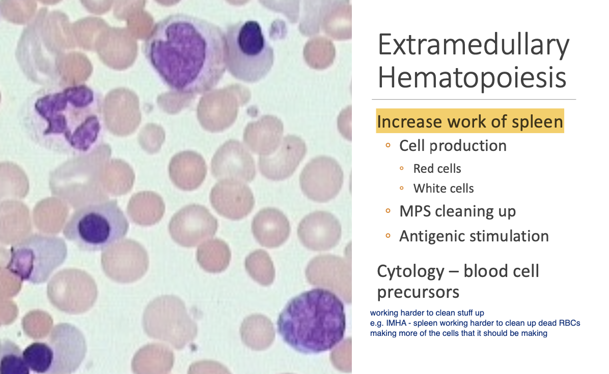
EMH occurs as the
workload of the spleen increases
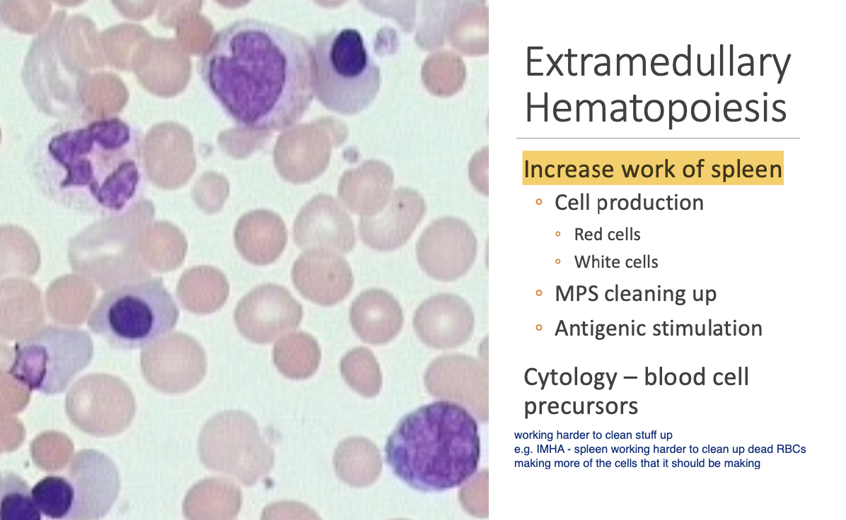
Workload of the spleen increasing can be due to increased (4)
removal of normal cells.
activity of the mononuclear-phagocytic system.
activity of the lymphoid tissues.
blood cell production.
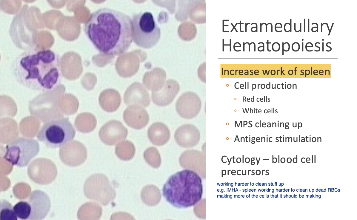
EMH on U/S (2)
Dogs - Diffuse splenomegaly that is hypoechoic.
Cats - diffuse nodular.
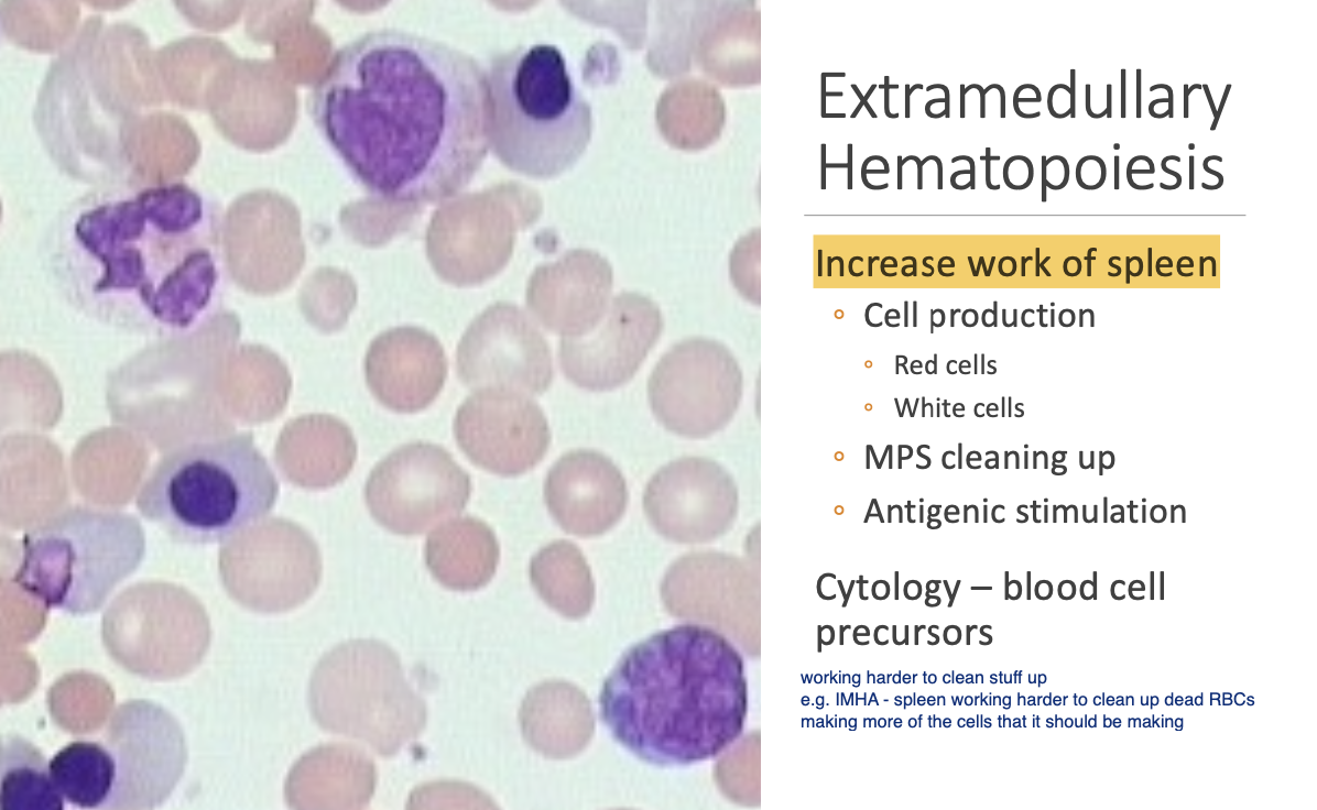
Aspirate results for EMH (2)
increased number of medium to large lymphocytes and plasma cells (hyperplasia).
increased number of blood cell precursors on splenic aspirates nRBCs peripherally may be seen.
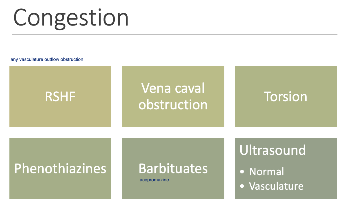
Splenic congestion can be seen w/ (2)
acepromazine.
barbiturates.
Portal HT.
Venous HT - R sided HF and CaVC occlusion.
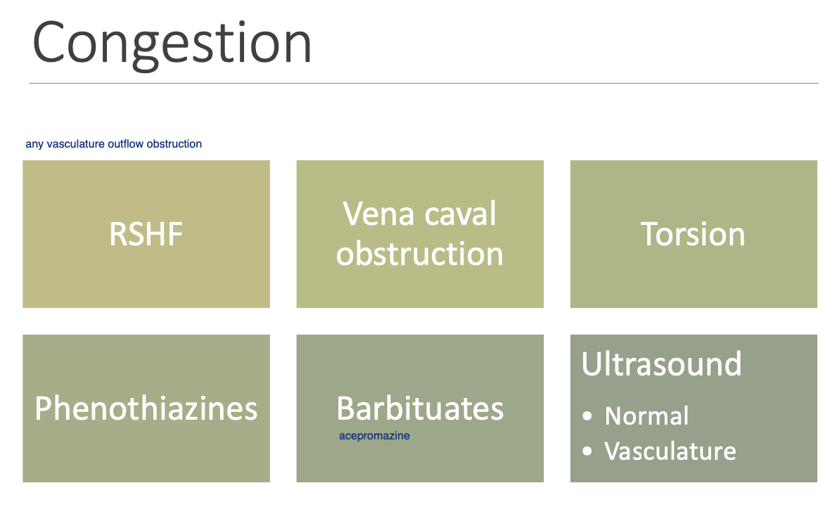
Splenic congestion on U/S
normal echotexture, but may have distended vasculature.
Splenic torsion spp trend
rare overall, but more common in dogs
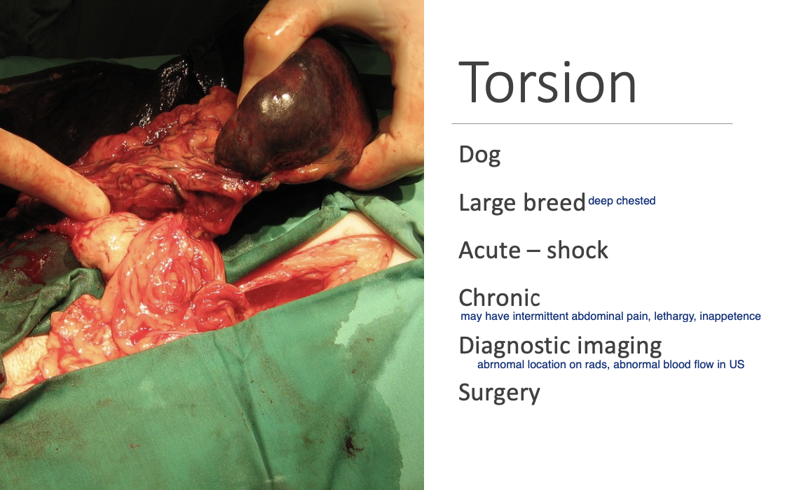
CS of dogs w/ splenic torsion (4)
shock.
anorexia.
V.
Abdominal pain.
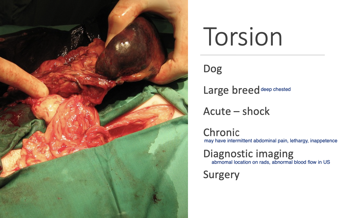
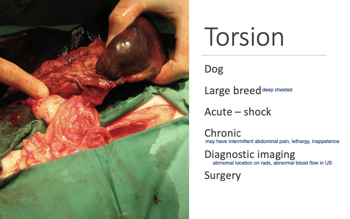
Torsion can rarely be
chronic and intermittent.
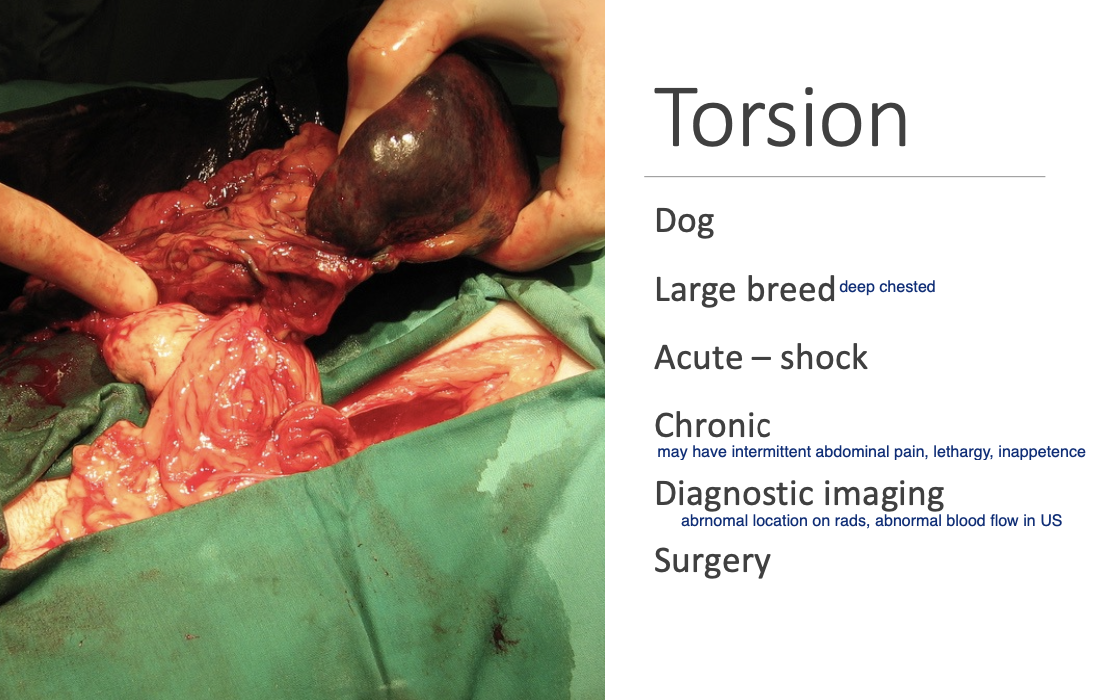
Rad findings of splenic torsion
decrease detail in the abdomen secondary to abdominal effusion.
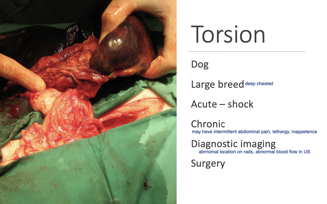
U/S findings of splenic torsion (2)
Abnormal location.
Diffuse enlargement.
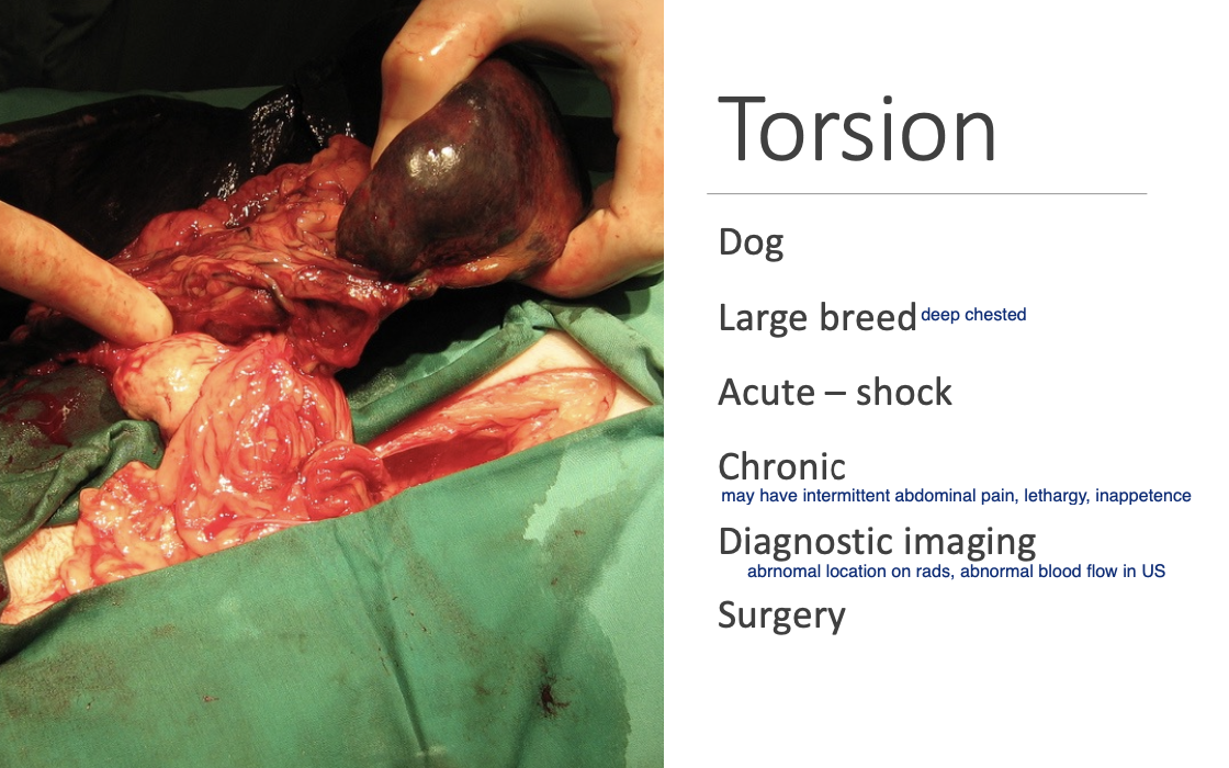
Tx for splenic torsion
Stabilization then Sx.
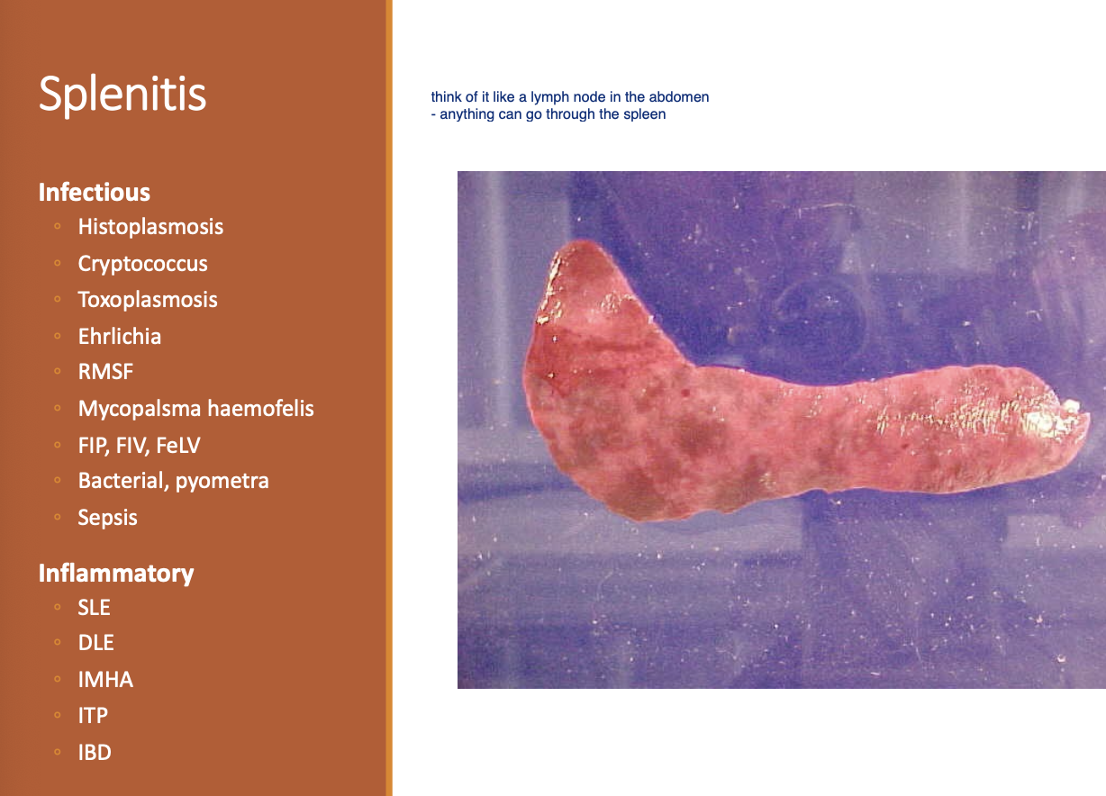
Splenitis def
Inflammation of the spleen.
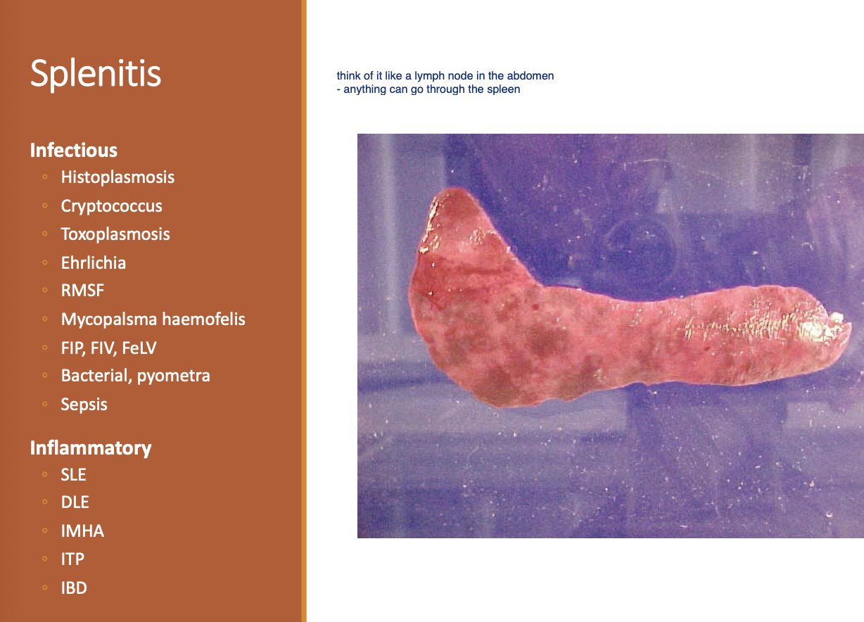
Splenitis may result in
diffuse splenomegaly
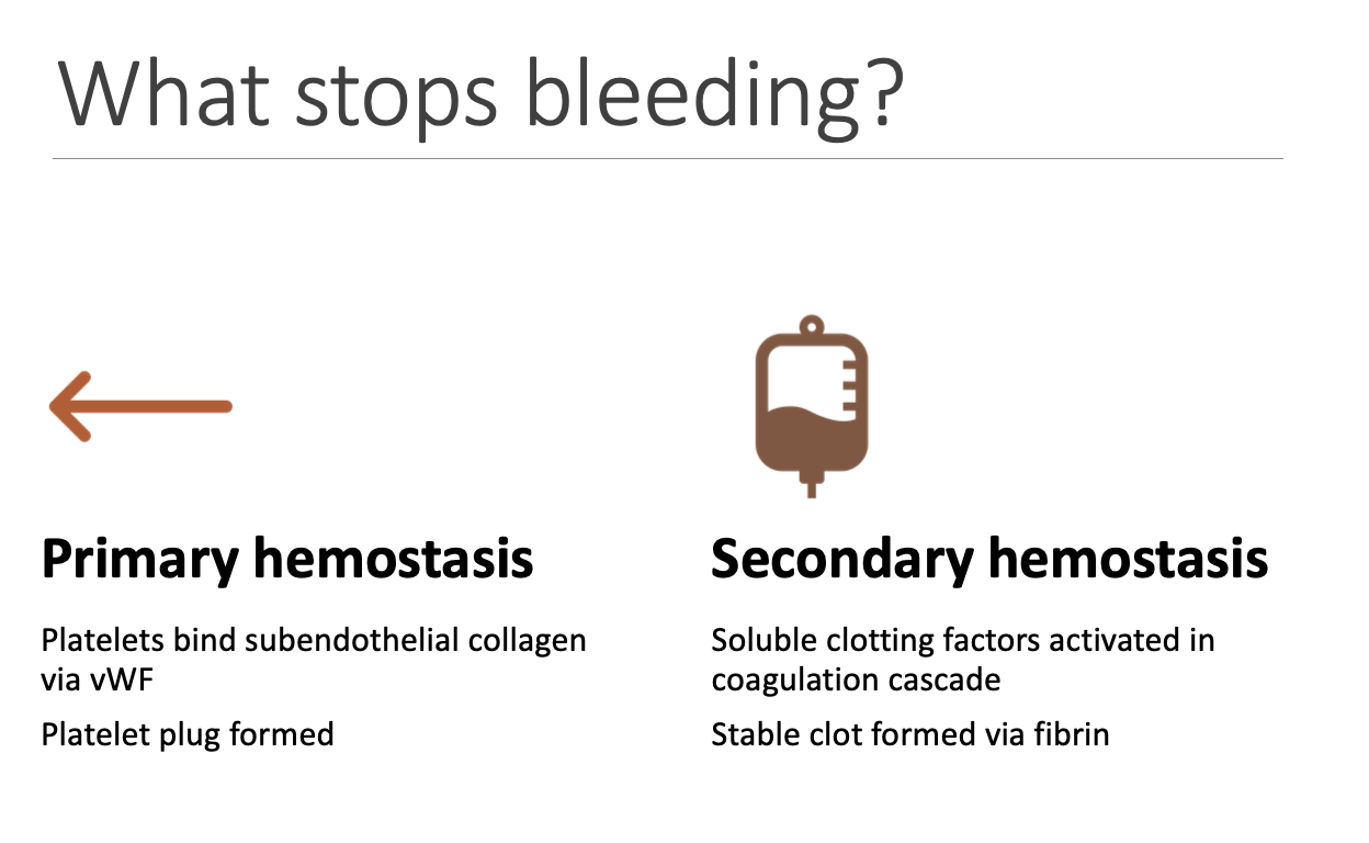
What is formed during each phase of hemostasis (2)
Primary - platelet plug.
Secondary - Stable clot via fibrin.
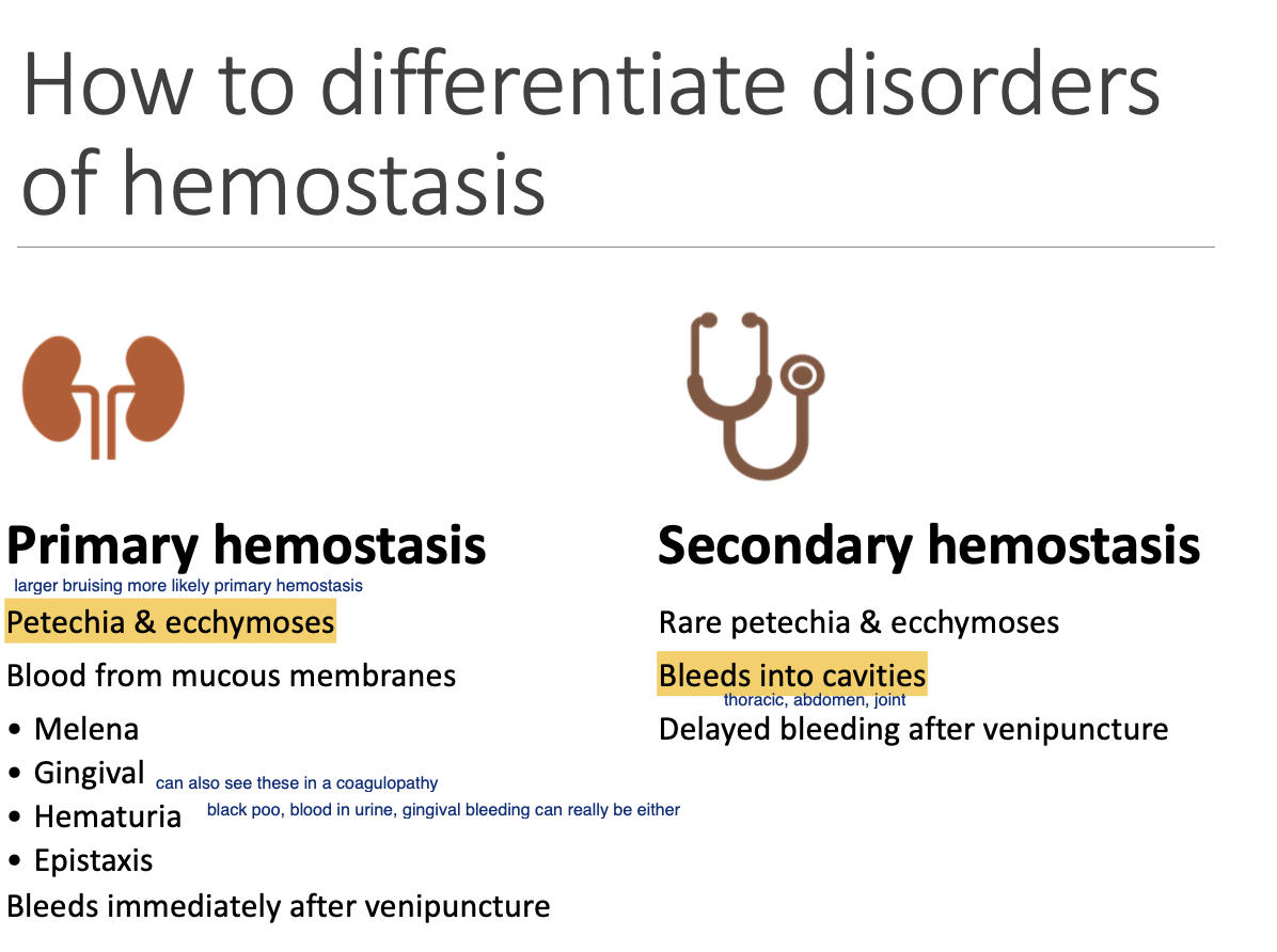
Primary hemostasis CS (3)
Petechia and ecchymoses.
Blood from mm (can be either).
Bleeding immediately after venipuncture.
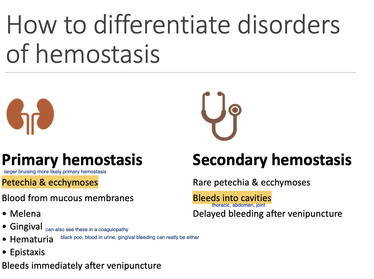
Secondary hemostasis CS (3)
RARE petechia and ecchymoses.
Bleeds into cavities.
Delayed bleeding after venipuncture.
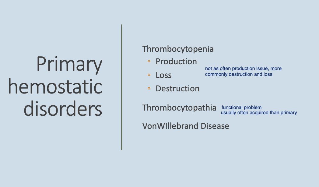
Primary hemostasic disorders (3)
Thromboyctopenia.
Thrombocytopathia.
vWD.
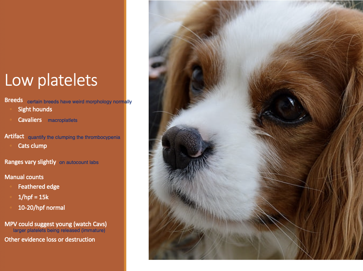
Thrombocytopenia Etiologies- non-path (2)
Breeds - Cavaliers and Sight hounds.
Artifact.
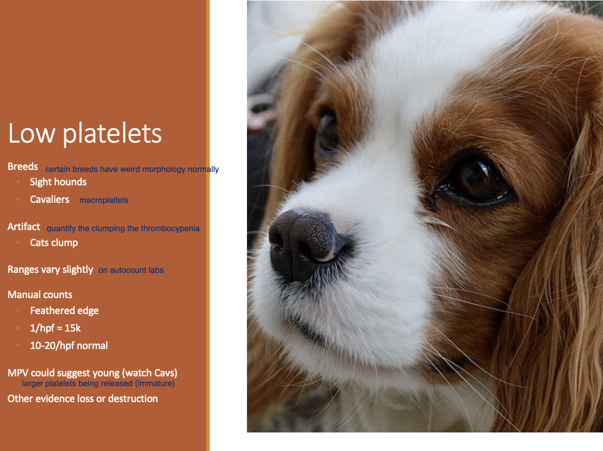
Manual count of platelets (2)
Feathered edge.
Normal = 10-20/hpf.
BMBT tests for
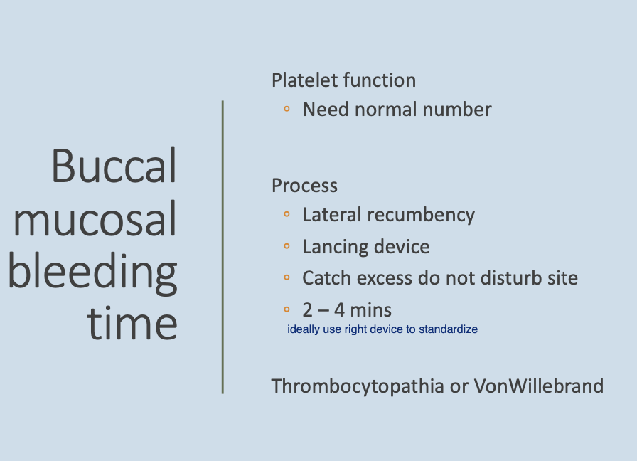
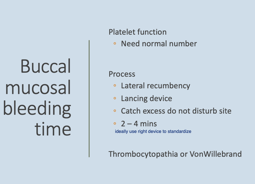
thrombocytopathia (platelet function) or vWD - need to have a normal platelet number
Process for BMBT (4)
Lateral recumbency.
Lancing device.
Catch excess while not disturbing site.
2-4m.
Platelet fxn analyzer is used for dx of
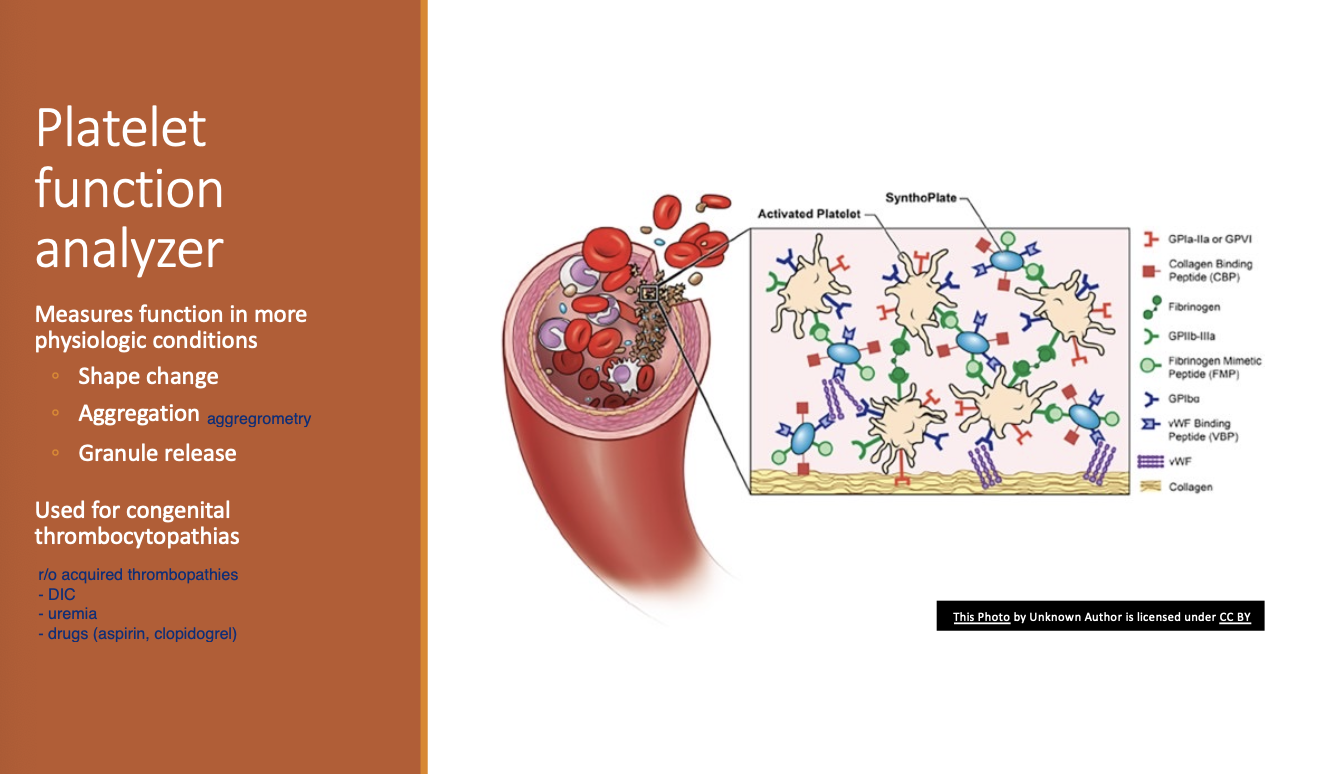
congenital thrombocytopathia
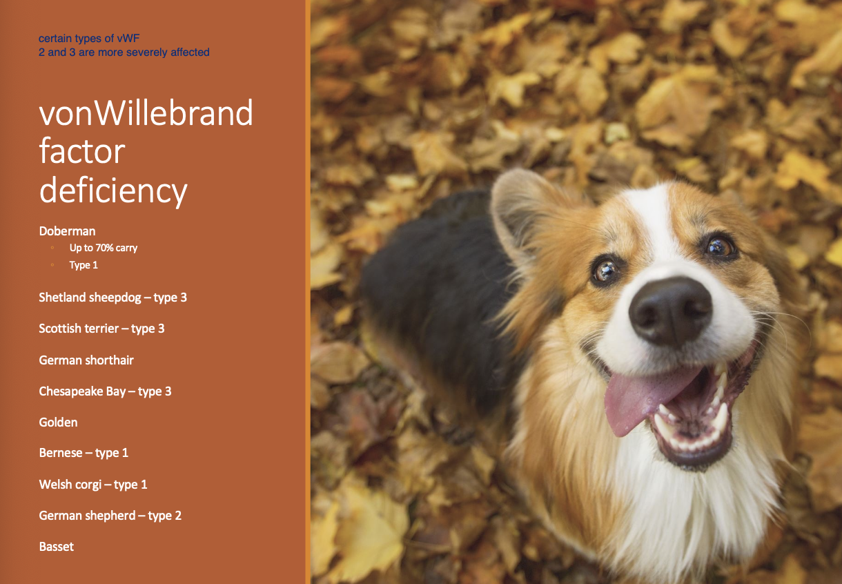
vonWillebrand factor deficiency breeds
Doberman - up to 70% carry, type 1
Shetland sheepdog
Scottish terrier
German shorthair
Chesapeake bay
Golden
Bernese
Welsh corgi
GSD
Basset
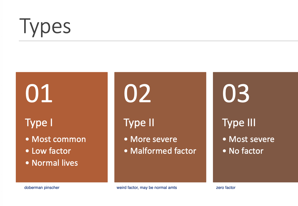
vWD Type 1 (3)
Most common form.
Low quantity of factor.
Live normal lives.
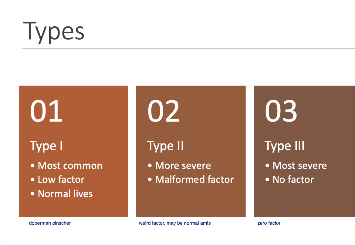
vWD Type 2 (2)
More severe form.
Malformed factor, but has normal quantity.
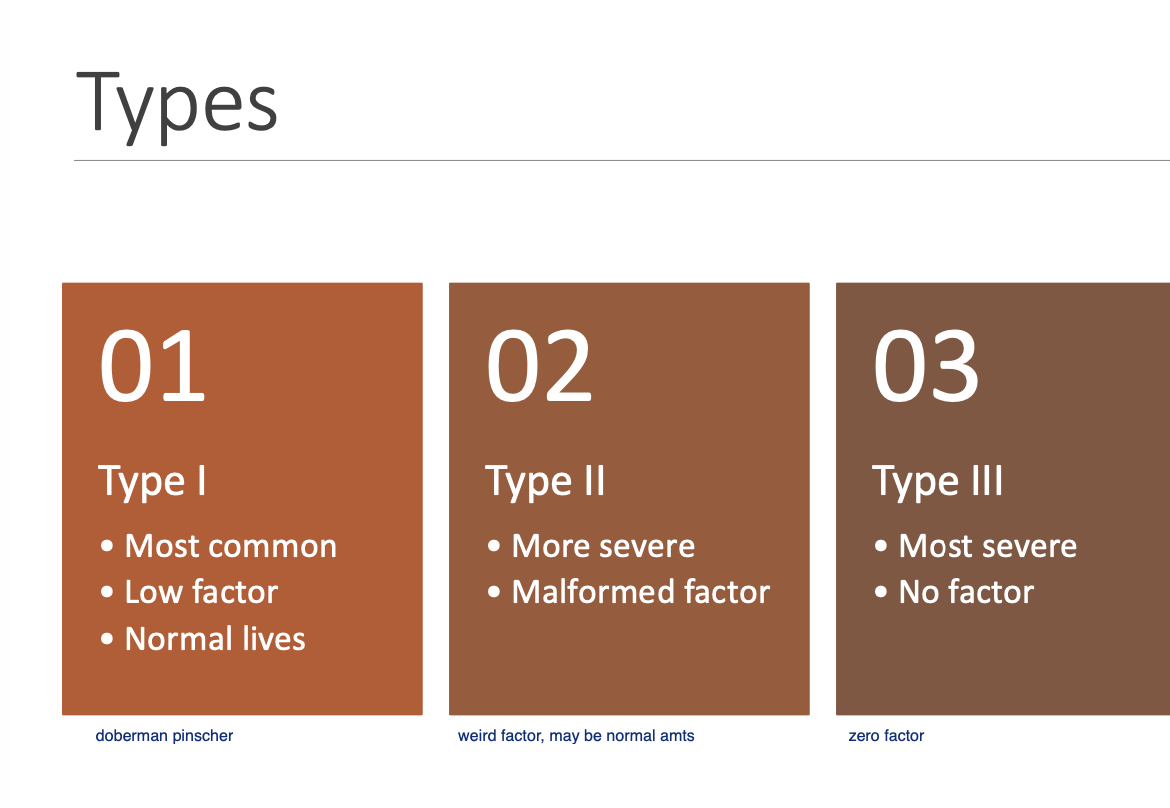
vWD Type 3 (2)
Most severe form.
No factor produced.
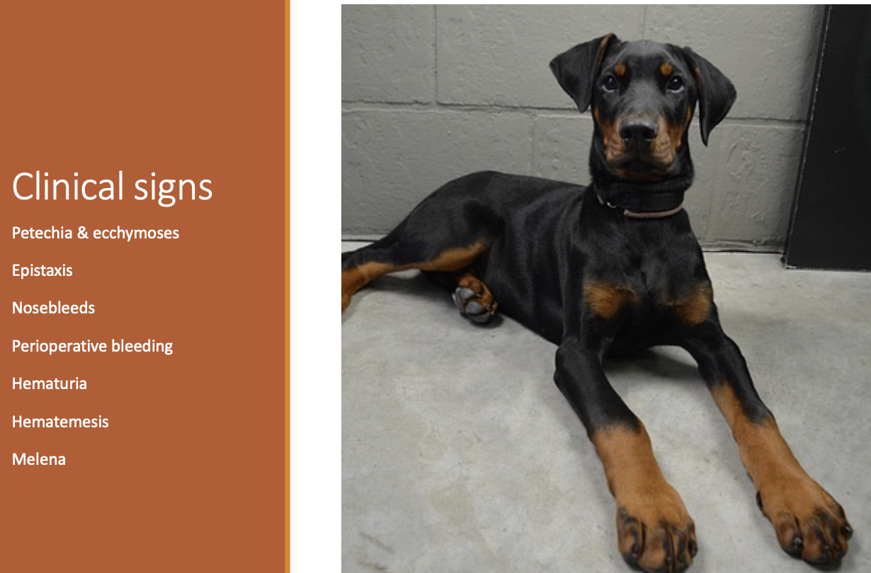
CS of vWD (3)
Petechia and ecchymoses.
Epistaxis.
Peri-op bleeding.
(Hematuria, Hematemesis, and Melena)
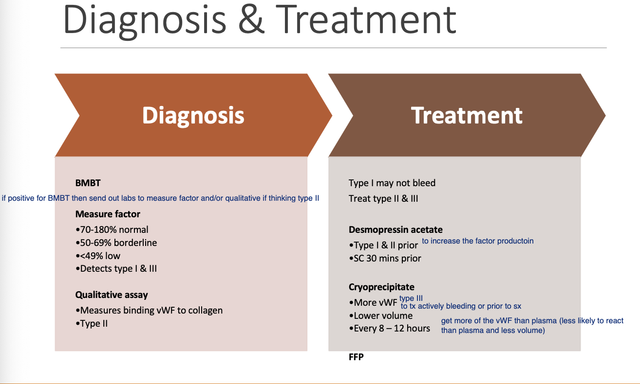
Dx of vWD (2)
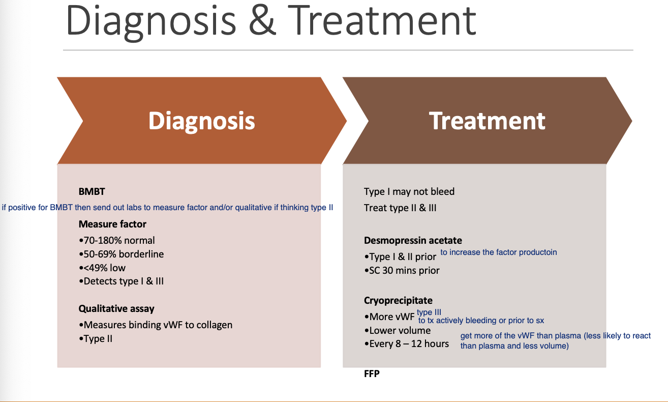
BMBT
Measure factor - detects Type 1 and 3.
Qualitative assay - detects Type 2 (measures binding vWF to collagen).
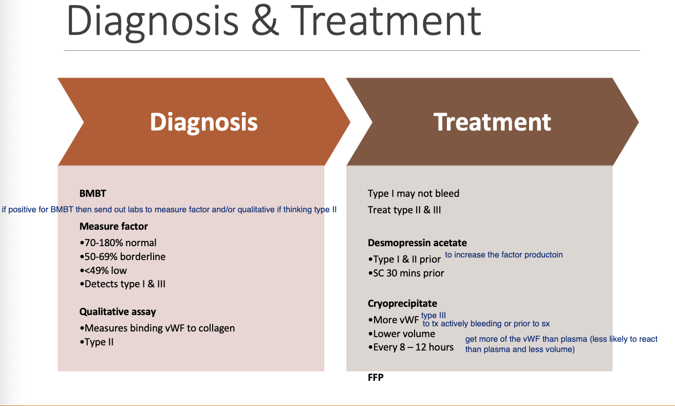
Tx for vWD - general concept based on type
Type 1 - may not bleed.
Type 2 and 3 - Tx.
Tx for vWD (3)
Desmopressin acetate.
Cryoprecipitate.
FFP.
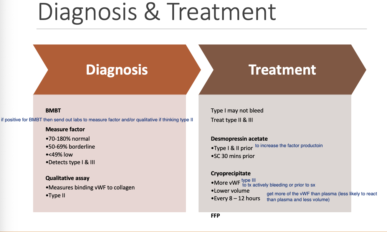
Desmopressin acetate in tx of vWD
Type 1 and 2 given SC 30m prior to procedures.
Cryoprecipitate in tx of vWD
Has more vWF at lower volume. Given q8-12h.
Platelet dysfxn can be
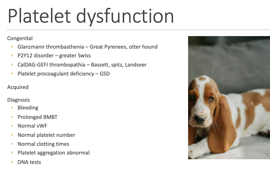
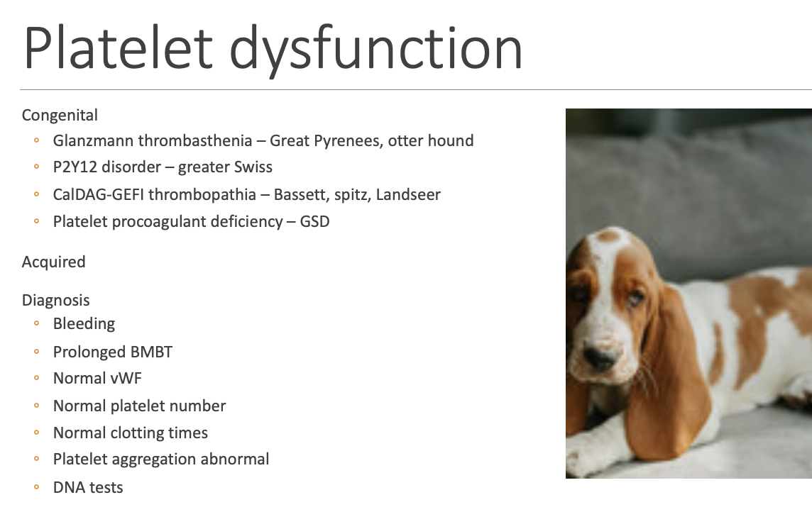
congenital or acquired.
Dx of platelet dyfxn (4)
Bleeding w/ prolonged BMBT.
Normal vWF, platelet numbers, clotting times.
Abnormal platelet aggregation.
DNA tests.
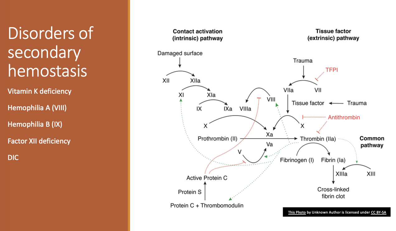
Disorders of secondary hemostasis includes (4)
Vit K deficiency.
Hemophilia A or B.
Factor 12 deficiency.
DIC.
Etiologies of Vitamin K deficiency (4)
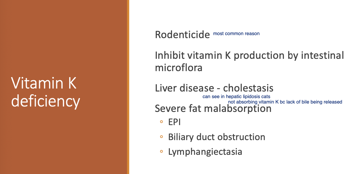
Rodenticide.
Inhibited Vit K production by intestinal microflora.
Liver dz - cholestasis.
Severe fat malabsorption (EPI, Biliary duct obstruction, Lymphangiectasia).
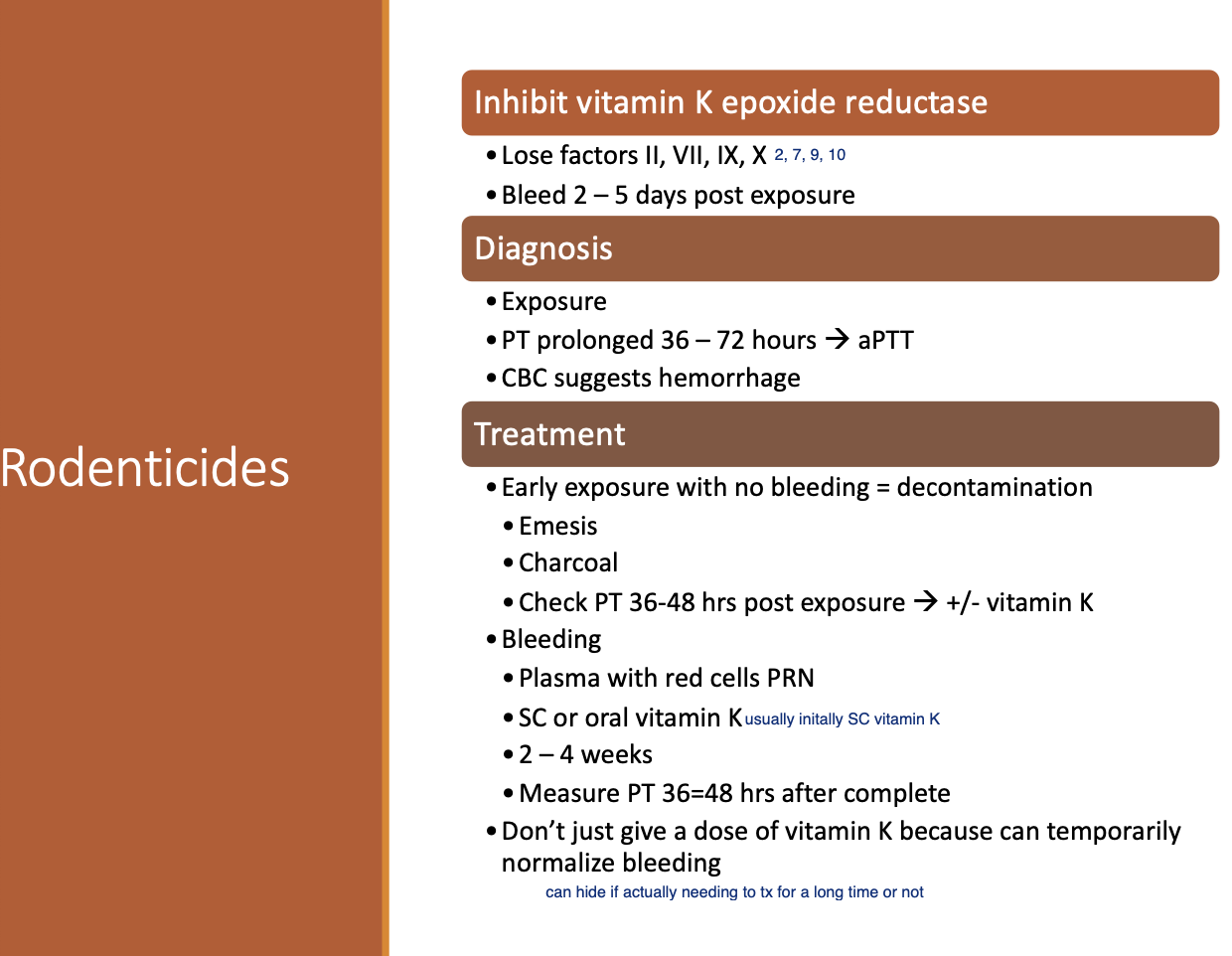
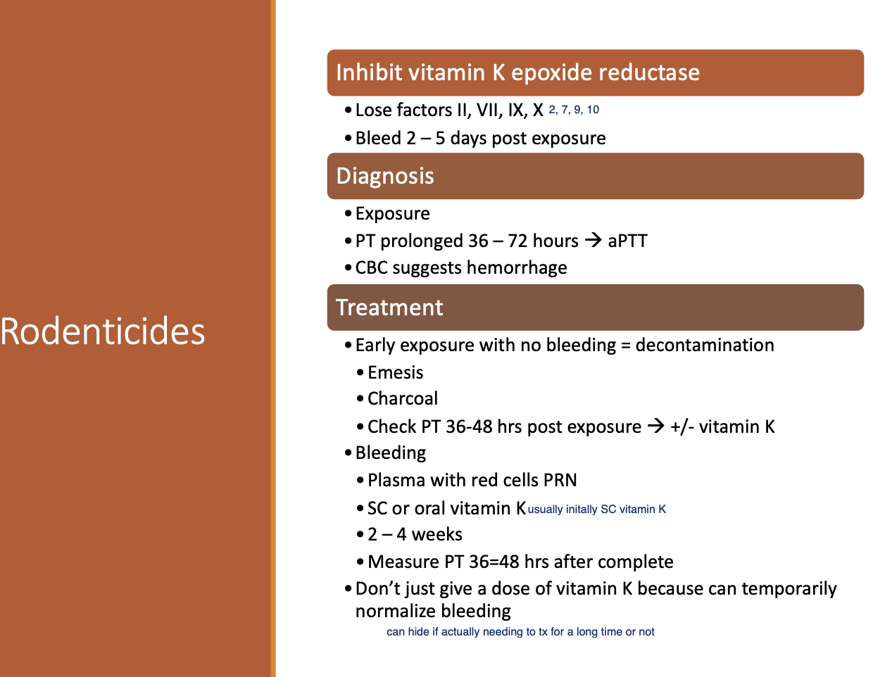
Pathogenesis of Rodenticide
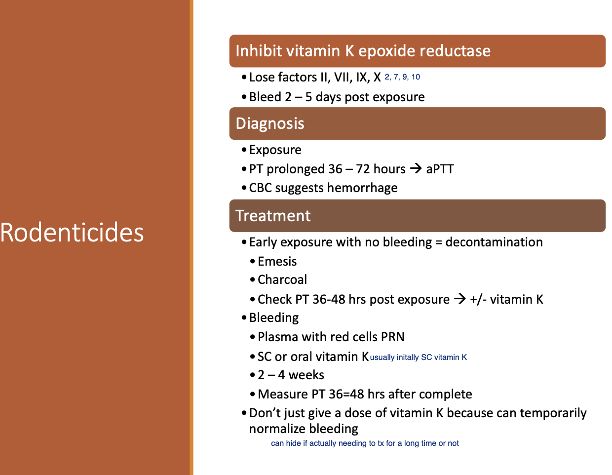
Inhibits Vitamin K epoxide reductase resulting in loss of factor 2, 7, 9, and 10.
Dx of Vit K deficiency (3)
Exposure to rodenticide.
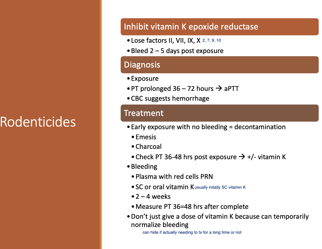
PT prolonged (36-72h) followed by PTT.
CBC suggesting hemorrhage.
Tx for Vit K deficiency - Early exposure w/ no bleeding (2)
Decontamination w/ emesis and charcoal.
Check PT at 36-48h post-exposure +/- Vitamin K.
Tx for Vit K deficiency w/ bleeding (3)
Plasma w/ red cells PRN.
SC or PO vitamin K x 2-4wks.
Measure PT 36-48 after completion
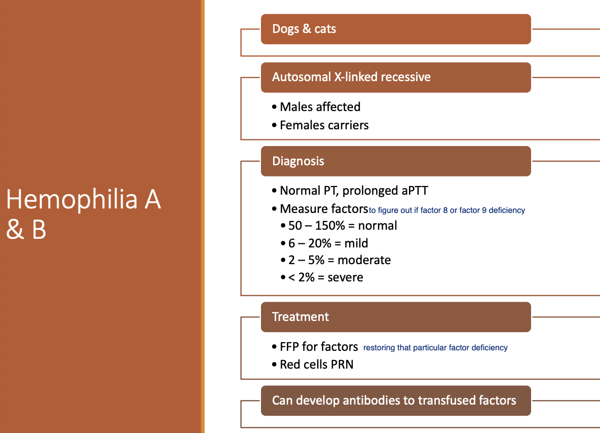
Hemophilia A and B inheritance
Autosomal X-lined recessive - males are affected
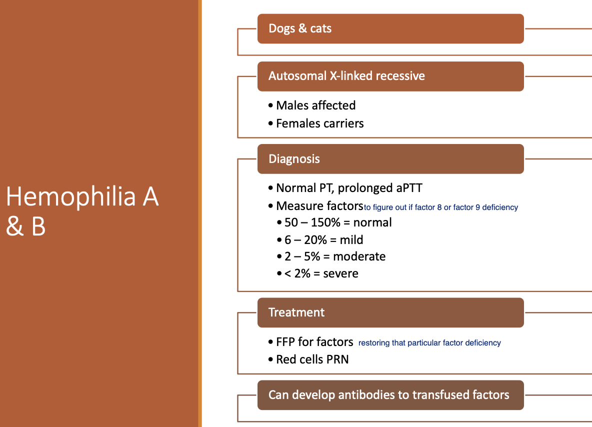
Dx of Hemophilia A and B (2)
Normal PT and prolonged PTT.
Measure factors.
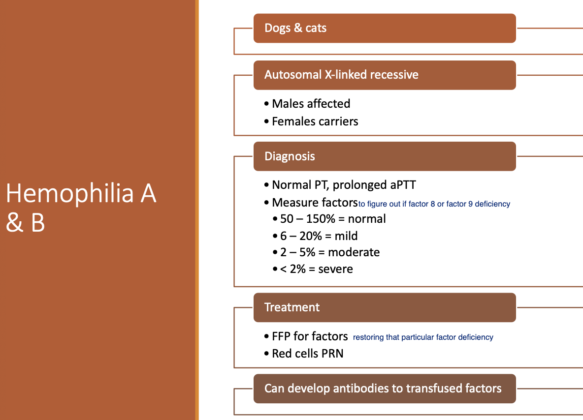
Tx for Hemophilia A and B (2)
FFP for factors.
Red cells PRN.
(Can develop Ab's to transfused factors)
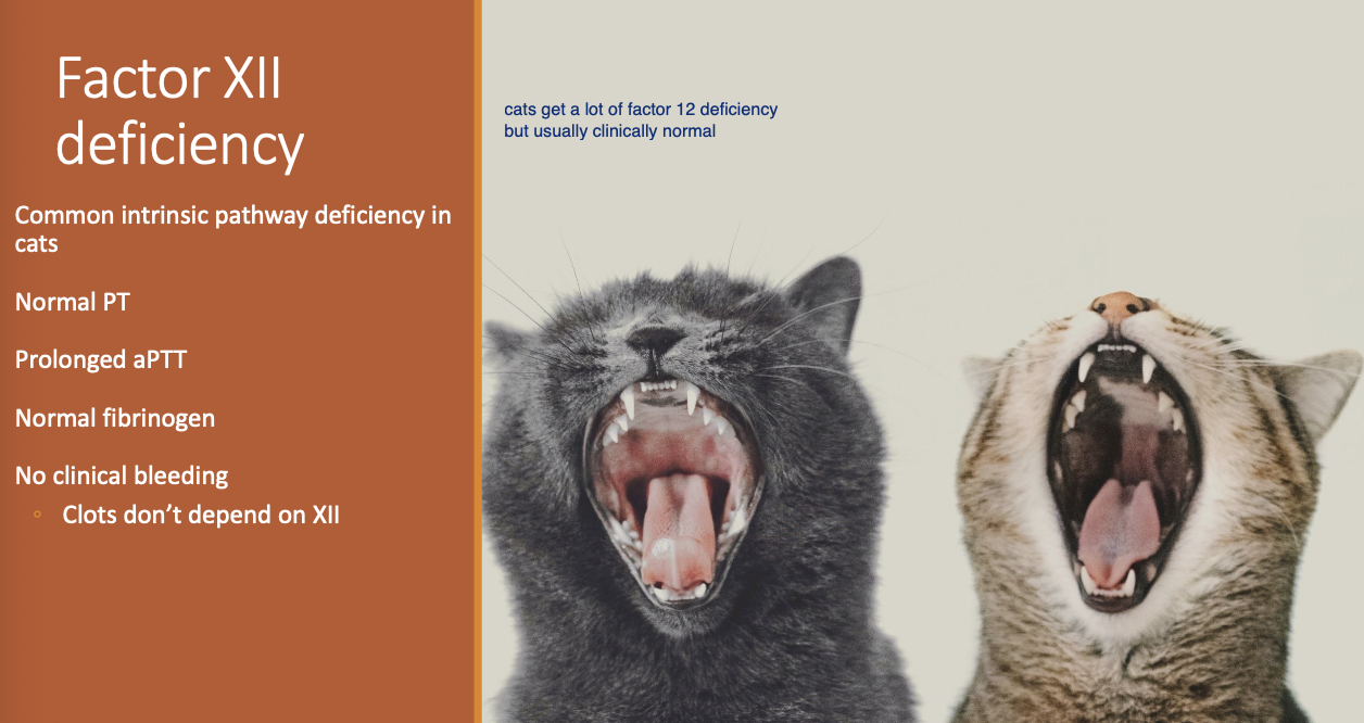
Factor XII deficiency is a common
intrinsic pathway deficiency in cats
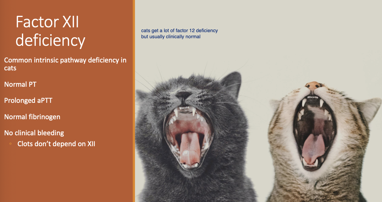
Dx for Factor XII deficiency (3)
Normal PT and prolonged PTT.
Normal fibrinogen.
No clinical bleeding b/c clot does not depend on 12 (may have prolonged bleeding w/ hemorrhage).
IMT etiology general (2)
Idiopathic - more severe.
Associated or secondary.
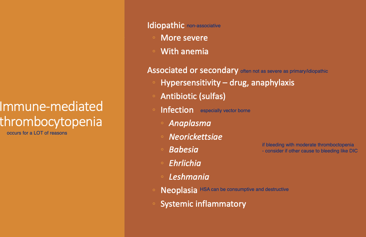
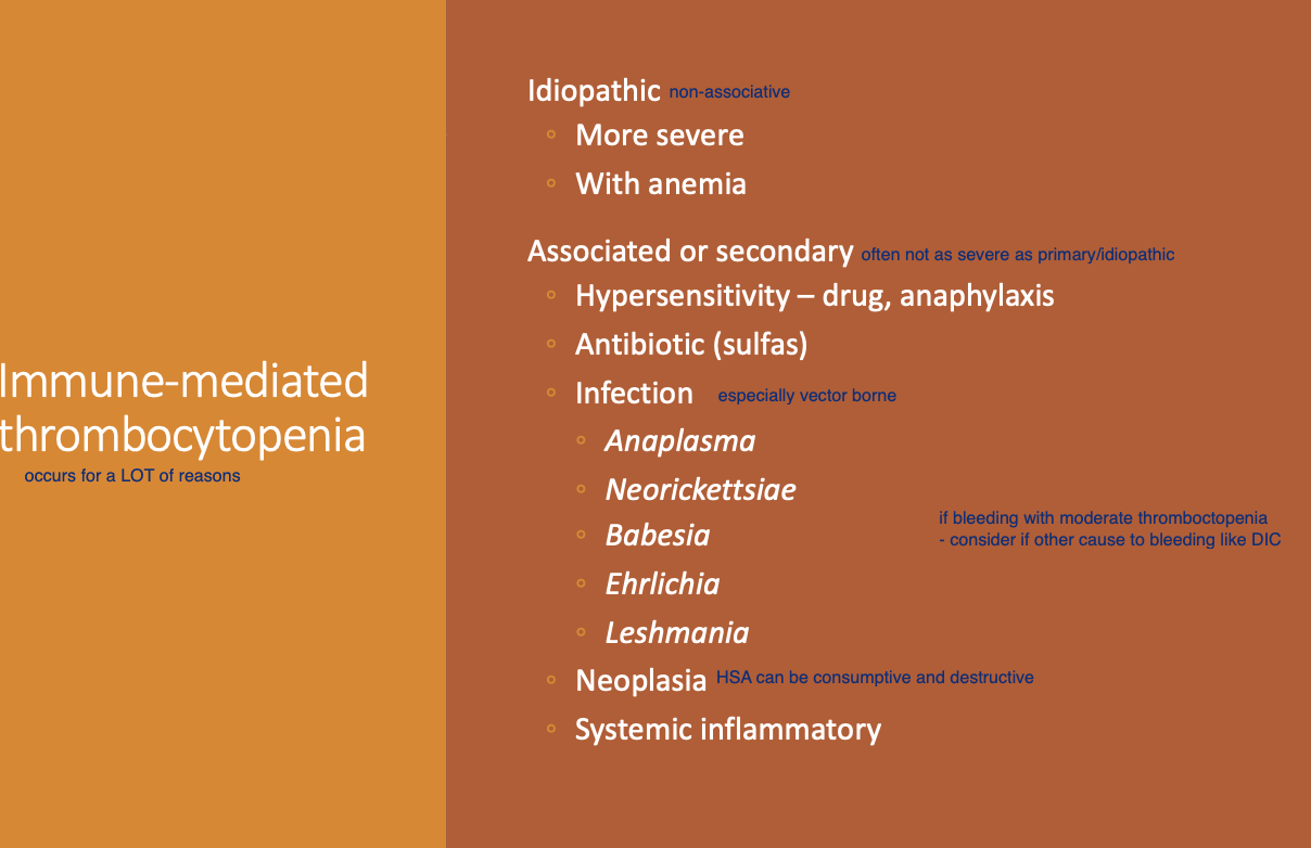
IMT etiologies - Secondary (5)
HS - drugs and anaphylaxis.
ABX - sulfa.
Infection.
Neoplasia.
Systemic inflammatory.
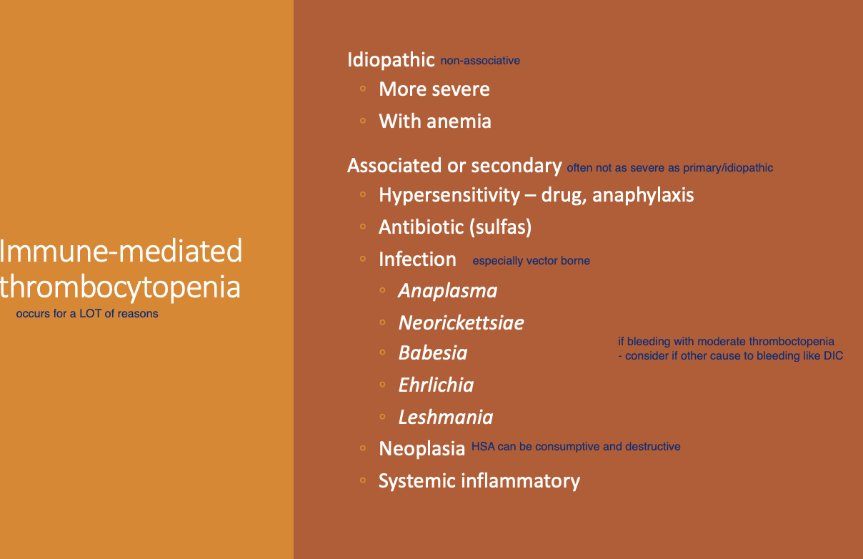
IMT - infectious etiologies (5)
Anaplasma.
Neorickettsiae.
Babesia.
Erlichia.
Leshmania.
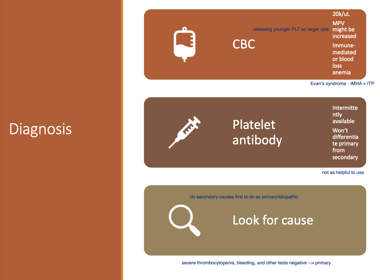
Dx for IMT (4)
Platelets <20k.
MPV may be increased.
Immune mediated or blood loss anemia.
Platelet Ab - intermittently available and does not differentiate primary v. secondary.
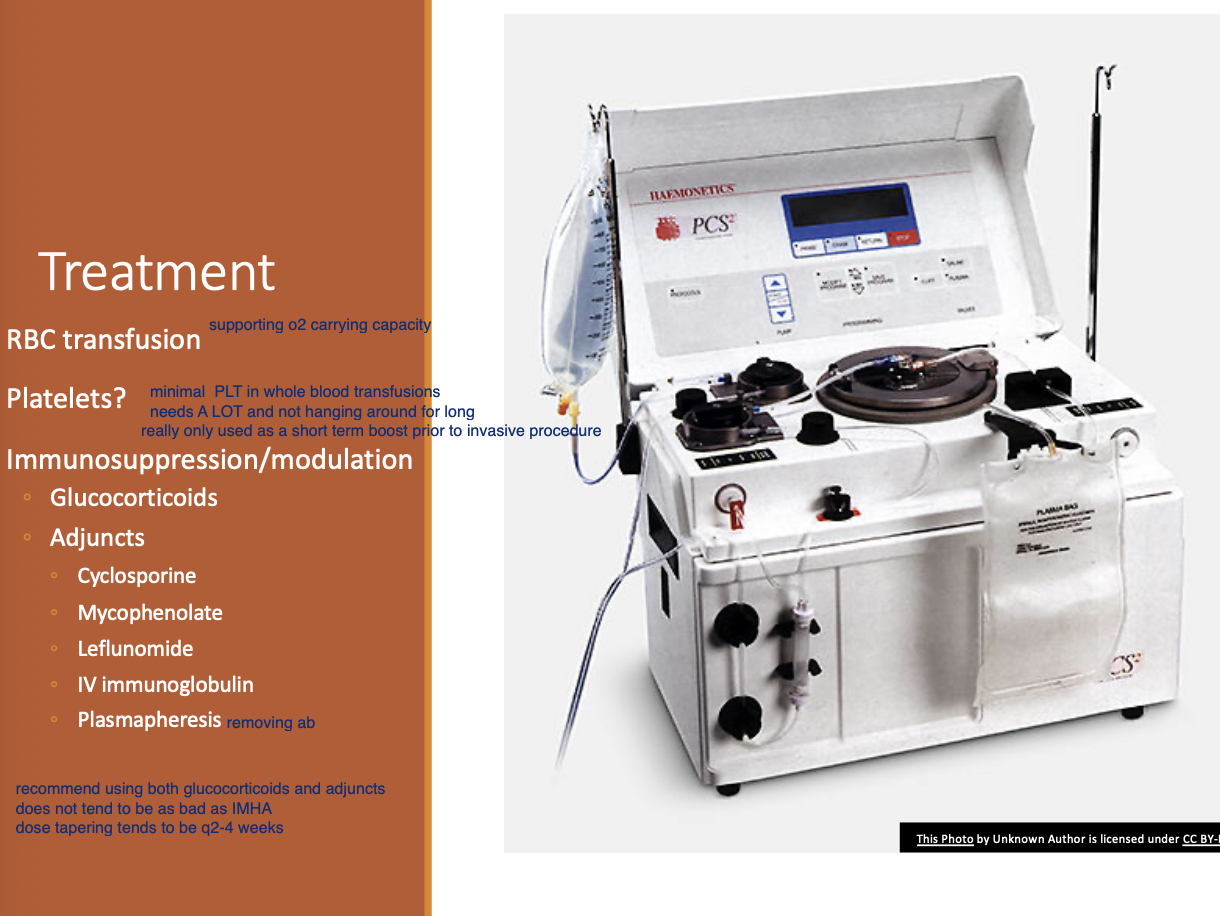
Tx for IMT - options (4)
RBC transfusion if anemic.
Glucocorticoids + adjunct.
Vincristine - premature platelet release (helpful in actively bleeding, unstable P).
Romiplastin - thrombopoietin receptor agonist.
Tapering for IMT (2)
GC: Taper q2wks w/ normal platelets by 25%
Adjunct: stop or taper once off pred.