Behavioural Neuroscience
1/31
There's no tags or description
Looks like no tags are added yet.
Name | Mastery | Learn | Test | Matching | Spaced | Call with Kai |
|---|
No analytics yet
Send a link to your students to track their progress
32 Terms
What have we learnt from comparative neuroanatomy.
Animals intelligence is not proportional to the size of the brain. Also relative size does not equal intelligence.
The more neurones within the brain, the greater the number of synaptic connections between neurones, the greater the coplexity of function the brain can support.
Animals that have particular types of skills have relatively larger brain areas dedicated to that particular function.
What were the propossed advantages of simulating the brain in the Human Brain Project?
Reduce the need for animal experiments
Study diseases in unprecedented in silico experiments
Improve the validation of data and experiments with computational validation.
Describe the major division of the nervous system.
CNS: Brain and spinal cord
PNS: Extensions of nerves out of the brain and spinal cord, that relay information to and from the CNS.
Within the PNS:
Somatic NS: receives sensory information from the sensory organs and controls movement of skeletal muscles. Voluntary movement
Remember Afferent is from periphery to the brain, efferent from the brain to the periphery.
Autonomic NS: connects central system to non-voluntary muscles and glands. Includes PSNS, SNS and ENS (supports digestion).
The ENS has its own reflexes and sense and can act autonomously (without CNS). NEarly every neurotransmitter found in the brain is also found in the gut. Plays a major role in emotion and stress.
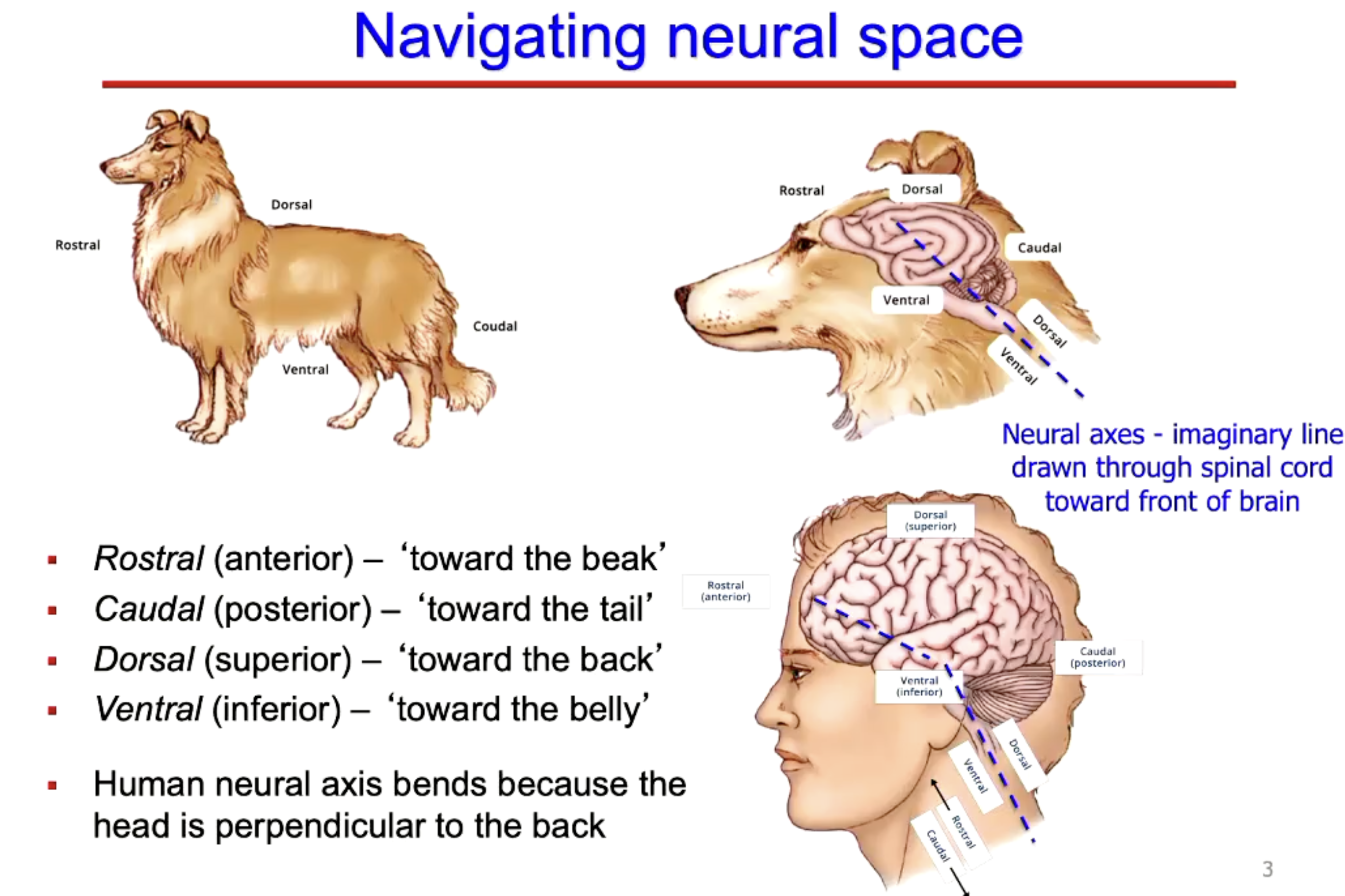
Describe the neuroaxis.
Rostral is anterior
Caudal (posterior
Dorsal (superior
ventral (inferior).
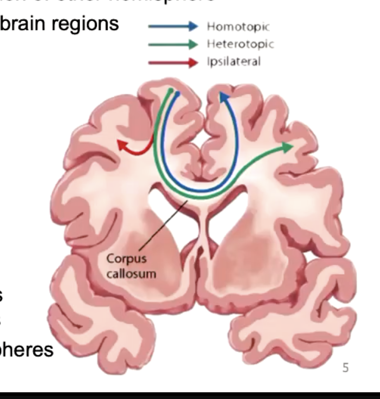
What is the Corpus callosum?
Consists of large bundle of axons that connect two hemispheres.
Green and plue is contralateral connections (opposite sides), red is ipsilateral (same side).
Homotopic connects complementary regions of other hemisphere.
Heterotropic communicates to different brain regions.
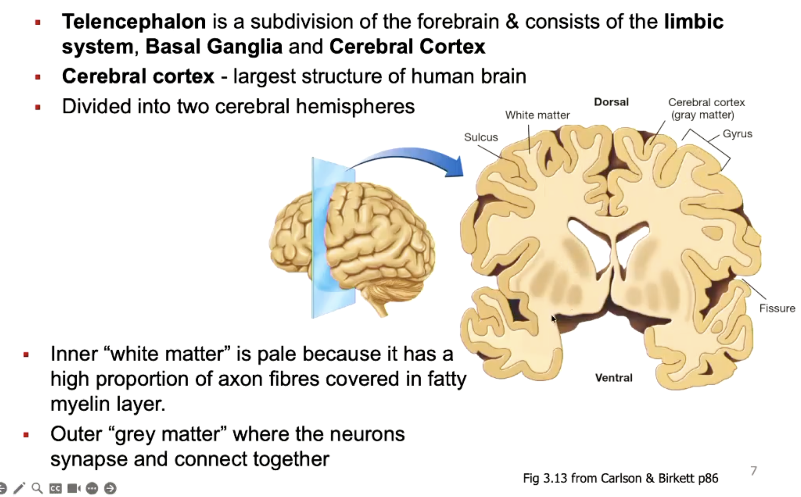
Describe the telencephalon.
Subdivision of the forebrain and consists of the limbic system, basal ganglia and cerebral cortex.
Divided into two cerebral hemisphers.
White matter is pale because contains myelinated axons (fatty).
Grey matter is where the neurones synapse and connect together.
Describe the basal ganglia.
The nuclei of the basal gangli (includes caudate nucleus and Putament) are responsible for controlling involuntary movement, particularly aspects that are highly autonomised or involuntary (such as walking)
BG dysfunction in Parkinson’s.
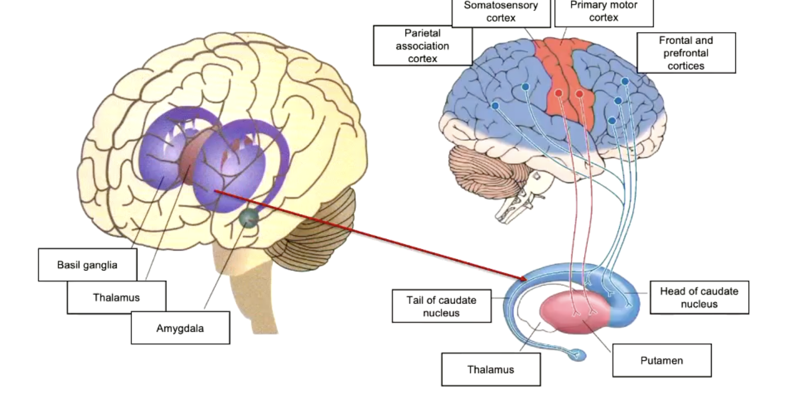
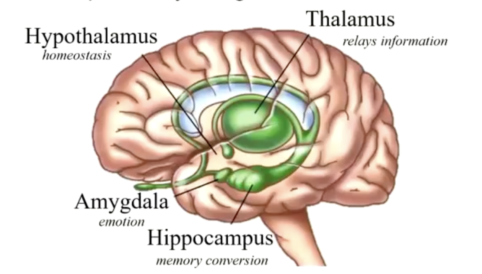
Describe the limbic system.
Includes the hypothalamus,thalamus, hippocampulus, amygdala, BG and was previously though to be the emotion circut.
Now we know the amygdala plays role in emotions, but hippocampus and parts surrounding the cortex are involved in learning and memory.
Thalamus: major relay station for sensory inputs to cerebral cortex.
Divided into several nuclei.
Sensory inputs in, relaying information back out of it.
Hypothalamus:
Controls the ANS and endocrine system.
Regualtes survival behaviours.
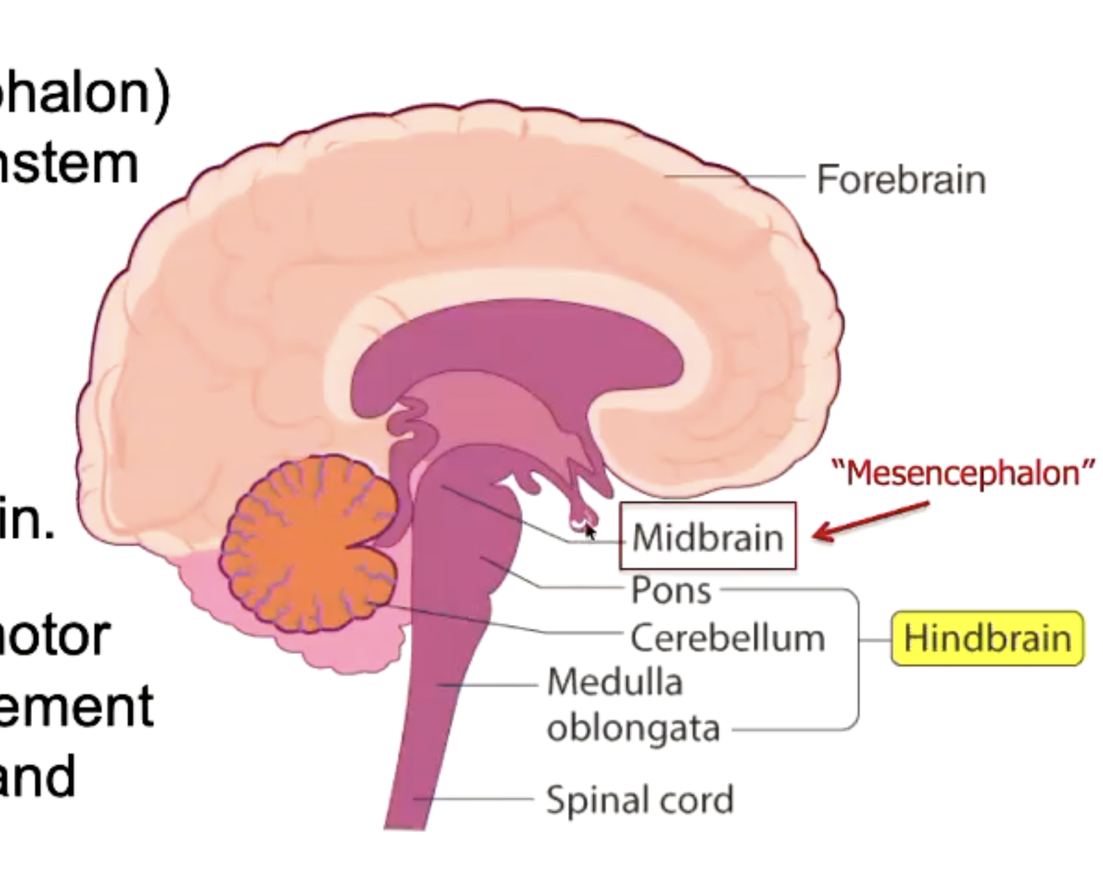
Describe the midbrain (mesencephalon)
The midbrain and hindbrain are located within the brainstem.
The MB is at topmost region of brainstem and sits directly above the hindbrain.
It connects the pons and cerebellum with the forebrain.
Plays an important role in motor movement, particularly movement of the eye and in auditory and visual processing.
Subsdivisions of forebrain, midbrain and hindbrain?
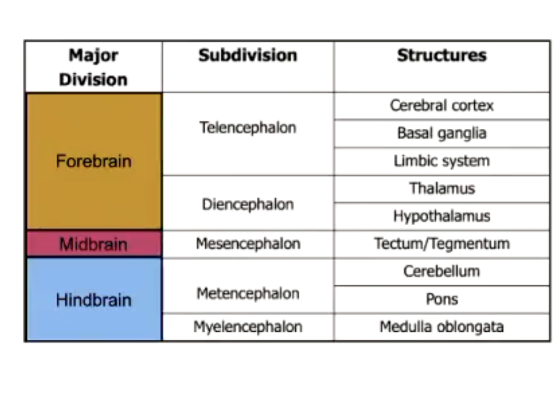
Describe the metencephalon.
Includes the cerebellum which receives info from the visual, auditory, somatosensory and vestibular (balance) systems helps coordination of movement.
Damage to the cerebellum causes problems with walking and leads to jerky, poorly coordinated movements and problems maintaining balance.
The pons lies on the ventral surface of the brainstem. It contains several nuclei important in regulating sleep and arousal; it also relays info from the cerebral cortex to the cerebellum.
Describe the myelencephalon.
Medulla oblongata.
Links the hindbrain to the spinal cord and contais neurones important for autonomic functions like respiration and heart rate.
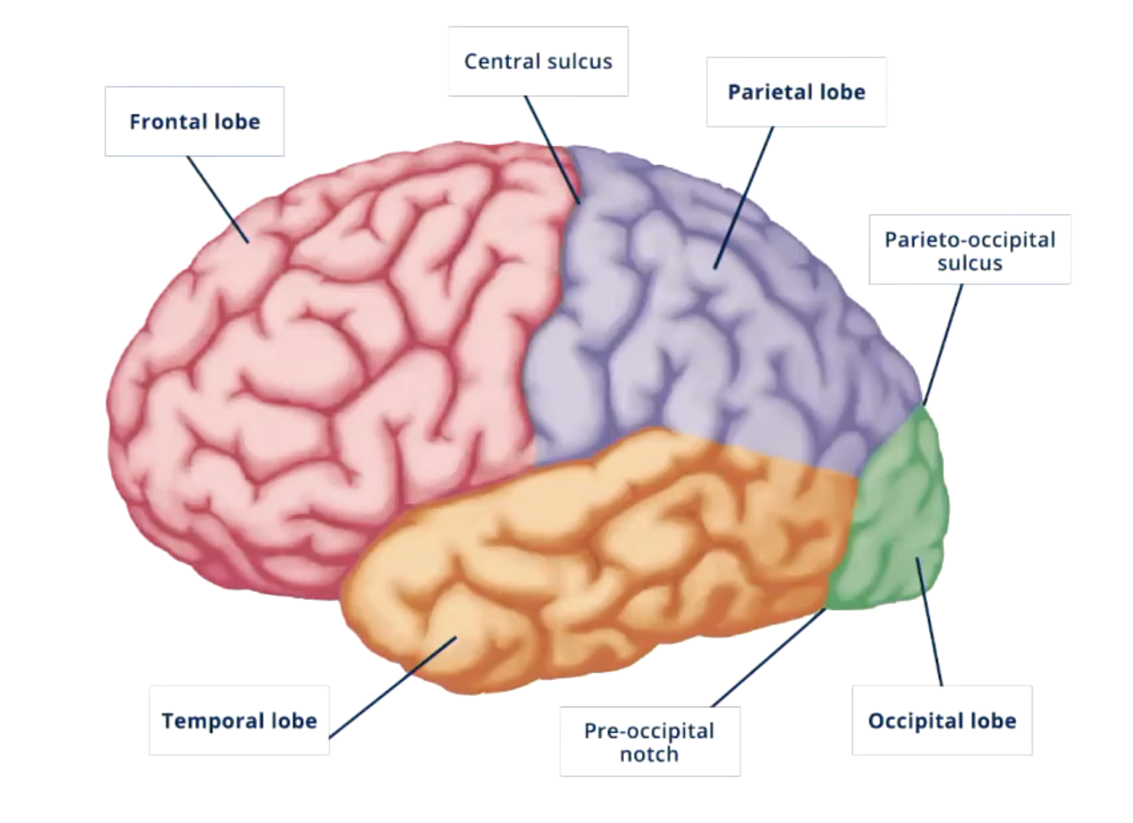
Describe the lobes of the cerebral cortex.
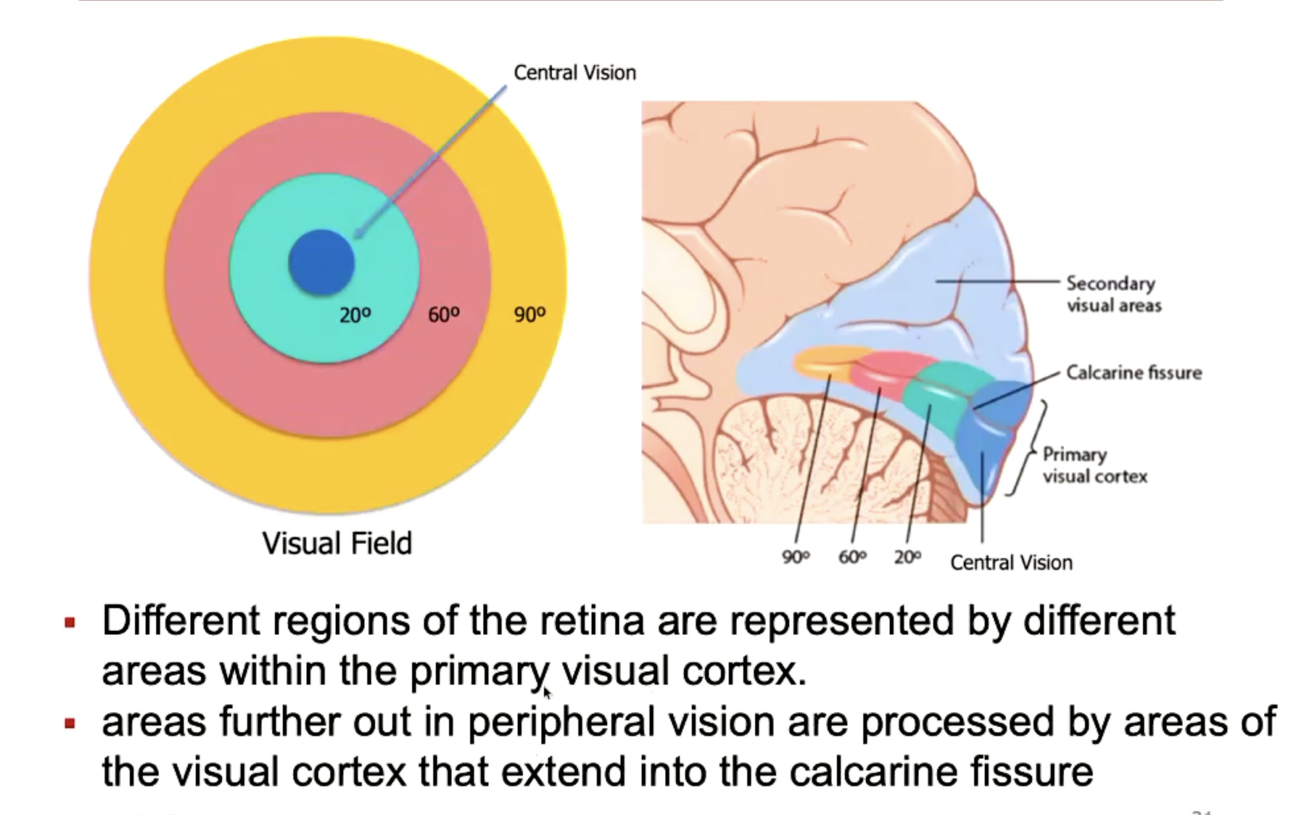
Describe the primary visual cortex.
Occupues the medial and lateral parts of the occipital lobe at the posterior of the brain.
Receives sensory info from retina.
The left and right visual fields are projected to the contralateral hemisphere (right visual field comes from left hemisphere).
Light stimulus from the external environment from both visual fields stimulate the corresponding area of the retina within the eye.
Different regions of the retina are represented by different area within the primary visual cortexz. The central vision has the alrgest region dedicated to the PVC, more neurones within this area opposed to outer regions.
Within the PVC, neurones show orientation selectivity. A straight vertical line leads to most neurones firing, opposed to horizontal line having the least, and specific neurones fire for only specific orientations.
Describe parietal lobe.
Involved in attention and spatial awareness.
Sits on dorsal surface of the cortex and is referred to as part of the dorsal stream and the “where pathway”.
Temporal lobe.
Involved in auditory processing.
Also involved in more complex visual processing (faces and complex object recognition).
Sits on the ventral surdace of the cortex and is part of the ventral stream and “what pathway”.
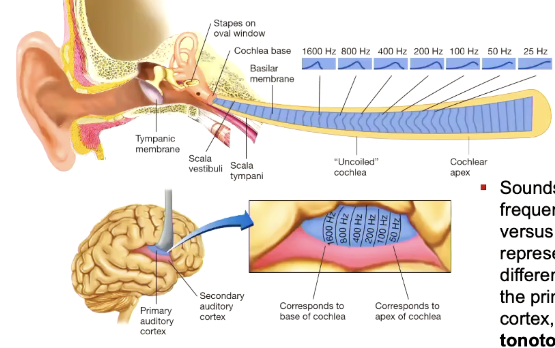
Describe the primary auditory cortex
Occupies superior part of the temporal lobe, as well as a patch of the cortex that is burried within the Sylvian fissure. It receives auditory sensory info from the cochlear.
Sounds of different freq are represented by different areas within the PAC, forming a tonotopic amp.
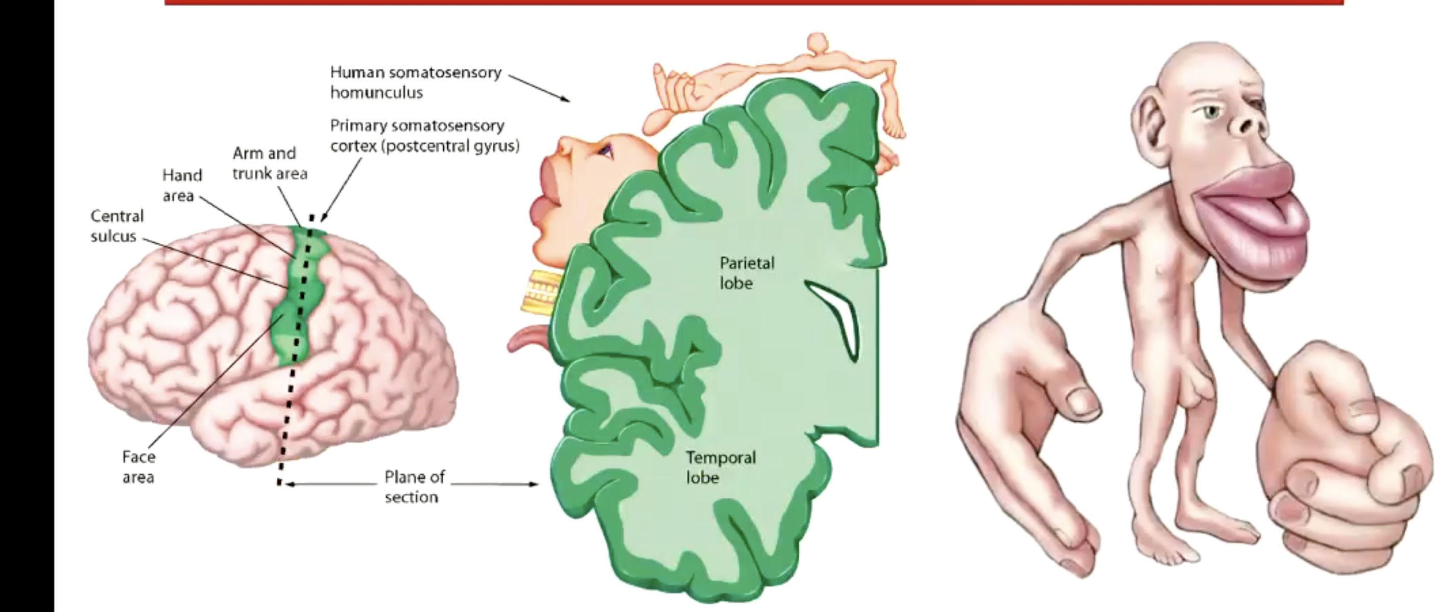
Describe the primary somatosensory cortex.
Located in the postcentral gyrus.
Receives sensory info from the skin.
Different regions of skin surface, represented by different areas along the strip of cortex, forming somatotopic map.
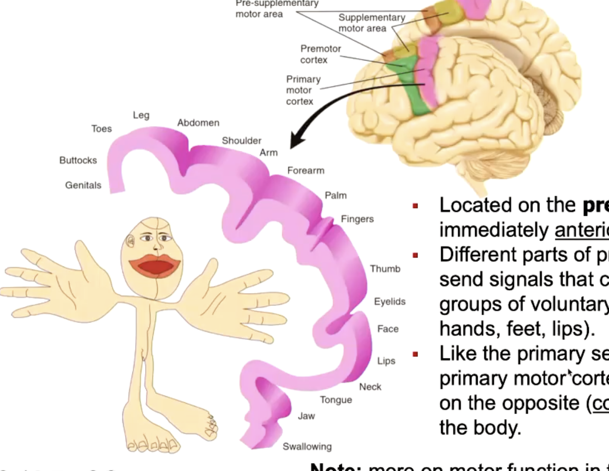
Describe the primary motor cortex.
Locates in precentral gyri.
Different parts of PMC send signals that control different groups of voluntary muscles.
The PMC controls muscles on contralateral side of body.
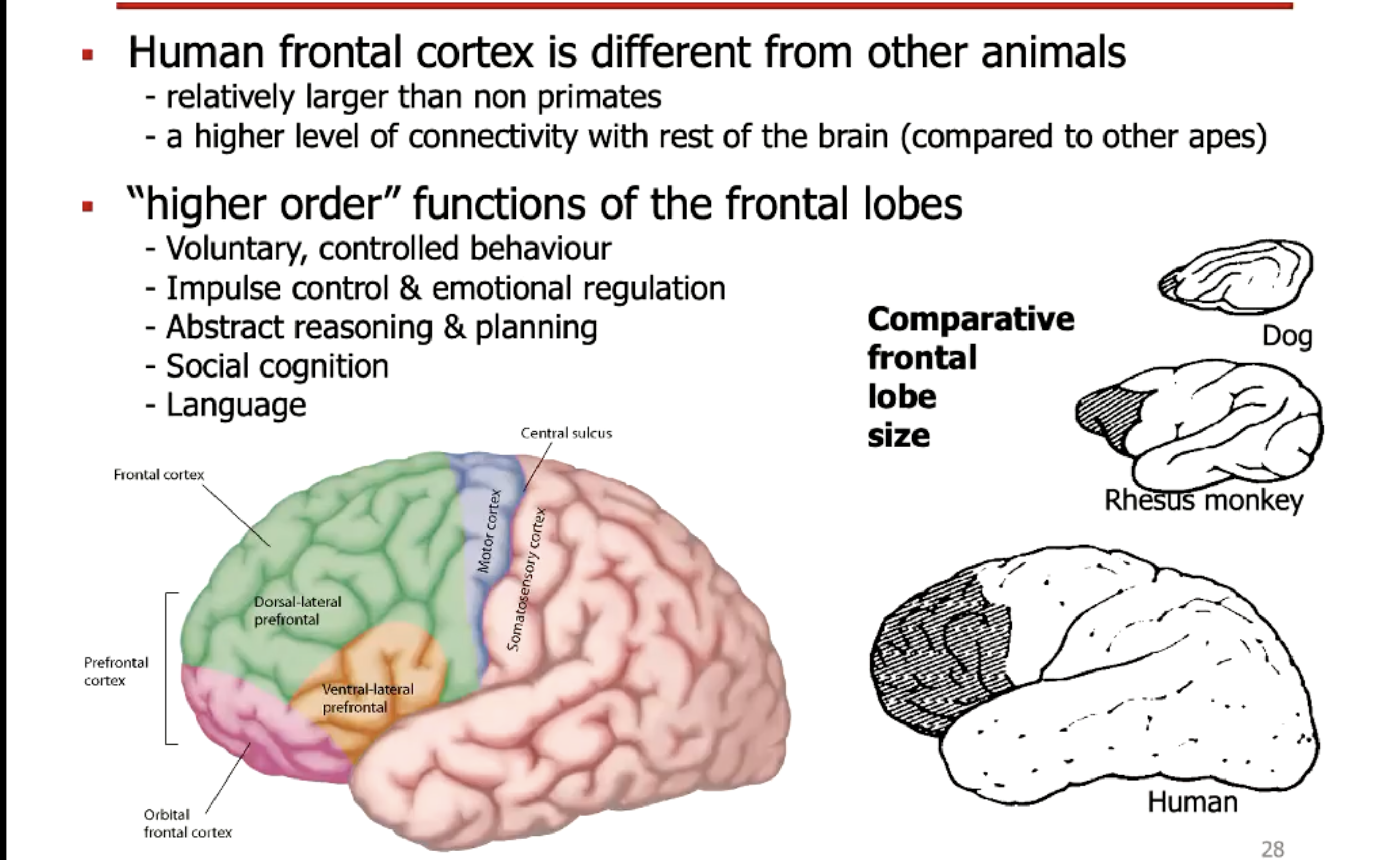
Frontal lobe.
Voluntary, controlled behaviour.
Impuse control and emotional reg
Abstract reasoning and planning
Social cognitoin and language.
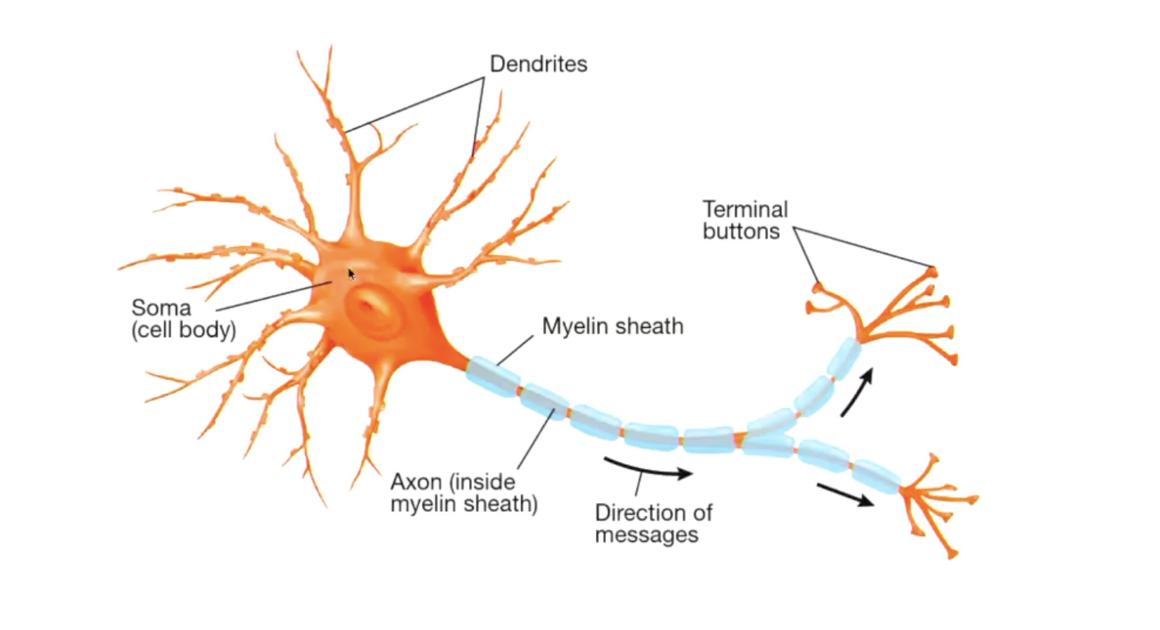
Describe structure of neurone.
Describe the generation of action potentials.
Caused by a change in flow of ions across the neurone’s PM, Na+, Cl-, K+ pump.
Depolarisation: Membrane potential (-70mV) reaches threshold potential (-55mV), leading to rapid opening of V-gated Na+ channels, increasing membrane conductance to Na+, leading to influx of Na+ into the cell. Leads to depolarisation to around +30mV. V-gated K+ channels remain close.
Repolarisation: V-gated Na+ become inactive due to inactivation gate blocking channel. V-gated K+ open, leading to K+ efflux as K+ leaves the cell. This leads to decrease in voltage.
Hyperpolarisation: Due to slow closing of V-gated K+ channels, it leads to membrane potential falling closer to equilibrium potential of K+.
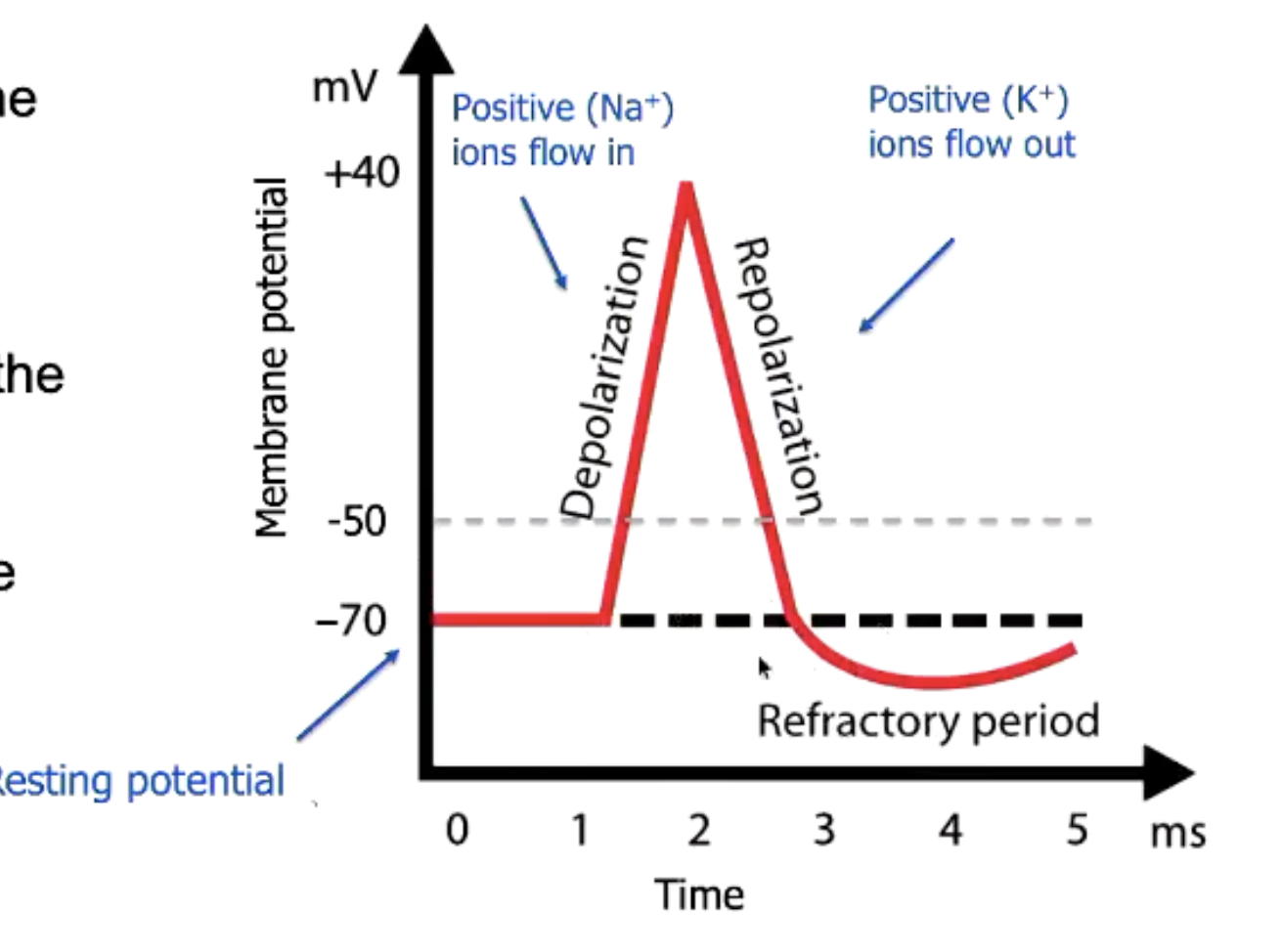
What are the refractory periods?
During and immediately after an action potential there are periods when it is hard to generate new action potentials.
Absolute refractory period: time where V-gated Na+ channel is open and inactive. Due to it being inactive, new action potential cannot be generated.
Relative refractory period: possible to generate new action potential, but it is harder to reach threshold potential due to the fact that V-gated K+ channel is open.
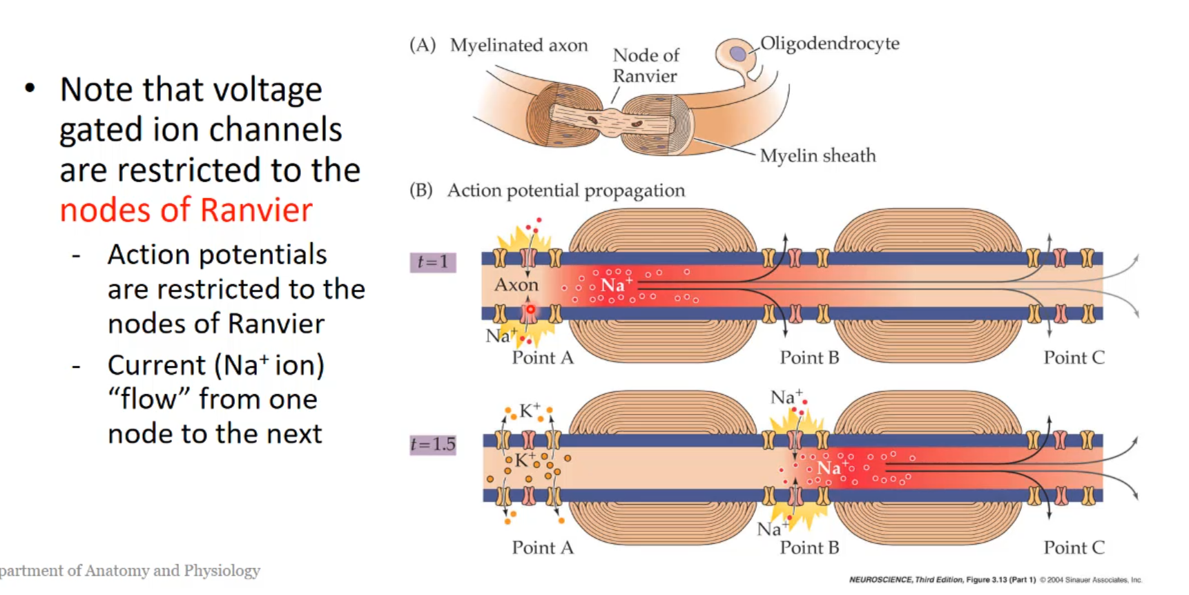
Describe the function of myelin.
Action potentials ‘jumps’ from myelinated section to myelinated section, where the current is pushed into the Node of Ranvier due to the generation of subsequent action potentials. This is called the “jumping transmission”(saltatory transmission). This is how transmission is fast at a myelinated transmission.
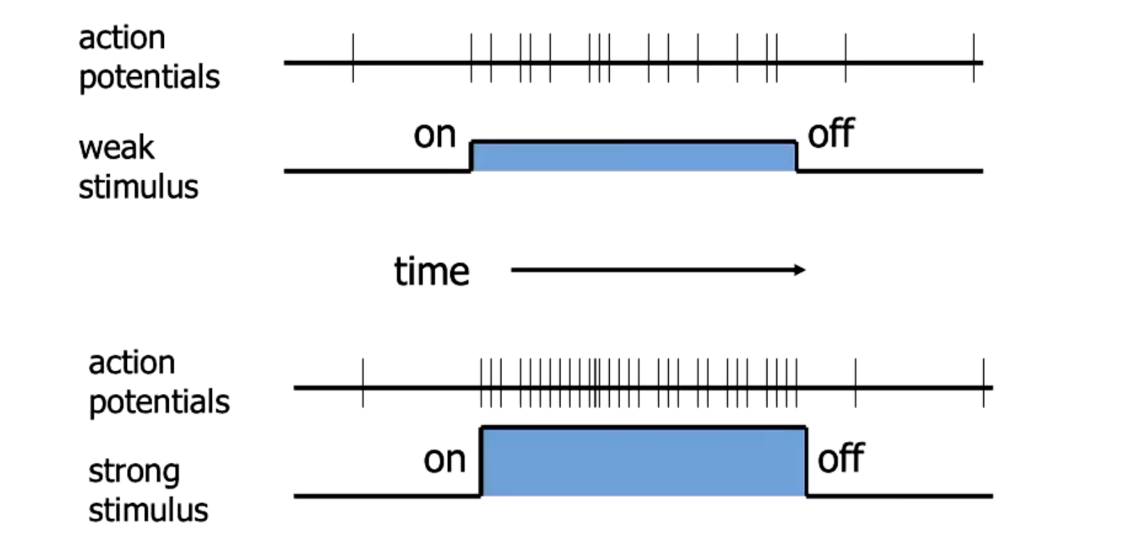
Describe the rate law of action potentials.
The neurone firing is all or none (no different intensities), so frequency of firing determines the strength of the neural signal.
Strong stimulus leads to reaching threshold potential faster, more frequent APs.
Describe neurotransmitter release.
At the axon button of the presynaptic neurone, there are neurotransmitter filled vesicles and present and primed to be released.
Across the synaptic cleft, the postsynaptic neurone contains receptors for the neurotransmitter.
Action potential depolarise the axon terminals PM of the presynaptic neurone. This leads to the opening of V-gated Ca2+ channels, allowing Ca2+ to enter the cell.
The incease of [Ca2+] in the intracellular environment, facilitates exocytosis. This causes neurotransmitters to enter the synpatic cleft via exocytosis.
Neurotransmitter passively diffuses across synaptic cleft to bind to receptors on postsynaptic neurone.
Describe neurotransmitter reuptake.
The synapse has the capcity to recycle and reuse neurotransmitters after they have been released via endocytosis.
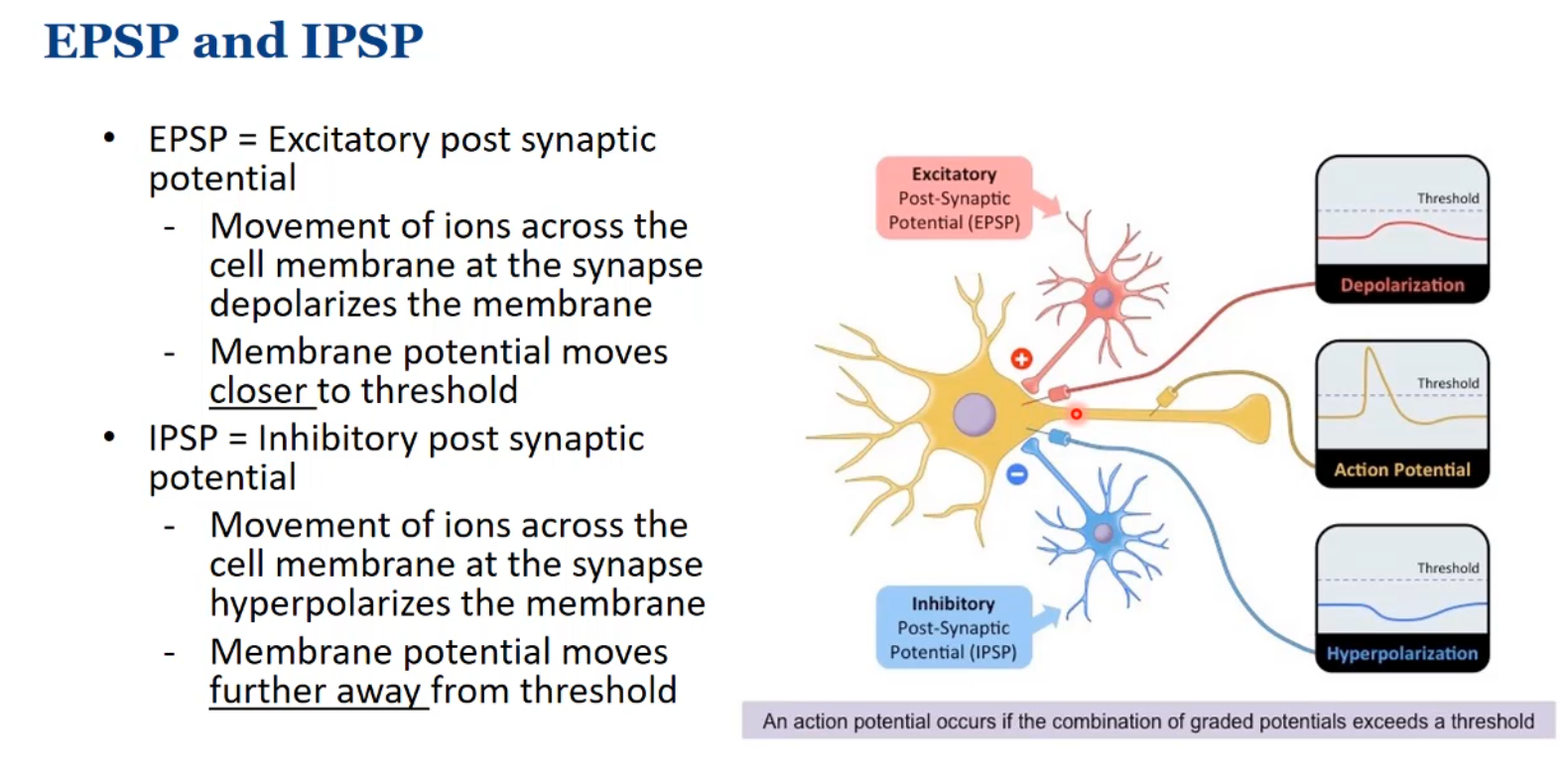
Describe EPSPs and IPSPs
Glutamate is the primary excitatory neurotransmitter.
GABA is the primary inhibitoruy neurotransmitter.
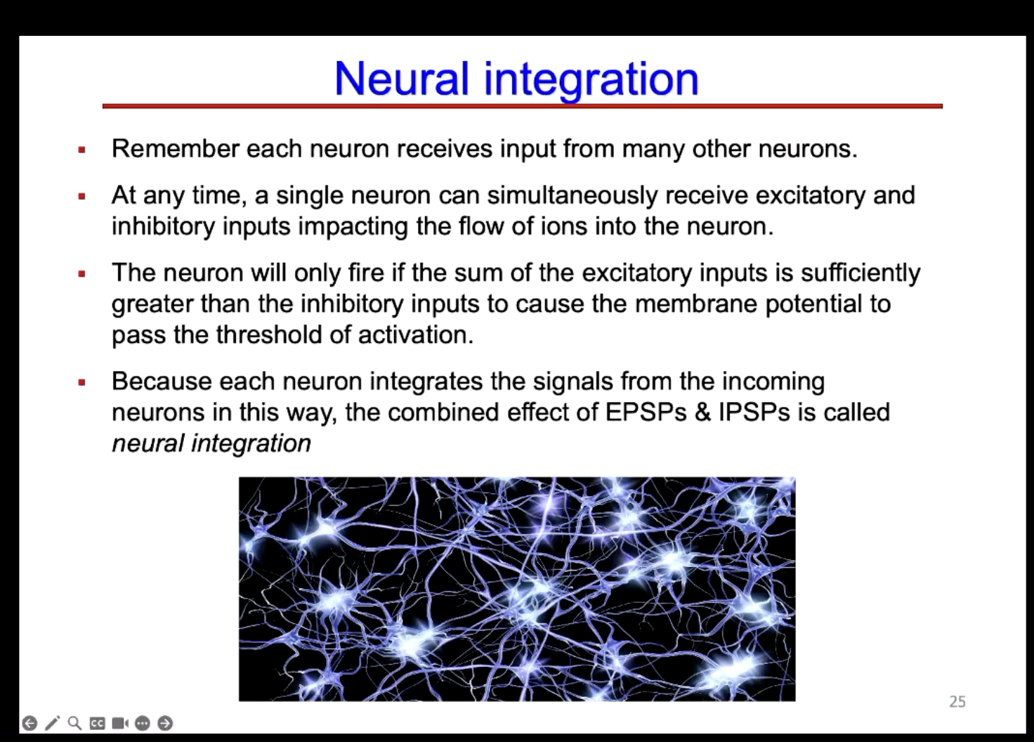
They can summate. The combined effect of EPSPs and IPSPs is called neural integration.
Describe neural integration.
Temporal summation.
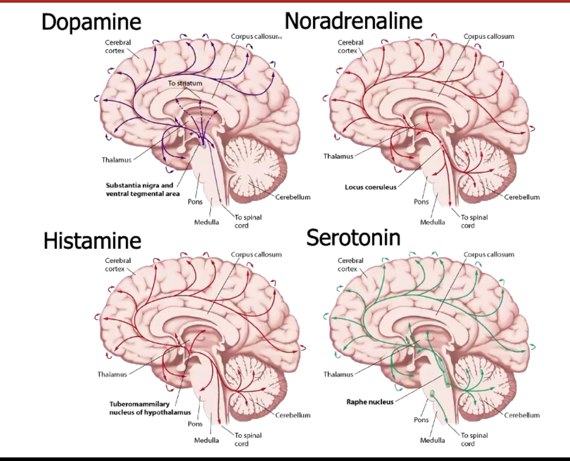
Describe neuromodulators.
Relatively small number of neurones for the neuromodulators, but have long projections. This allows for coordination of brain activity.
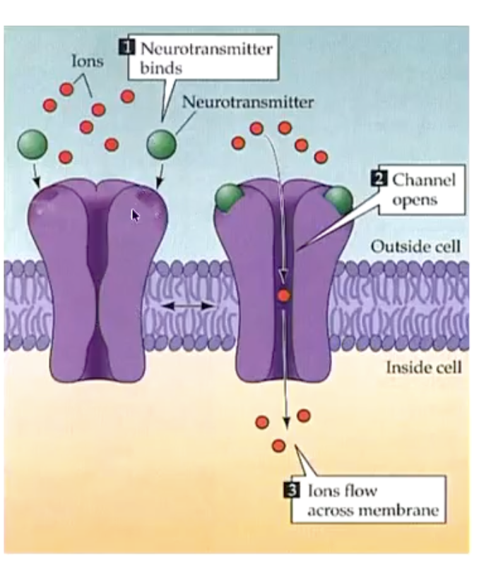
Describe the activity of neurotransmitter at receptors.
Neurotransmitters do not typically enter the post-S neurone.
TO cause an effect on the PS neurone, the chemical message is receivied by attaching to the bindingsite for the receptor, which may open something like an ion channel.
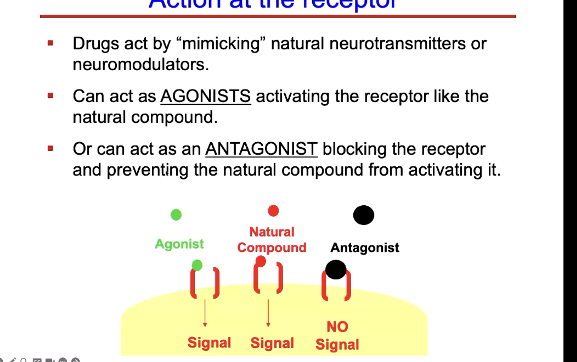
Describe the action at a receptor that can be caused by a drug.
Drugs can impact every stage of neurotransmitter function from synthesis to release to receptor binding.
Drugs impact psychological processes only because they mimic the same biological responses triggered by naturally occuring substances. Psychological evenets that directly impact the biological process requires neurones to fire and chemical messages to be sent across neurones.