exam 3 full
1/167
There's no tags or description
Looks like no tags are added yet.
Name | Mastery | Learn | Test | Matching | Spaced | Call with Kai |
|---|
No analytics yet
Send a link to your students to track their progress
168 Terms
What are the two functional classifications of the nervous system based on consciousness?
-Somatic Nervous System (Consciously controlled)
-Autonomic Nervous System (Nonconsciously controlled)
What are the main characteristics of the somatic Nervous system ?
Skeletal muscles
Consciously controlled system
Somatic sensory neurons: Stimuli induced (i.e., special senses)
Somatic motor neurons: Voluntary initiation of skeletal muscle movement
What are the main characteristics of the Autonomic Nervous System ?
Smooth and cardiac muscle
Nonconscious controlled systems.
Visceral Sensory: Sensory system that initiates response of ANS
Autonomic motor: Involuntary initiation of smooth and cardiac muscles or glands
What is the function of the ANS ? (Autonomic System)
It is to maintain homeostasis
How much number of motor neurons do somatic motor units have ?
one
How much number of motor neurons do Autonomic motor units have ?
two
What are the two divisions of the Autonomic Nervous System ?
-Sympathetic
-Parasympathetic
What are the functions of the Sympathetic Division ?
Increases metabolic and alertness activities
Regulates the state of overall elevated activity and attention (“flight or fight response)
blood pressure and heart rate increase
What are the functions of the Parasympathetic Division ?
Maintains homeostasis
Conserves and replenishes energy
“Rest and digest” division
Where do all sympathetic preganglionic neurons originate ?
Lateral Horn of the T1 – L2 spinal cord
What are the four pathways regulated by sympathetic neurons ?
Spinal Nerve Pathway
• Postganglionic sympathetic nerve pathway
• Splanchnic nerve pathway
• Adrenal medulla pathway
What are the two internal fluid cavities of the eye ?
Anterior Cavity (Aqueous humor)
Posterior Cavity (Vitreous humor)
What accessory structures of the eye protect foreign objects from entering the eye ?
Eyebrows
Eyelashes
Eyelids
What accessory structure of the eye keep exposed surface of the eye moist ?
Lacrimal apparatus
Which accessory eye structure provides superficial covering of the anterior and posterior
exposed surface.
Conjuctiva
What are the three layers (tunics) of the eye ?
Fibrous
Vascular
Retina
What are the three main divisions of the ear ?
Inner ear
Middle ear
External Ear
What are the structures in the external ear ?
Auricle
External meatus acoustic
Tympanic membrane
What is the structure and function of the Auricle ?
Most external portion
skin-covered, cartilaginous funneled shape structure
that protects the inner workings of the ear and to direct sounds inwards
What is the structure and function of the acoustic meatus ?
Hollow tube that moves sounds inwards.
What is the structure and function of the tympanic membrane ?
Connected from external acoustic meatus
vibrates transmitting sounds energy into the middle and inner ear.
Aka Eardrum
What are the structures in the middle ear ?
Tympanic cavity
Auditory ossicles
Auditory Tube
What is the structure and function of the Tympanic Cavity ?
Houses the auditory ossicles
maintains an opening to the outside through the nasopharynx called the
Auditory Tube (Eustachian Tube)
What are the structure/function of the Auditory ossicles ?
made up of three bones which vibrate to amplify the sound going into the inner ear (Malleus, Incus, Stapes)
protected by two muscles the stapedius and tensor tympani which restrict the bones movement when
loud sounds are detected
What are the structures in the inner ear ?
Bony labyrinth
Membranous lambyrinth
What is the bony labyrinth and its three compartments?
Semicircular canals
Vestibule
Cochlea
What is the membranous labyrinth ?
Resides inside the bony labyrinth
membrane-lined tubes filled with fluid
house the receptors for equilibrium and hearing.
What fluids are found in the bony labyrinth?
perilymph fluid
What fluids are found in the membranous labyrinth ?
endolymph and perilymph fluid.
What are the olfactory organs components?
• Mucous layer
• Olfactory Epithelium
• Lamina Propria
• Cribriform Plate
• Olfactory Bulb
What are the three specialized cells in the olfactory epithelium?
Olfactory receptor cells
Supporting cells
Basal cells
What are the functions of the Olfactory Receptor cells in the olfactory epithelium?
Detects odors
What are the functions of the supporting cells in the olfactory epithelium?
Sandwich the olfactory receptors and sustain receptors
What are the functions of the basal cells in the olfactory epithelium?
Continually replace Olfactory Receptor Cells
What is the purpose of the olfactory epithelium?
Lines the superior part of nasal cavity
What is the function of the lamina propria in the olfactory organs ?
house the Olfactory glands that are responsible for producing the mucous as well as the blood supply and nerves
What is the structure of the Olfactory Receptor cells?
Bipolar Neurons with one dendrite and an unmyelinated axon.
Chemoreceptors that detect odors.
Axons form bundles of the Olfactory Cranial Nerve (CN I)
What are the three type of cells that make up our taste buds ?
Gustatory cells
Supporting cells
Basal cells
What are the functions of the Gustatory cells in Taste Buds ?
detect tastants (taste-producing molecules and ions). Short lived
Regenerated every 7 to 9 days.
Starting at age 50 regeneration declines and our ability to
distinguish taste decline.
What are the functions of supporting cells in taste buds ?
Sustain Gustatory cells
What are the functions of the Basal cells in taste buds ?
Neural stem cells that continually replace Gustatory cells
what is the nervous system composed of?
conductive calls and supportive cells
What are the major components of the Central Nervous System (CNS)?
brain and spinal cord
what is the central nervous system?
the command and control center of the nervous system
what does the peripheral nervous system do
projects info to and receives info from the central nervous system
what are the anatomical components of the peripheral nervous system
cranial and spinal nerves and ganglia
what are the special characteristics of neurons
high metabolic rate
extreme longevity
nonmitotic
excitable
conductivity

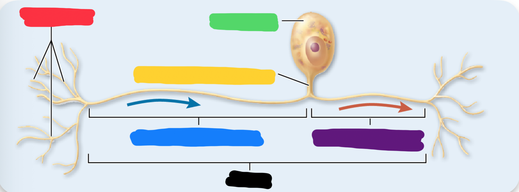
what is the blue blank pointing to on the neuron?
dendrites
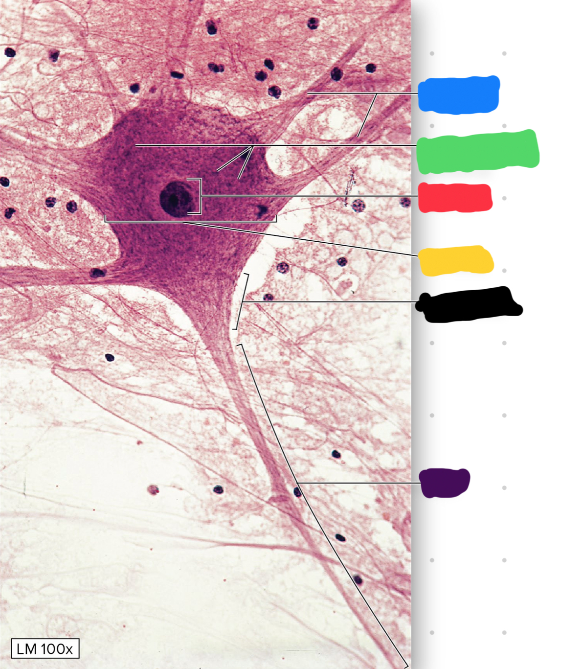
what is the green blank pointing to on the neuron?
chromatophilic substance

what is the red blank pointing to on the neuron?
nucleus

what is the yellow blank pointing to on the neuron
cell body

what is the black blank pointing to on the neuron
axon hillock

what is the purple blank pointing to on the neuron
axon
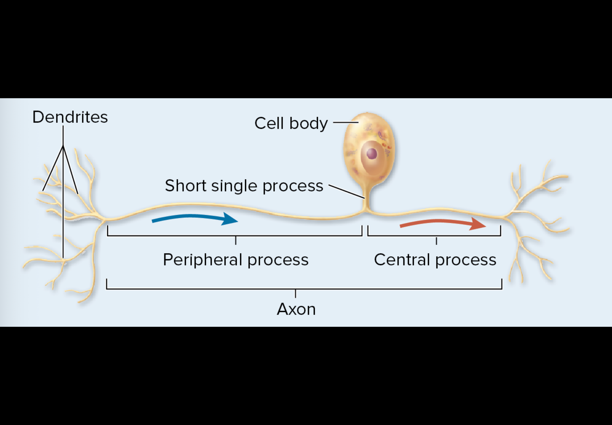
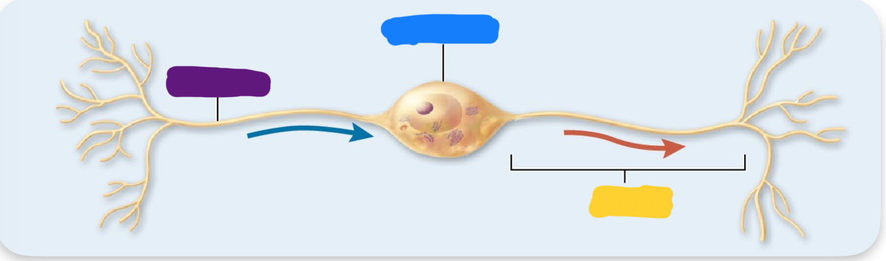
which type of neuron is this: (polar)
unipolar
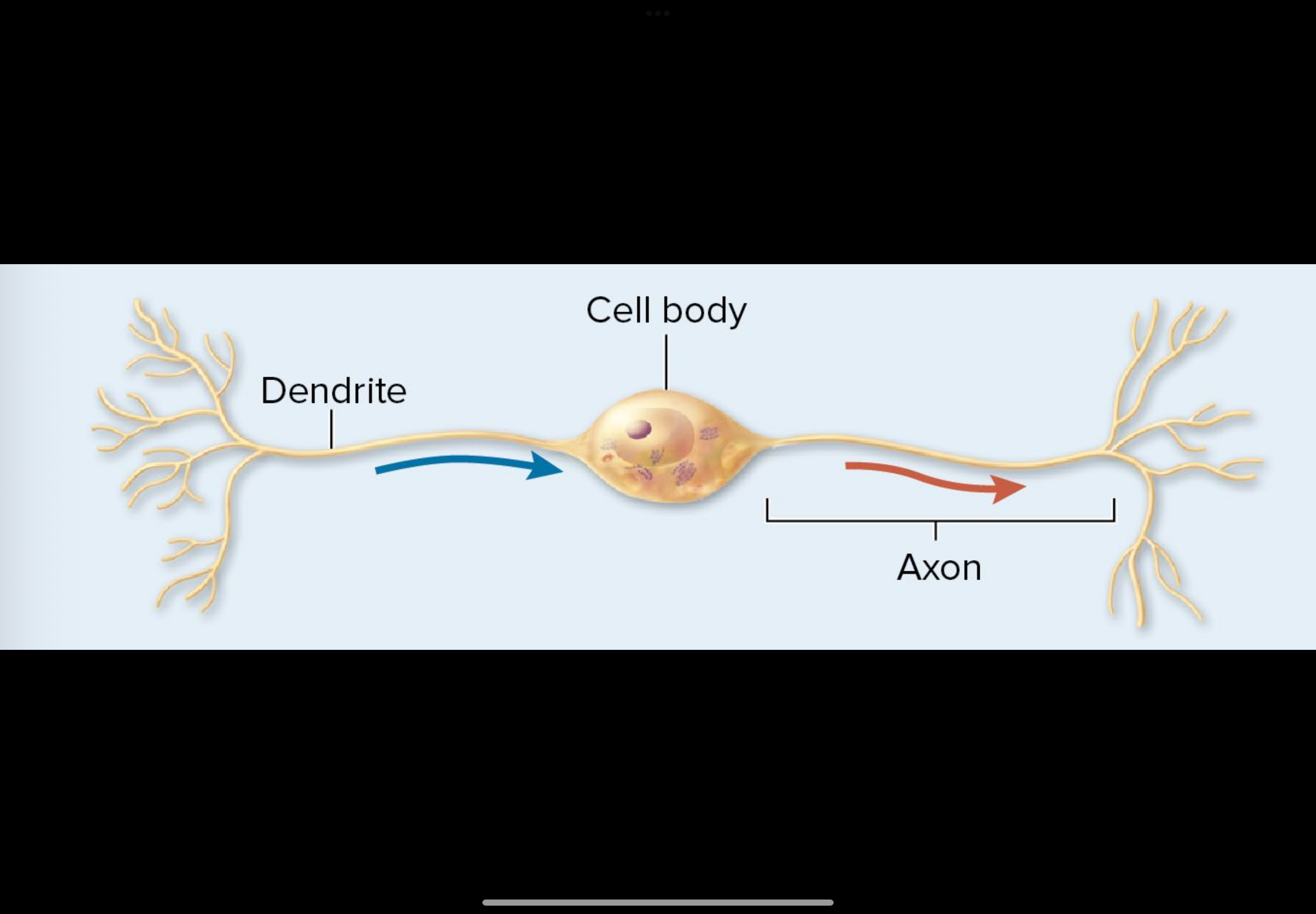
which type of neuron is this: (polar)
bipolar
which type of neuron is this: (polar)
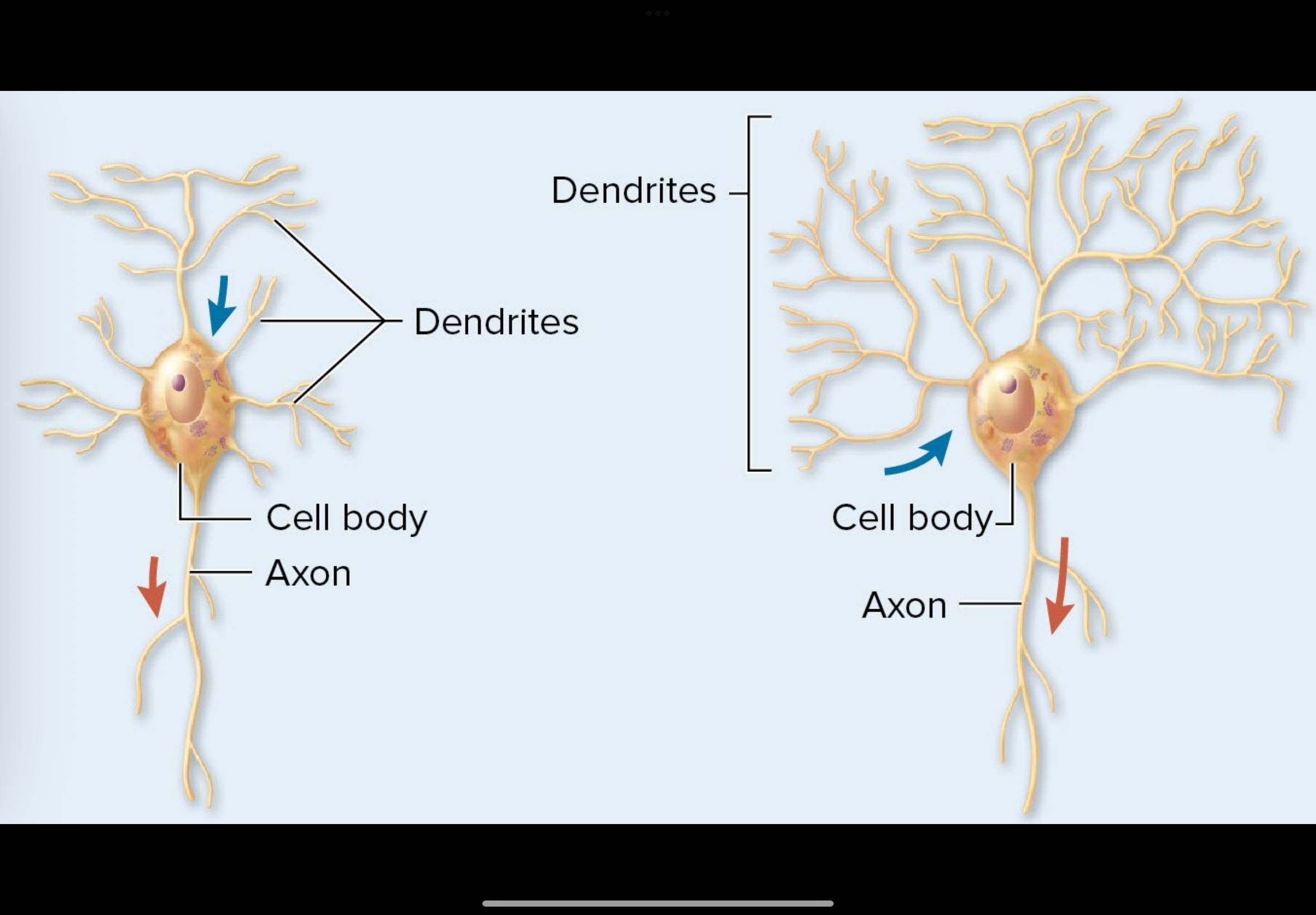
multipolar

what is the red pointing to on the unipolar neuron
dendrites

what is the blue pointing to on the unipolar neuron
peripheral process

what is the green pointing to on the unipolar neuron
cell body

what is the black pointing to on the unipolar neuron
axon

what is the yellow pointing to on the unipolar neuron
short single process
what is the purple pointing to on the unipolar neuron
central process

what is the purple pointing to on the bipolar neuron
dentrite

what is the blue pointing to on the bipolar neuron
cell body
what is the yellow pointing to on the bipolar neuron
axon

what is the red pointing to on the multipolar neuron
cell body
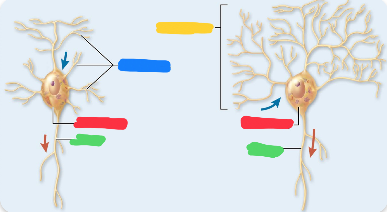
what is the blue pointing to on the multipolar neuron
dendrites

what is the yellow pointing to on the multipolar neuron
dendrites

what is the green pointing to on the multipolar neuron
axon
how are neurons classified functionally
by the direction the nerve pulse travels relative to the CNS (central nervous system)
what neuron classification carries impulses from sensory receptors into central nervous system?
sensory (afferent)
what neuron classification carries impluses away from the central nervous system to muscles or glands?
motor (efferent)
what classification of neuron carries impluses within the central nervous system, typically multipolar neurons.
interneuron(associate)
what neuron classification makes up 99% of neurons found in the body
interneuron (associate)
what does neuroglia do
supports cells that are non-conductive
what type of neuroglia is large, has branching cells that form the blood brain barrier?x
astrocytes
which type of neuroglia has cells with few branches that form myelin
oligodendrocytes
which type of neuroglia has tiny cells with complex branches and are phagocytes
microglia
which type of neuroglia is ciliated and secretes the cerebrospinal fluid
ependymal cells
which type of neuroglia is myelin producing
neurolemmocyte
which type of neuroglia isolates and nourishes cell bodies in ganglion
satellite cells
what is the main role of the axon?
to send nerve pulses
what is a nerve impulse
the rapid movement of an electrical charge along the axon plasma membrane
electrons are electrically excitable (true/false)
true
what allows for the speeding up of the nerve impulse along the axon
myelination
how is oligodendrocyte different from neurolemmocyte is myelination process?
oligodendrocytes can myelin ate multiple axons white neurolemmocyte can only do one axon.
what is the movement of a nerve impulse from one node to another node
saltatory conduction
what is the process In unmyelinated axons the nerve impulse travels in one pass
continuous conduction
what is the most common synapses
axodendritic synapses
which synapses is the one where synaptic knobs of pre-synaptic neuron interaction with dendrites of post synaptic neuron
axodendritic synapse
which synapses is the one where synaptic knobs of pre-synaptic neuron interaction with cell body of post synaptic neuron
axosomatic synapse
which synapses is the one where synaptic knobs of pre-synaptic neuron interaction with synaptic knob of post synaptic neuron
axoaxonic synapse
what are the two distinct types of neural tissue which make up the brain an spinal cord
gray matter and white matter
which matter houses motor neuron and interneuron cell bodies, dendrites, terminal arborizations, and unmyelinated axons
gray matter
which matter Contains myelinated neurons, which
give the tissue its color
white matter
what is the outer layer in cranial meninges, closest to the skull?
dura mater
what is the middle layer in cranial meninges?
arachnoid mater
what is the inner layer in cranial meninges?
pia mater