PPOM 1 Week 7 LEC 59-68
1/519
Earn XP
Description and Tags
Together, we can stop this. Spread the word.
Name | Mastery | Learn | Test | Matching | Spaced | Call with Kai |
|---|
No analytics yet
Send a link to your students to track their progress
520 Terms
(59) Cerebrovascular Disease (CVD)
Medical condition that includes all disorders in which an area of the brain is temporarily or permanently affected by ischemia or bleeding and one or more of the cerebral blood vessels are involved in the pathological process
(59) Stroke or Cerebrovascular Accident (CVA)
Medical condition where there is lack of blood flow to a part of the brain causing cell death or infarction and results in acute focal neurological symptoms
(59) Ischemic stroke
Medical condition in which arterial blood supply to brain tissue is impaired because of occlusion of an upstream artery, ultimately resulting in infarction; more common
(59) Hemorrhagic stroke
Medical condition caused by bleeding into the brain by the rupture of a blood vessel; less common
(59) Infarction
Cell death due to lack of blood flow (leading to lack of oxygen aka hypoxia), occurs after roughly 5 min
(59) Histology of ischemic infarction 12-24 Hours since the ischemic stroke:
Eosinophilic cytoplasm + pyknotic nuclei (red neurons)
(59) Histology of ischemic infarction 24-72 Hours since the ischemic stroke:
Necrosis + neutrophils
(59) Histology of ischemic infarction 3-5 days since the ischemic stroke:
Macrophages (microglia)
(59) Histology of ischemic infarction 1-2 weeks since the ischemic stroke:
Reactive gliosis (astrocytes) + vascular proliferation (rebuilding blood flow to affected area); liquefactive necrosis
(59) Histology of ischemic infarction >2 weeks since the ischemic stroke:
Resolution of Liquefactive Necrosis leaving behind Cystic Cavity, which becomes Glial scar
(59) Thrombotic Stroke
Type of Ischemic Stroke in which clot occurs within the arteries of the cerebrum (brain arteries) itself; can be due to Atherosclerosis or Lacunar Stroke (due to Lipohyalinosis)
(59) Embolic Stroke
Type of Ischemic Stroke in which clot (embolus) comes from arteries outside the brain/another part of the body; can be Cardiogenic or non-cardiogenic
(59) Global Ischemia
Type of Ischemic Stroke that occurs in watershed zones
(59) Watershed zones
Regions in the brain situated at the border zones between major cerebral arteries, particularly the anterior, middle, and posterior cerebral arteries
(59) Cortical Watershed Zones
Outer cortical areas where ACA, MCA, and PCA territories meet
(59) Subcortical Watershed Zones
Deep brain regions, typically between the deep vascular territories of the MCA and ACA
(59) Atherosclerosis
A buildup of fat and lipids (aka cholesterol) inside the walls of blood vessels; common sites include Carotid bifurcation and MCA
(59) Stenosis
Narrowing of the blood vessel typically due to plaque buildup; increases pressure
(59) Amaurosis Fugax
Symptom of transient monocular or binocular vision loss due to Atherosclerotic clot in central retinal artery
(59) Lacunar Stroke
Acute stroke due to occlusion of small, penetrating arteries (e.g., lenticulostriate artery); often presents with upper and lower extremity weakness
(59) Lipohyalinosis
Medical condition of degeneration/thickening of the walls of small blood vessels in the brain; common sites include Internal capsule, Basal ganglia, Pons, Corona radiata
(59) Atrial Fibrillation
Medical condition where the upper chambers of the heart beat irregularly and rapidly causing stasis of blood; increases chance of creating a clot
(59) Embolic shower
Type of cardiogenic embolic stroke in which clot coming from heart (due to atrial fibrillation) causes strokes in multiple regions of the brain (eg both left and right sides)
(59) Paradoxical embolus
Type of Non-cardiogenic embolic stroke in which DVT from venous circulation ends up in the arterial circulation; bypasses pulmonary circulation due to Patent foramen ovale/atrial septal defect and ends up in brain
(59) Fat embolism
Type of Non-cardiogenic embolic stroke in which Fat globule (from yellow bone marrow) enters systemic circulation after long bone fracture; presents with respiratory failure, focal neurological deficits & petechial rash
(59) Amniotic fluid embolism
Type of Non-cardiogenic embolic stroke in which Amniotic fluid enters mother’s blood stream during or shortly after birth
(59) Hemorrhagic Transformation
Medical condition in which body (trying to restore blood flow to area after ischemic stroke) raises blood pressure which causes further damage
(59) Hemorrhagic infarction
Type of Hemorrhagic Transformation in which there is petechial bleeding into infarct without mass effect
(59) Parenchymal hemorrhage
Type of Hemorrhagic Transformation in which there is more severe bleeding with mass effect, potentially life threatening.
(59) CT without contrast
Diagnostic method which should be first step if suspecting a stroke
(59) Tissue Plasminogen Activator (tPA)
protein enzyme that plays a crucial role in the breakdown of blood clots; used for treating ischemic stroke
(59) Transient Ischemic Attack
Brief, reversible episodes of focal, non-convulsive ischemic neurologic disturbance; Associated with cerebral atherosclerosis and/or amaurosis fugax
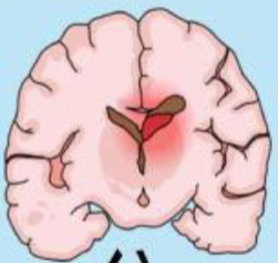
(59) (65) Intracerebral hemorrhage (ICH)
Type of Hemorrhagic stroke with bleeding into the brain parenchyma. Secondary headache
(59) (65) Subarachnoid hemorrhage (SAH)
Type of Hemorrhagic stroke with bleeding into the subarachnoid space, where cerebrospinal fluid (CSF) circulates. Secondary headache
(59) (65) Cause of Subarachnoid Hemorrhage
Most commonly due to trauma, followed by ruptured aneurysms (e.g. berry aneurysms).
(59) (65) Presentation of Subarachnoid Hemorrhage
Sudden onset of a severe “thunderclap” headache (“worst headache of my life”), neck stiffness, photophobia. Can rapidly loose consciousness
(59) (65) Imaging of Subarachnoid Hemorrhage
Blood in the subarachnoid space on CT
(59) (65) Cause of Intracerebral Hemorrhage
Often due to hypertension, cerebral amyloid angiopathy, anticoagulation therapy, or trauma; Compression of pain-sensitive structures by the hematoma
(59) (65) Presentation of Intracerebral Hemorrhage
Focal neurological deficits depending on the hemorrhage location, often with sudden onset; Headache, vomiting, altered consciousness
(59) Imaging of Intracerebral Hemorrhage
Hyperdense area on CT, usually within the brain tissue
(59) Cerebral Amyloid Angiopathy (CAA)
Medical condition of cerebrovascular disorder caused by accumulation of cerebral amyloid-β (Aβ) in tunica media and adventitia of leptomeningeal and cortical vessels of the brain; resultant vascular fragility tends to manifest in normotensive elderly patients as lobar intracerebral hemorrhage
(59) Charcot-Bouchard aneurysms
Microaneurysms of lenticulostriate arteries due to uncontrolled chronic hypertension; often presents with upper/lower extremity weakness, blood in basal ganglia
(59) Berry Aneurysm
small, thin-walled blisters protruding from arteries of circle of Willis or its major branches
(59) Traumatic or Sulcal SAH
Most common cause of SAH; Classically presents with blood in the sulcus of the brain due to tear of small arteries travelling in the subarachnoid space
(59) Aneurysmal SAH
Most common spontaneous SAH, commonly due to berry aneurysms of the Circle of Willis. Presents as classic “thunderclap headache”
(59) Arteriovenous Malformation (AVM)
cause of spontaneous SAH, but can also result in ICH; collection of dysplastic dilated blood vessels (neither arteries nor veins) wherein arterial blood flows directly into draining veins without the normal interposed capillary beds. usually congenital, appear grossly as a “tangle” of vessels
(59) Depressed Skull Fracture
Fracture of skull compresses upon brain parenchyma or cortical vessels
(59) Basilar Skull Fracture
Fracture of orbital roof, sphenoid bone, or petro-mastoid portion of temporal bone – usually follows impact to occiput or sides of head
(59) Primary brain injury
occurs at time of trauma (cortical contusions, lacerations, bone fragmentation, diffuse axonal injury, and brainstem contusion)
(59) Secondary injury
develops subsequent to the initial injury. Includes injuries from intracranial hematomas, edema, hypoxemia, ischemia
(59) Direct Parenchymal Injury
Type of Primary brain injury that features Contusion (bruise caused by blunt trauma) due to blow to surface of brain transmitted through skull
(59) Coup/Contre-Coup Injury
Type of Primary brain injury in which Blow to head may result in contusion at point of contact (coup injury) or on brain surface diametrically opposite it (contrecoup injury); “brain rattling in head”
(59) Concussion
A mild traumatic brain injury (mTBI) caused by a direct or indirect blow to the head. Results in temporary neurological dysfunction without structural damage visible on imaging
(59) Acute Symptoms of Concussion
Headache, dizziness, confusion, nausea, blurred vision
(59) Delayed Symptoms of Concussion
Mood changes, memory issues, sleep disturbances
(59) Diffuse Axonal Injury (DAI)
Traumatic High speed brain injuries damaging deep white matter regions; stretching or shearing of brain tissue may occur from marked changes in angular acceleration (e.g., blast injuries), even in absence of physical impacts of skull
(59) Epidural Hematoma
Medical condition of bleeding located between the skull and the periosteal layer of dura
(59) Cause of Epidural Hematoma
Often due to trauma, particularly with skull fractures that damage the middle meningeal artery
(59) Presentation of Epidural Hematoma
“Lucid interval” followed by rapid deterioration; symptoms include headache, vomiting, and potential loss of consciousness
(59) Imaging of Epidural Hematoma
Lens-shaped (biconvex) on CT scan
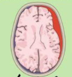
(59) Subdural Hematoma
Medical condition of bleeding located between the dura and arachnoid mater
(59) Cause of Subdural Hematoma
Usually from tearing of bridging veins, often in the elderly and chronic alcoholics: those with brain atrophy
(59) Presentation of Subdural Hematoma
Symptoms may develop over hours to weeks; includes headache, confusion, and focal neurological deficits
(59) Imaging of Subdural Hematoma
Crescent-shaped (concave) bleed on CT scan
(59) Shaken baby syndrome
A severe form of child abuse, Brain bangs against skull causing tearing of bridging veins, leading to Subdural Hematoma
(59) Intraventricular Hemorrhage
Medical condition of bleeding located within the brain’s ventricular system
(59) Cause of Intraventricular Hemorrhage
Can result from extension of intracerebral hemorrhage or trauma, commonly seen in preterm infants: germinal matrix hemorrhage
(59) Presentation of Intraventricular Hemorrhage
Variable; may cause symptoms of hydrocephalus if blood obstructs CSF flow
(59) Imaging of Intraventricular Hemorrhage
Blood in the ventricular spaces on CT
(59) Brain Herniation
Medical condition that occurs when something inside the skull produces pressure that moves brain tissues
(59) Transtentorial (Uncal) Herniation
Type of brain herniation that involves medial portion of temporal lobe, Uncus, herniating downward through the tentorium cerebelli and compressing ipsilateral oculomotor nerve: ipsilateral fixed and dilated pupil (“blown pupil”)
(59) Transforaminal (Tonsillar) Herniation
Type of brain herniation in which the cerebellar tonsils herniate down through the foramen magnum. Causes compression of the brainstem and medulla oblongata, disrupting vital functions such as respiration and cardiovascular regulation
(59) Subfalcine (Cingulate) Herniation
Type of brain herniation in which the cingulate gyrus herniates down below the falx cerebri; Most common type of herniation. Can cause headaches due to increased intracranial pressure, and contralateral leg weakness
(60) Neurodegenerative Diseases
Medical conditions that mostly affect gray matter; feature progressive loss of neurons and associated secondary changes in white matter tracts, as well as accumulation of protein aggregates (inclusions) resistant to degradation
(60) (61) (62) Alzheimer’s Disease
Medical condition of neurodegenerative disease affecting cerebral cortex via cortical atrophy; Loss of brain cells, presents with dementia and progressive loss of cognitive function/impaired memory
(60) (62) Pathogenesis of Alzheimer’s Disease
Accumulation of two proteins (Aβ and tau) in specific brain regions; amyloid (neuritic) plaques of aggregated Aβ (A-beta) peptides, and neurofibrillary tangles of microtubule binding protein tau. Mutation in presenilin genes, decreased acetylcholine synthesis
(60) Biomarkers of Alzheimer’s Disease
increased phosphorylated tau and reduced Aβ in the CSF
(60) Neurofibrillary tangles and dementia:
Number of neurofibrillary tangles correlates better with the degree of dementia than does the number of neuritic plaques
(60) Neuritic plaques
focal, spherical collections of dilated, tortuous, neuritic processes usually around a central amyloid core, surrounded by clear halo
(60) (62) Neurofibrillary tangles
tau containg bundles of filaments in cytoplasm of neurons which displace or encircle nucleus. Found in other neurodegenerative diseases - not specific for AD
(60) Frontotemporal Lobar Degeneration (FTLD)
Medical condition of focal degeneration of frontal and/or temporal lobes with alterations in personality, behavior, and language (aphasias) preceding memory loss
(60) Pick Disease
Medical condition in which brain shows severe, usually asymmetric “knife-edge” (thin) atrophy of frontal and temporal lobes, and intense gliosis in affected cortical regions
(60) (61) (62) Parkinson’s Disease
Medical condition of Neurodegenerative hypokinetic movement disorder caused by loss of dopaminergic neurons from substantia nigra. Clinical triad of tremor, rigidity, and bradykinesia (shuffling gait). Severity of motor syndrome proportional to dopamine deficiency
(60) Pathogenesis of Parkinson’s Disease
Protein accumulation and aggregation, mitochondrial abnormalities, and neuronal loss in the substantia nigra and other areas of brain; presence of Lewy Bodies
(60) Lewy Bodies
eosinophilic, round/elongated inclusions with dense core surrounded by pale halo; composed of abnormal aggregates of α-synuclein
(60) Huntington Disease
Medical condition of Autosomal dominant, progressive movement disorder with dementia caused by degeneration of striatal neurons
(60) Chorea
Sudden jerky, irregular movements with muscle contractions
(60) Athetosis
Twisting and writhing motions
(60) Dystonia
Sustained muscle contractions - usually result of repetitive motions
(60) Pathogenesis of Huntington Disease
polyglutamine trinucleotide repeat expansion causing toxic gain-of function mutation; Neuronal loss and severe atrophy with gliosis in the caudate and putamen. Motor symptoms usually precede cognitive impairment
(60) Choreoathetosis
Loss of striatal neurons (usually dampen motor activity), causes increased motor output
(60) Choreiform
Movement disorder of Huntington Disease, increased and involuntary jerky movements of all parts of the body; writhing movements of extremities
(60) Amyotrophic Lateral Sclerosis (ALS)
Medical condition of progressive disease - loss of upper motor neurons in cerebral cortex and lower motor neurons in spinal cord and brain stem. Results in weakness, increasing as disease progresses
(60) Pathogenesis of Amyotrophic Lateral Sclerosis (ALS)
mutations in gene encoding copper-zinc SOD1 on chrom 21; mutated SOD1 protein misfolds and aggregates, causing cell injury
(60) Clinical presentation of Amyotrophic Lateral Sclerosis (ALS)
Asymmetric weakness of hands, early – dropping of objects and difficulty with fine-motor tasks along with cramping and spasticity of arms and legs
(60) Prion Diseases
Medical condition of Rapidly progressive neurodegenerative diseases caused by aggregation and intercellular spread of misfolded (abnormal) prion protein (PrPsc); may be sporadic, familial, transmitted
(60) Pathogenesis of Prion Diseases
Abnormal prion protein of mainly beta-pleated sheets. Abnormal prion protein “recruits” normal prion to abnormal shape (secondary structure),allowing for “propagation”. clinical rapidly progressive dementia
(60) Creutzfeldt-Jakob Disease
Most common prion disease, rare disorder - Most cases show little or no cerebral atrophy – clinically rapid, progressive dementia
(60) Alpha-synuclein proteins are associated with:
Lewy Bodies, Parkinson’s Disease, Multiple System Atrophy (MSA)
(60) Amyloid Precursor Proteins are associated with:
Alzheimer’s Disease