3.2.4 - Cell recognition and the immune system
1/40
There's no tags or description
Looks like no tags are added yet.
Name | Mastery | Learn | Test | Matching | Spaced | Call with Kai |
|---|
No analytics yet
Send a link to your students to track their progress
41 Terms
What do lymphocytes do?
They distinguish between pathogens and self-cells and then destroy them
How do lymphocytes distinguish between self-cells and pathogens?
Each cell has a specific molecule on its surface and this is usually a protein therefore it can be identified by its 3D tertiary structure as it is complementary to the lymphocytes
What happens if a non-self cell is detected?
A response will be triggered to destroy the cell
What are 4 non-self cells?
Pathogens (eg. bacteria, fungi, viruses, etc.)
Toxins
Abnormal body cells (eg. cancer cells)
Cells from other organisms of the same species
What is an antigen? (2 marks)
A foreign protein that stimulates an immune response
Where are antigens located?
On the surface of cells
What is antigen variability?
When the pathogen mutates meaning the DNA changes therefore the shape of the antigen will change so the memory cells will only have a memory of the old antigen shape meaning that immunity is no longer effective
What is a phagocyte?
A type of white blood cell that can do phagocytosis
Where are phagocytes found?
In blood and tissues
What is phagocytosis? (1 mark)
A non-specific immune response to any non-self cells
Describe the steps of phagocytosis
The phagocyte will be attracted towards the pathogen by debris released by the pathogens
There are many receptor binding sites on the surface of the phagocyte and these will attach to the antigens on the pathogen
The phagocyte then changes shape to engulf the pathogen
Once engulfed the pathogen is contained ina phagosome
A lysosome within the phagocyte will fuse with the phagosome and release the enzyme lysozyme into the phagosome which hydrolyses and destroys the pathogen
The useful soluble products are absorbed and used by the phagocyte
What happens to the phagocytes after phagocytosis?
They become APC (antigen presenting cells) as the antigens are positioned on the cell surface
What is an APC (antigen presenting cell)?
Any cell that presents a non-self antigen on its surface
How do cytotoxic T cells destroy infected cells?
They release a protein called perforin which creates a pore in the cell membrane
This pore either allows water to enter causing the cell to burst or water to exit causing the cell to shrivel - both result in cell death
What do plasma cells do?
Produce antibodies
How are B-cells activated?
B-cells contain antibodies on their surface
Antigens in the blood collide with the complementary antibody on a B-cell which causes the B-cell to take in the antigen by endocytosis (engulf it)
It then presents the antigen on its surface and collides with a T helper cell activating it to go through clonal expansion and differentiation
They differentiate into plasma cells or memory B cells
If a pathogen reinfects the body what do B memory cells do?
Divide by mitosis into plasma cells to make large numbers of antibodies rapidly to destroy the pathogen
Describe the cell-mediated response
Phagocytosis occurs and produces an APC
T helper cells have receptors on their surface which bind to the antigens on APC
Once attached this activates the T helper cells to divide by mitosis to replicate and make many clones (clonal expansion)
The cloned T helper cells differentiate into different cells: some remain as T helper cells, some stimulate phagocytes to perform more phagocytosis and some become cytotoxic T cells
Name the parts of an antibody (5 points)
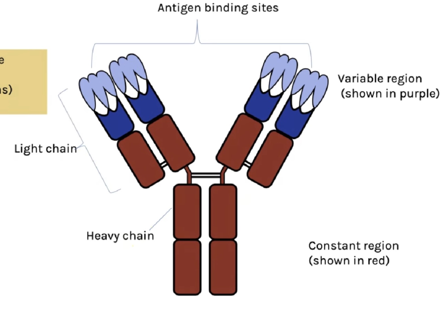
What is the protein structure of an antibody?
Quaternary structure
What is the function of the hinged section on an antibody?
To bend and allow flexibility so that it can easily attach to antigens
What is agglutination?
When antibodies bind to multiple antigens and clump together forming many antigen-antibody complexes
Why is agglutination effective?
Makes it easier for phagocytes to locate and destroy pathogens
What is passive immunity?
When antibodies are introduced into the body so no memory B cells or plasma are made
What is active immunity?
When you are exposed to the pathogen or its antigen and you become immune due to the production of antibodies and memory cells
What is the difference between artificial active immunity and natural active immunity?
Natural - You naturally get infected and your body creates antibodies and memory cells to become immune
Artificial - You are introduced to a weakened version of the pathogen or antigens via a vaccine
How do vaccines work?
Weak or dead versions of pathogen are introduced into the body
The antigens activate the B cells to go through clonal expansion and differentiation
B cells undergo mitosis and make large numbers of plasma cells which make antibodies and memory B cells which can divide into plasma cells when reinfected with the same pathogen
What is herd immunity?
If enough of the population are vaccinated the pathogen can’t spread easily amongst the population
What is an advantage of herd immunity?
It provides protection for those who haven’t been vaccinated
What are the 4 key structures in HIV?
Core
Capsid
Envelope
Protein attachments
What does the core contain?
RNA
The enzyme reverse transcriptase
What is the capsid?
Outer protein coat
What is the envelope?
Extra outer layer made of lipids taken from host
What are the protein attachments and what do they do?
Glycoproteins on the exterior of the envelope to enable the virus to attach to the host’s T helper cell
What cells does HIV infect?
T helper cells
How does HIV replicate in T helper cells?
HIV is transported in the blood until it attaches to a protein on the T helper cells via its attachment proteins
The HIV capsule fuses with the T helper cell membrane which enables the RNA and reverse transcriptase to enter
The enzyme copies the RNA into a DNA copy and moves into the T helper cell nucleus
mRNA of HIV and the host is transcribed in the nucleus creating viral proteins which are reassembled and move to the cell membrane and are released from the cell making a new viral particle
How does HIV make the immune system weaker?
It targets T helper cells which activate B cells therefore there is less antibodies
What is immunodeficiency?
When the body no longer has an effective immune response
What is a monoclonal antibody?
A single type of antibody that can be isolated and cloned
What’s the difference between an indirect ELISA and a direct ELISA test?
Direct = antigen attached to well of beaker first
Indirect = antibody attached to well of beaker first
Describe the process and positive result of a direct ELISA test
Add the sample to the base of the beaker
Wash and remove any unbound sample
Add a complimentary antibody to the antigen in the well
Wash and remove any unbound antibody
Add a secondary antibody (with an enzyme attached) that is complementary to the first antibody
Add the colourless substrate for the enzyme - when E-S complex forms, it produces a colour
The presence of colour indicates the presence of the antigen in the sample and the intensity indicates the quantity