Cardiovascular System
1/53
There's no tags or description
Looks like no tags are added yet.
Name | Mastery | Learn | Test | Matching | Spaced | Call with Kai |
|---|
No analytics yet
Send a link to your students to track their progress
54 Terms
keeps blood in motion
beats about 100,000 times per day
pumps about 1.5 million gallons of blood per year
about 3 gallons per minute
has 4 chambers
Heart
contains nutrients and oxygen
collects waste
blood
Where is the heart located?
Mediastinum
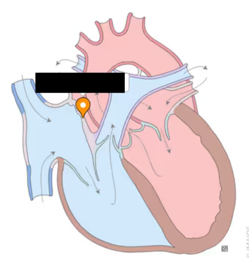
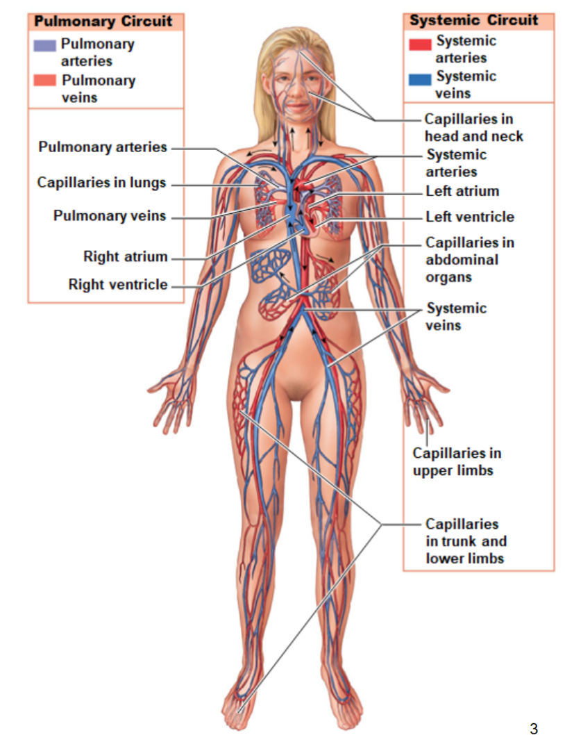
Heart pumps blood into two circuits
Pulmonary circuit, systemic circuit
depleted of oxygen
to/from the lungs
blood returning from the body
enters the right side of the heart
in the lungs oxygen gets added to the lungs
carbon dioxide gets removed
goes back to the heart and out the aorta
Pulmonary circuit
left side of heart
pumps blood to the body with oxygen
systemic circuit
Once blood leaves the heart into blood vessels
transport blood away from the heart’
most contain oxygenated blood that is ready to nourish cells
arteries
Once blood leaves the heart into blood vessels
transports blood toward the heart
veins
Once blood leaves the heart into blood vessels
interconnects arteries & veins
allow nutrients & waste transfer between body cells & blood
capillaries
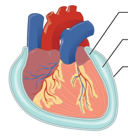
heart is surrounded by the ______
consists of two parts
outer fibrous ______
inner serous ______
pericardium
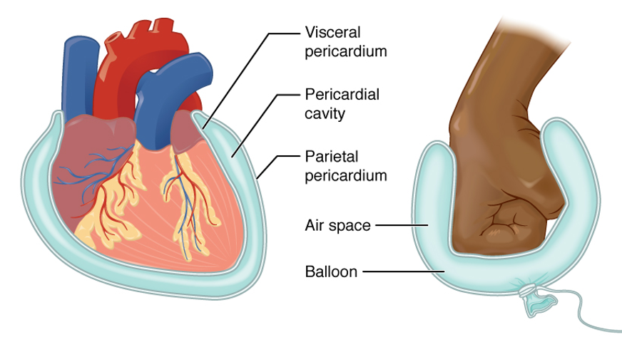
Consists of two parts
inner visceral layer = epicardium
attached to the surface of the heart
outer parietal layer = adjacent to the fibrous pericardium
Space between the layers is called the pericardial cavity & contains pericardial fluid
Serous pericardium
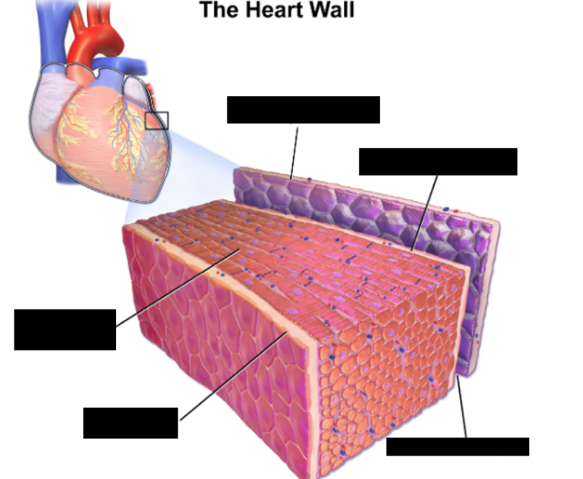
Walls of the heart consist of 3 layers
Epicardium, Myocardium, Endocardium
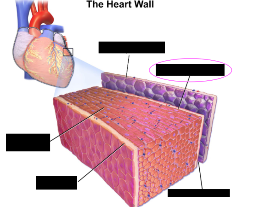
Wall of the heart
Outer layer of the heart
AKA the visceral layer
made up of connective tissue & has blood vessels
Epicardium
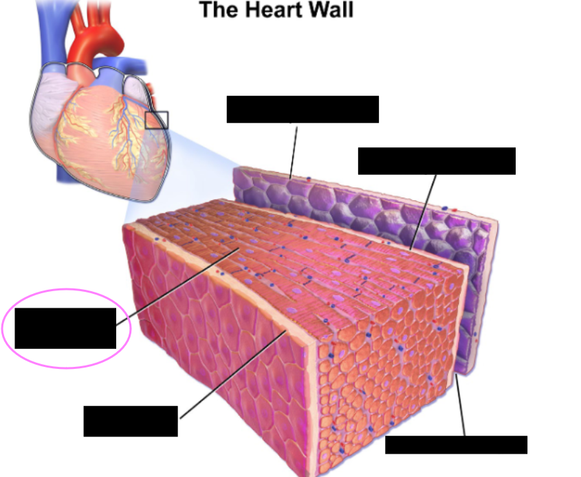
Wall of the heart
Middle layer
consist of cardiac tissue, including cardiac muscle cells
connective tissue, blood vessels & nerves
Myocardium

Wall of the heart
internal layer, endothelial surface
lining the inside of the chambers of heart
connective tissue
thinnest
Endocardium
Cardiac muscle Tissue
striated appearance
Sarcomeres
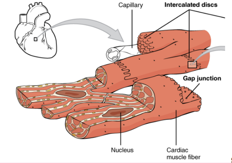
Branched morphology (shape)
Dependent on aerobic respiration to make energy
oxygen required
lots of mitochondria & myoglobin
has many blood vessels to nourish it
muscle cells contract without info coming from the CNS
cardiac muscle cells are tightly interconnected
intercalated discs
Cardiac Muscle tissue

Only have one nucleus
Branch shape
need lots of oxygen
a lot of mitochondria
contracts by itself without the NS (involuntary)
Cardiac muscle cells
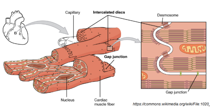
cell to cell junctions
binds the myofibrils of the cells together
Desmosomes
proteins that hold two cardiac muscle fibers together
really strong
gap junctions to allow cells to communicate directly → allow heart muscle cells to send action potentials without neurotransmitters
Intercalated discs
helps keep the heart its shape
mostly made up of connective tissue
every cardiac cells is wrapped in an elastic sheath & fibrous sheet
helps prevent overexpansion
helps spread out forces of contractions
cardiac skeleton
Heart has 4 chambers
Two _____
left and right
have muscle around them & when contract they push blood into the second set
Two _____
left and right
atria, ventricles
superior border of the heart
where the heart vessels enter and leave (connect together)
base
inferior/ bottom part of the heart
ventricles come to a point
apex
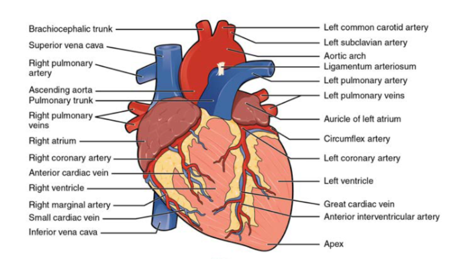
consist of the:
right atrium
right ventricle
left ventricle
anterior surface
consist of the:
left atrium and a small portion of the right atrium
posterior surface
left and right regions
positioned at the base of heart
both have thinner walls
both contain an expandable anterior portion called an auricle
atria
left and right regions
positioned at the apex of the heart
both have thicker walls
left is thicker than right (more cardiac muscle)
ventricles
the left and right atria are separated by?
interatrial septum
left and right ventricles is separated by?
interventricular septum
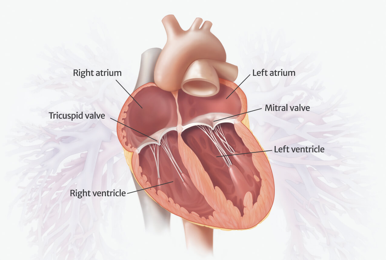
What separates the atria and the ventricles
atrioventricular valves
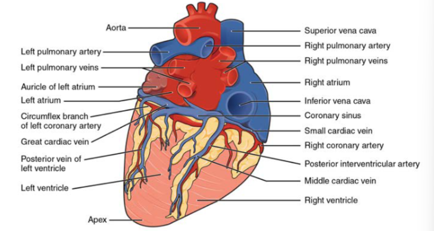
receives blood depleted of oxygen via the Superior vena cava, inferior vena cava, and coronary sinus
vena cavae bring blood from body back to heart
coronary sinus enters posterior side of the right atrium
right atrium
receives oxygen-poor blood
blood enters the right ventricle by passing thru the right atrioventricular valve aka tricuspid valve
blood leaves by passing thru the pulmonary valve (pulmonary semilunar valve)
leads to the pulmonary trunk then to the right & left pulmonary arteries
Right ventricle
This right valve has three flaps/cusps & 3 papillary muscles
connected to papillary muscles (smooth muscle) & chordae tendineae (cords that…)
prevent valve inversion when ventricles contract
tricuspid valve
receives oxygenated blood from lungs via right & left pulmonary veins
blood passes thru left atrioventricular valve
mitral valve/left AV valve
left atrium
Which veins are the only ones that carry oxygenated blood
right and left pulmonary veins
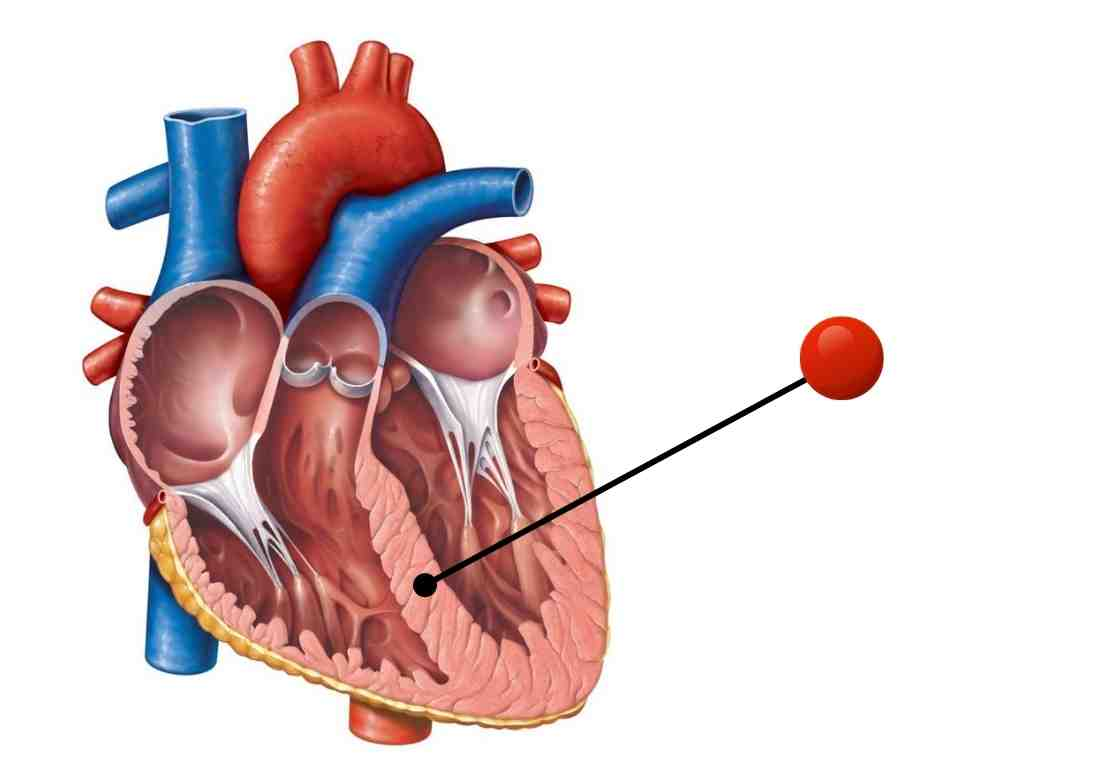
largest chamber in the heart & has the thickest (myocardium) wall
needed for strong contractions to pump blood thru entire circuit/ all of our blood pressure
it also has chordae tendineae connecting yo 2 cusps & 2 papillary muscles
left ventricle
When blood leaves the left ventricle which valve does it pass through?
AKA aortic semilunar valve
first blood enters the ascending _____
then travels to the _____ arch & down the ascending ___ & to all body parts
aortic valve
thinner wall
weaker contraction
right ventricle
thicker wall
powerful contraction
left ventricle
Heart gets oxygen from this
supply blood to
the cardiac muscle via _____
right and left _____ arteries
drain cardiac venous blood from lungs & into right atrium
coronary circulation
made up of arteries & veins that transport blood between the heart & the lungs
to add oxygen and remove waste/ co2
travels short distances
blood pressure is lower
walls of the arteries are thinner → less smooth muscle
pulmonary circuit

made up of arteries & veins that transport oxygenated blood between the heart & al other tissues
often travels longer distances
blood pressure is higher
walls of the arteries are thicker → more smooth muscle
systemic circuit
vessels that interconnect arteries and veins
capillaries
contain 30-35% of the blood volume
arteries and capillaries
contain 65-70% of the blood volume
veins
Blood vessels that act as blood reservoirs (storage)
more elastic/stretchy
vein
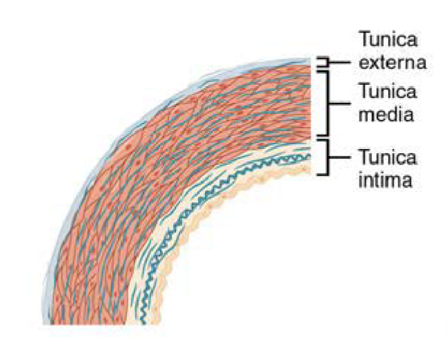
The walls of the blood vessels (except capillaries) consists of 3 layer (tunics)
give tremendous strength 💪
wall are thick & even contain their own blood vessels
intima, media, externa
deepest layer
endothelium
tunica intima
middle layer
smooth muscle
involved in vasoconstriction (walls contract and lumen gets smaller) & vasodilation (walls relax and lumen widens)
tunica media
superficial layer
AKA adventitia
fibers (collagen/connective tissue) of this wall anchor the blood vessels
tunica externa
inside blood vessel
where blood flows
lumen
large vessels up to 2.5 cm in diameter
contain elastic connective tissues in media & intima
tolerate pressure changes during cardiac cycle
can store pressure
ex:
aorta
brachiocephalic trunk
pulmonary trunk
elastic arteries