ruminant anatomy and physiology
1/24
There's no tags or description
Looks like no tags are added yet.
Name | Mastery | Learn | Test | Matching | Spaced | Call with Kai |
|---|
No analytics yet
Send a link to your students to track their progress
25 Terms
anatomy of the rumen
-where relative to the cecum
-how does it connect to the oesophaus
how is it divided
left hand side- putting a stethoscope by the paralumbar fossa, you can hear the rumen
cecum is to the right
1st chamber after leaving the oesophagus
connects to the oesophagus by the cardiac sphincter
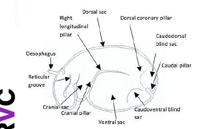
fissures (macroscopic invaginations into the lumen of the rumen) called pillars divide the rumen
biggest fermentation chamber
rumen
function
layers
type of epithelium
no gastric secretions
fermentation and mechanical secretions
papillae to increase surface area
host bacteria for digestion of cellulose (no cellulase)
non glandular stratified squamous epithelium, lamina propria, submucosa, muscularis interna
layer of keratin for physical protection and thick tunica muscularis
regurgitation
what contracts
how is food drawn into the oesophagus
where is the food moved into and how
reticulum and dorsal sac of the rumen contract
simultaenously the animal breathes in with a closed glottis reducing pressure in the thoracic cavity and the oesophagus
increased rumen pressure and decreased oesophageal pressure draws food into the oesophagus
it is moved into the oral cavity through antiperistaltic contraction
motility of the rumen
what are ruminoreticular movements regulated by which nerve
where do these fibers of the nerve originate
what monitors what
how is it stimulated
reticuloruminal movements are centrally regulated by CNX (vagus nerve)
dorsal vagal nucleus of the brainstem
afferents from the lumen of the ruminoreticulum monitor distension, ingesta consistency, pH, VFA conc
simulation through distention (stretch receptors), ingesta consistency, pH, VFA conc
primary and secondary movements of the rumen
where does primary start
what type of contraction and what order
what does primary do
primary frequency
what type of ontraction is secondary and what does this do
where does it start and where to
secondary frequency
where can secondary be assesed
primary
contraction across the ruminal wall which starts at the reticulum- wave of contraction
biphasic reticular contraction (2nd most powerful) followed by contraction of the dorsal then ventral sac
mixes ingested food with microbes facilitating the production of VFAs which cross the rumen wall into the bloodstream
some gases produce cannot cross the wall and must be expelled
1-3 x a min
reticular contraction every 1 min
secondary
caudal cranial wave of contraction pushes gas back to the oesophagus to be removed via the mouth
starts in the caudalventral blind sac to dorsal
approx once every 1-2 min
can be assessed on paralumbar fossa
implication of CNX damage
motility of the rumen is compromised
gas accumulation as fermentation is still occuring
bloating which expands the rumen wall limiting abdominal and diaphragmic contraction- affects breathing
which gases are absorbed by the ruminal wall after anaerobic digestion
VFAs
propionate
butyrate
acetate
these produce energy when metabolised in the liver
anatomy of the reticulum
which chamber
which side and is it dorsal or ventral
glandular or non glandular
function
2nd chamber
ventral left hand side
close to rumen and oesophagus
non glandular
mechanical digestion and secretion
histology of the reticulum
stratified squamous epithelium, lamina propria, lamina muscularis mucosae
papillae arranged in a honeycomb structure
anatomy of the omasum including how it is connected to the reticulum
3rd chamber
ventral right hand side
connected to the reticulum through reticulo omasal orifice
circular
no gastric secretion
have folds called omasal laminae for absorption of water and nutrients
anatomy of the abomasum
last chamber
chemical digestion
glandular secretions
similar to carnivores
covered by gastric pits and ruggae
has a lesser and greater curvature
simple columnar epithelium
have gastric pits which secrete hcl, mucus
middle right hand side, close to omsum
important in young for milk difestion
greater and lesser omentum
blood supply to the stomach
celiac artery to all of foregut (splenic, hepatic, gastric)
splenic artery- spleen
anastomosis
gastric groove
gastric/reticular/oesophageal groove is present in newborn ruminants
muscular channel taking milk from the oesophagus into the abomasum bypassing the rumen, reticulum and omasum
composed of 2 layers of muscle
when the animal is extending its neck to reach the mothers teat this triggers the groove to contract allowing milk to pass from the oesophagus into the abomasum where it is digesting by chemically
can be used to administer oral antibiotics
pancreas
next to duodenum
exocrine and endocrine funcions
enlarged in ruminants
major and minor papilla
liver
position
always on ventral RHS
4 lobes
big gall bladder
spleen
LHS
recieves blood from the splenic artery
elongated
falls on top of the rumen
abdominal musculature
cutaneous trunk line
external oblique
internal oblique
transverse abdominus and rectus abdominus
abdominal muscles have a aperneurosis that forms the linea alba along the ventral midline
thin muscular layer called the cutaneous trunk line which contracts to repel flies from the body
hardware disease- traumatic reticuloperitonitis
animal ingests nails, wire
reticulum is punctured
ingesta and bacteria can leak into the peritoneal cavity
peritonitis and adhesions in the abdomen
can even lead to reticulopercarditis
issues arising after pregnancy
enlarged uterus
abdominal organs are compressed
after giving birth uterus no longer enlarged creating empty space for organs to move around
abomasum can move from to left or right- displaced abomasum
gas gets entrapped inside abomasum and ifi it moves it cannot pass into intestine
should adjust diet to prevent
longitudinal pillars
separate the dorsal sac from the ventral sac
cranial pillar
separates the cranial sac (or ruminoreticular atrium) from the ventral sac
caudal pillar
divides the rear of the rumen into the caudoventral blind sac and caudodorsal blind sac
coronary pillars
where is the origin
what does it separate
originate from the caudal pillar and further separate the caudal blind sacs from the main dorsal and ventral sacs
where does water absorption occur in the omasum
base of the omasal laminae which do not contain a keratinised epithelium