AMD Classification and Management - Posterior Segment and Ocular Disease Spring 2026
1/100
There's no tags or description
Looks like no tags are added yet.
Name | Mastery | Learn | Test | Matching | Spaced | Call with Kai |
|---|
No analytics yet
Send a link to your students to track their progress
101 Terms

Subretinal drusenoid deposits (retincular pseudodrusen) (Pic)
Subretinal drusenoid deposits (retincular pseudodrusen) (Pic)
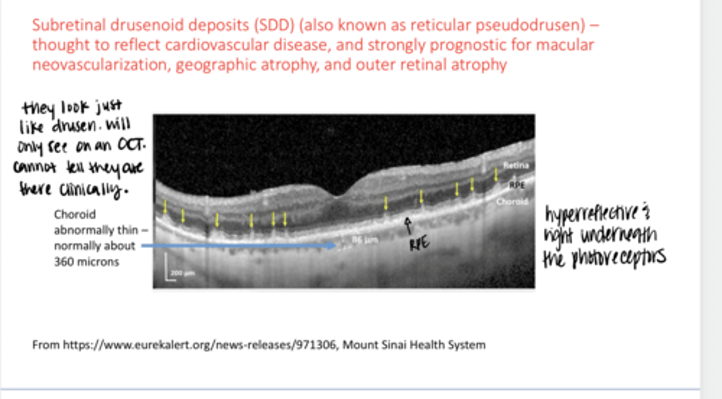
The more subretinal drusenoid that are present, the more likely for what else to be present (systemically)?
reflects cardiovascular disease

The more subretinal drusenoid that are present, the more likely for what else to be present (in the retina)?
macular neovasc, geographic atrophy, and outer retinal atrophy

Can you tell that Subretinal drusenoid deposits are there clinically?
No

How to find Subretinal drusenoid deposits?
on OCT & look at them very carefully

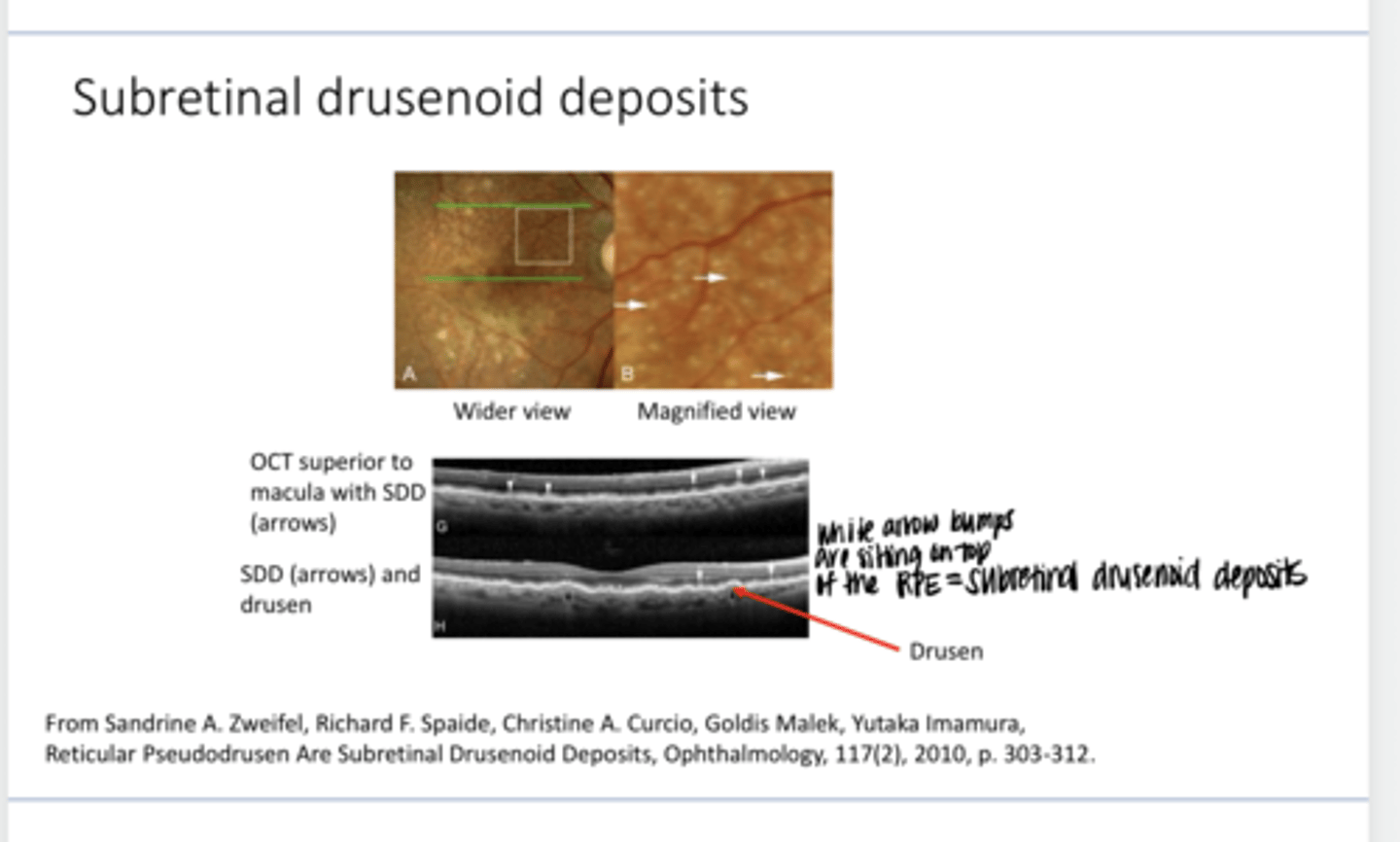
Subretinal drusenoid deposits look the same as what on OCT?
as drusen

Does Subretinal drusenoid deposits being present change your management plan for a patient?
No

Subretinal drusenoid deposits are a (good/bad) prognostic sign?
bad

Subretinal drusenoid deposits sit on top of the _____
RPE
Subretinal drusenoid deposits are (hyper/hypo) reflective on OCT
hyperreflective

With Subretinal drusenoid deposits you can get _____ thinning & atrophy
choroidal

Is thinning of the choroid a really bad sign?
Yes -- you no longer have perfusion to the RPE

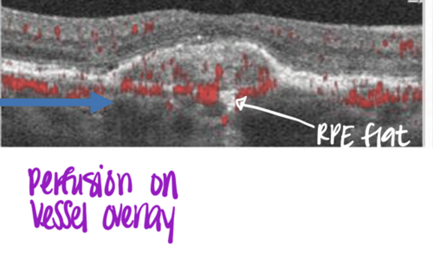
Subretinal drusenoid deposits - -on OCT (Pic)
Subretinal drusenoid deposits -- on OCT (Pic)
Is there a need to detect AMD EARLIER than we are?
Yes
A study published in 2017 showed that ophthalmologists and optometrists miss AMD ____% of the time
25
____% of the AMD that was missed was in the intermediate-stage disease
30
_______ may not appear until well after pathophysiological changes associated with AMD occur
Drusen
What tests may work for earlier detection of AMD?
OCT, FAF, dark adaptometry, MPOD
What are the tests currently used to diagnose Dry (Nonexudative) AMD?
1) Clinical examination
2) Amsler grid
3) Fundus photography
What is the MOST IMPORTANT thing to diagnose Dry (Nonexudative) AMD?
Clinical examination
What is the Amsler grid looking for a presence of?
Leaking choroidal Neovascularization
What is the Amsler grid's sensitivity to detecting macular disease?
detects 50% of the time
What is fundus photography useful for?
keeping track of any AMD changes
EXAM QUESTION:
AMD Grading/Management Scale from the AREDS Study
What are the 4 major risk factors for AMD?
1) Large drusen >125um
2) Intermediate Drusen 63-125um
3) Pigmentary abnormalities (hyperpigmentation, hypopigmentation, noncentral geographic atrophy (does not involve the fovea)
4) Bilaterality
AMD Grading/Management Scale from the AREDS Study
What will the risk factors of AMD eventually lead to?
severe vision loss
AMD Grading/Management Scale from the AREDS Study
More points in the grading scale = ?
RIsk of progression at 5 years to advanced AMD is higher (choroidal Neovasc or central/foveal geographic atrophy)
EXAM QUESTION:
SUMMARY: What are the most important risk factors for the development and progression of AMD from the clinical examination?
1) Age
2) Smoking
3) Large Drusen
4) Pigment Abnormalities
5) Extensive intermediate size drusen
6) Bilaterality
7) Advanced AMD in one eye
8) MPOD lower?
9) FAF findings?
10) Genetic testing?
11) Dark adaptometry?

EXAM QUESTION: What are the findings for early or mild AMD?
unlimited small drusen, less than 5 intermediate drusen
**independently for each eye

EXAM QUESTION: What are the findings for intermediate or moderate AMD?
5-20 intermediate drusen (or intermediate RPE clumps) less than 4 large drusen (or large RPE clumps), RPE Window Defects but less than geographic atrophy

EXAM QUESTION: What are the findings for advanced or late stage AMD (including serous detachment)?
More than 20 intermediate drusen, 4 or more large drusen, geographic atrophy, choroidal neovascularization, nonexudative macular neovascularization

What are the AMD RED FLAGS?
-large numbers of intermediate drusen
-any amount of large drusen
-any significant (intermediate or larger) pigmentary changes

EXAM QUESTION: Are pigmentary changes considered early or mild AMD?
No

EXAM QUESTION: Which are worse?
RPE Window Defects or RPE Clumping
RPE Window Defects are worse than Pigment Clumping

EXAM QUESTION: Intermediate or advanced AMD requires what?
more imaging -- OCT, FAF
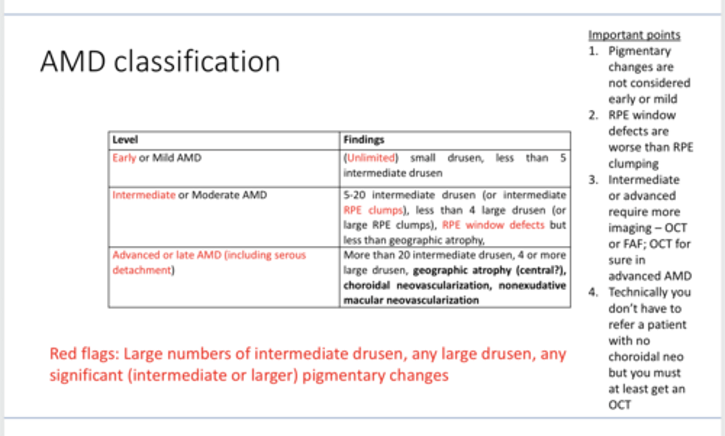
EXAM QUESTION: Do you have to refer a patient with AMD if they DO NOT have choroidal neovascularization? What do you AT LEAST have to do for these patients?
No technically, but you have to at least get an OCT
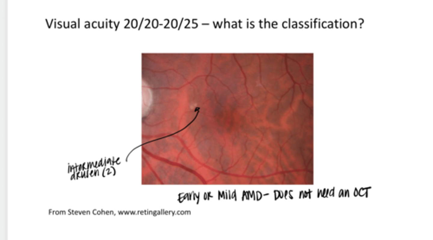
Visual Acuity is 20/20-20/25. What is the classification of this AMD (see pic)?
Early or Mild AMD
**2 intermediate drusen present

Would you do an OCT on this patient?
No -- no evidence of choroidal Neo
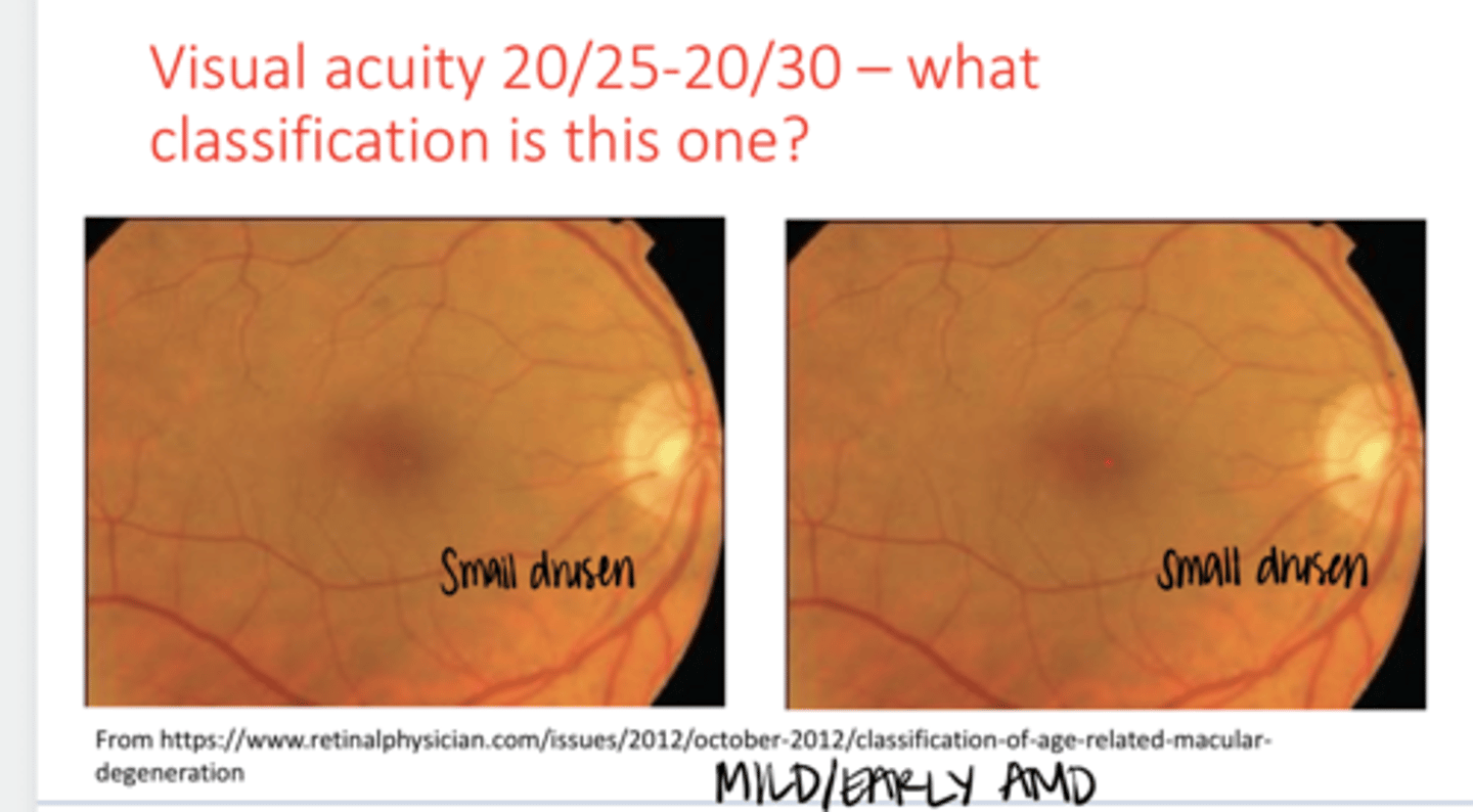
Visual Acuity is 20/25-20/30. What is the classification of this AMD? (See Pic)
Early or Mild AMD
**only small drusen & no pigmentary changes

Visual Acuity is 20/25-20/40. What is the classification of this AMD? (See Pic)
Intermediate/Moderate AMD
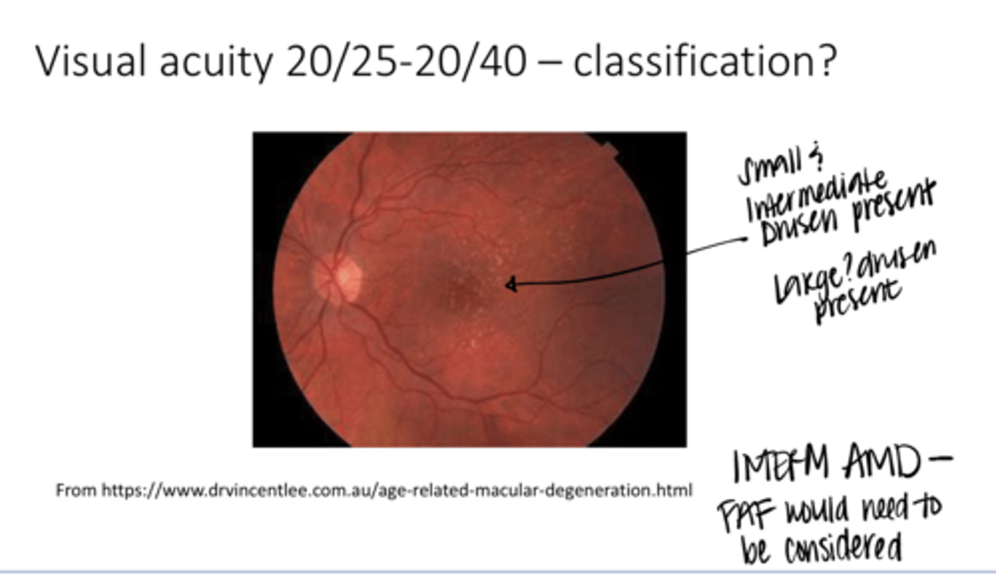
Should you do an OCT on this patient?
You do not have to -- consider an FAF
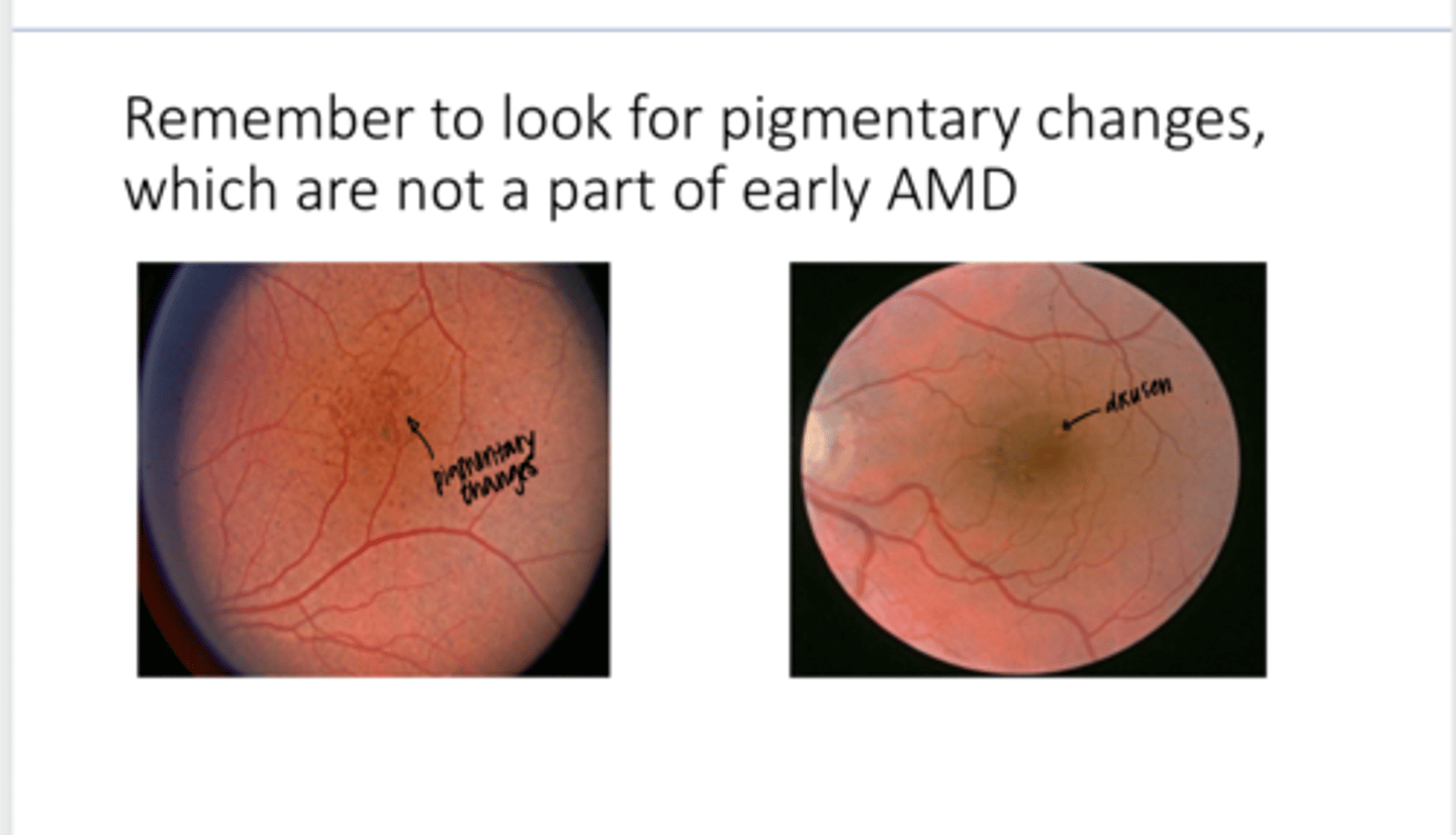
REVIEW: Are pigmentary changes a part of EARLY AMD according to the table Fogt gave us?
No

Visual Acuity is 20/40-20/60. What is the classification of this AMD? (See Pic)
Intermediate/Moderate AMD; on the border of Advanced AMD
**small and intermediate drusen present
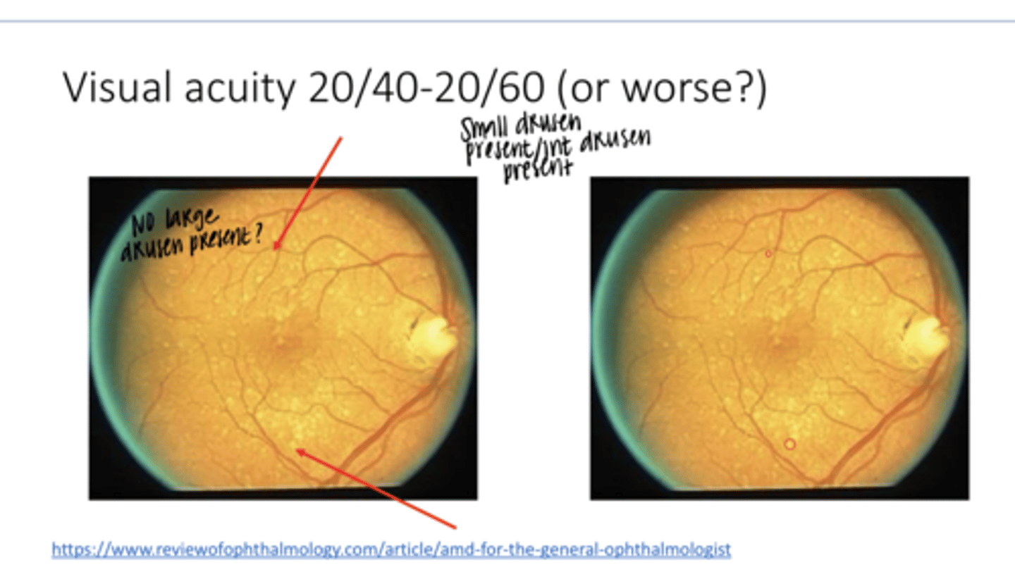
Should you do an OCT on this patient (see pic)?
Yes
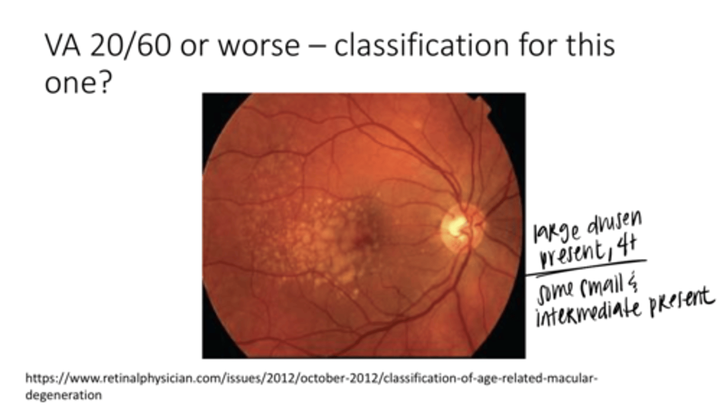
Visual Acuity is worse than 20/60. What is the classification of this AMD? (See Pic)
Advanced AMD
**small/intermediate/More than 4 large drusen

Does this patient need an OCT?
Absolutely Yes
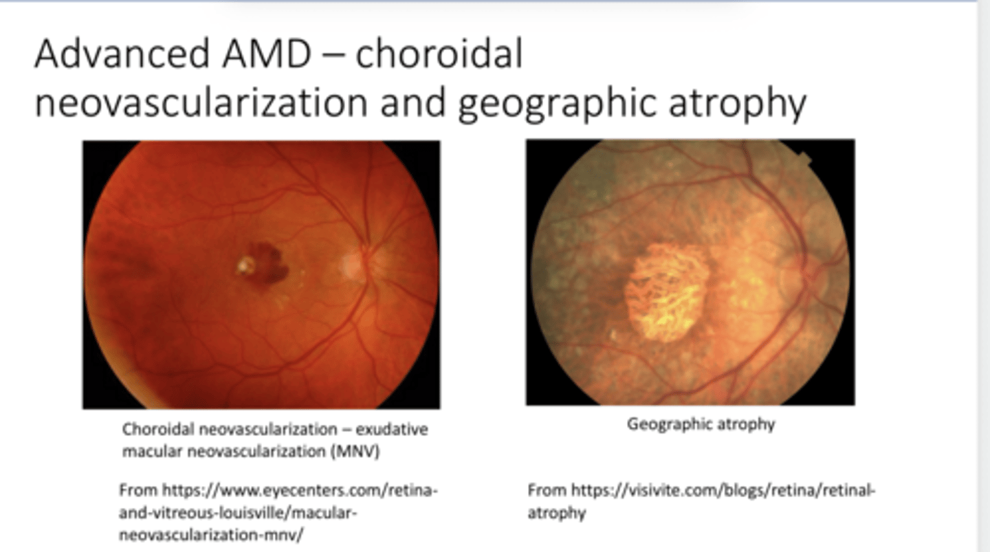
Advanced AMD w/ Choroidal Neovasc & Geographic Atrophy (Pic)
Advanced AMD w/ Choroidal Neovasc & Geographic Atrophy (Pic)
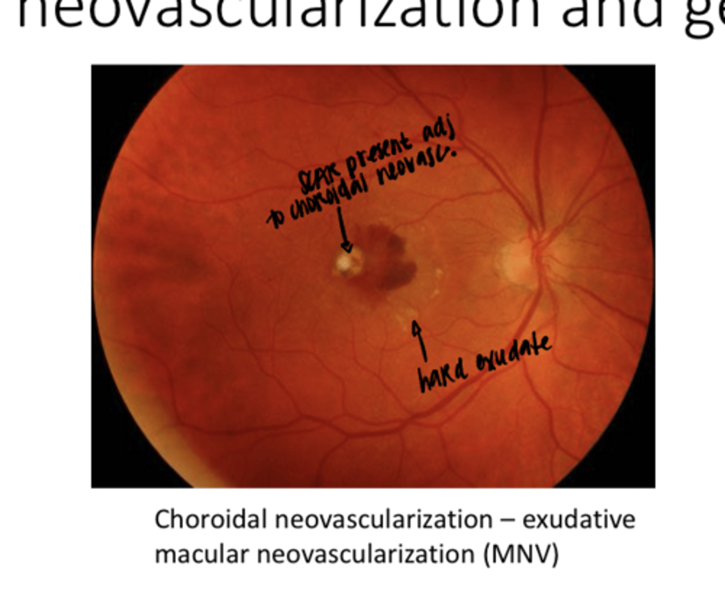
Choroidal Neovascularization -- WET AMD -- Exudative Macular Neovascularization (Pic)
Choroidal Neovascularization -- WET AMD -- Exudative Macular Neovascularization (Pic)

Geographic Atophy -- Worse Case of Dry AMD (Pic)
Geographic Atophy -- Worse Case of Dry AMD (Pic)
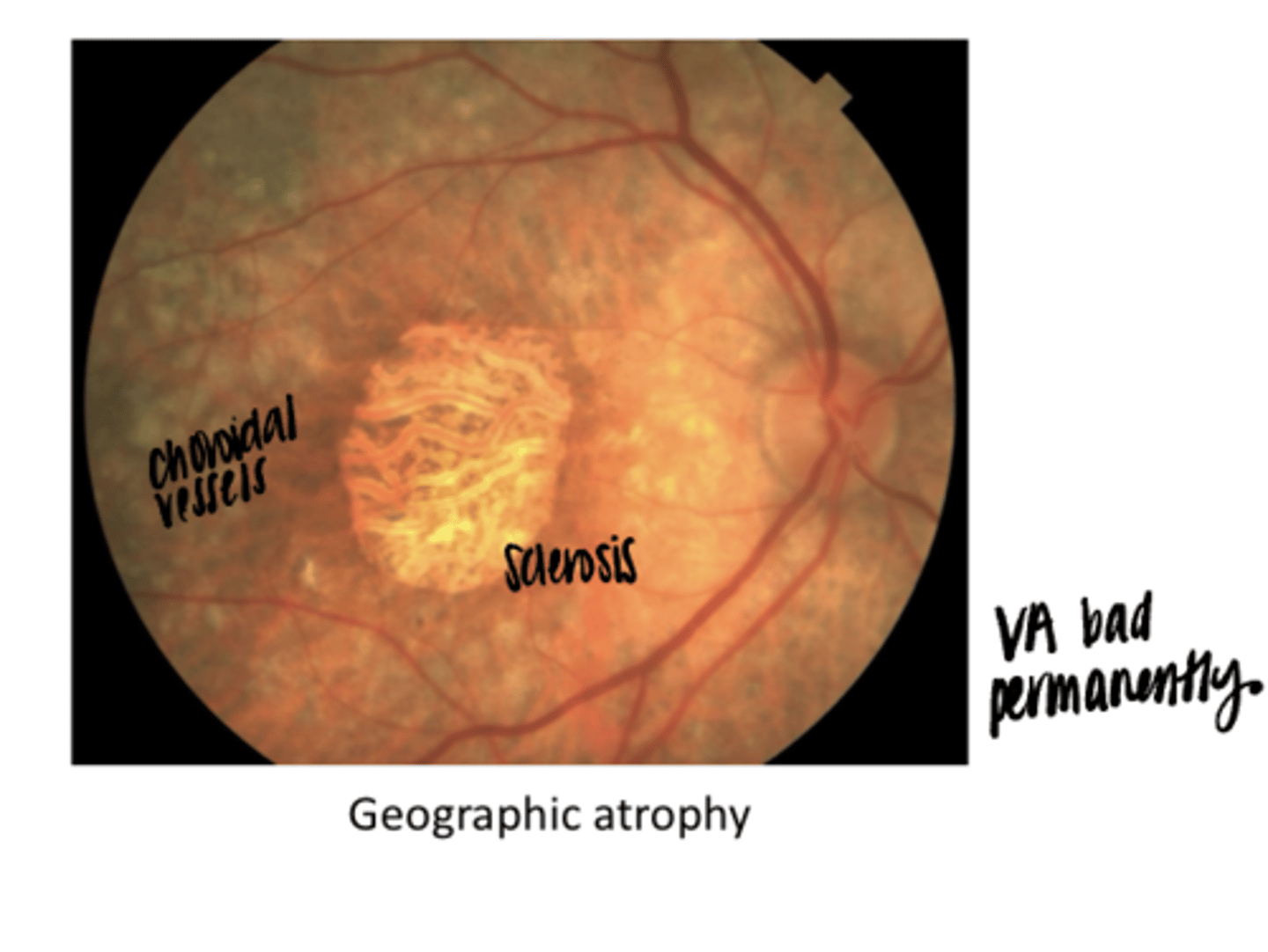
What is present in this pic?
Choroidal vessels and sclerosed vessels
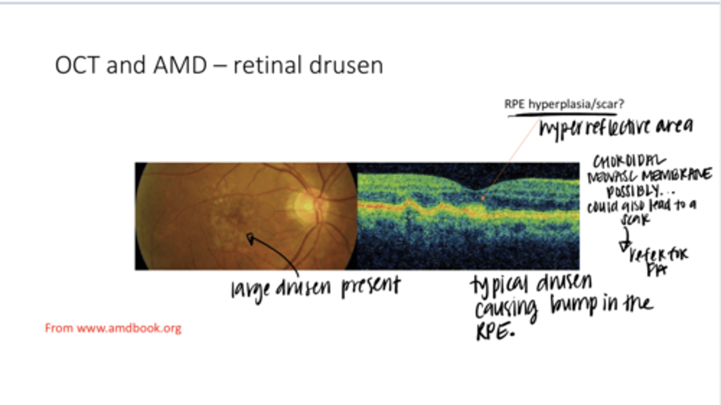
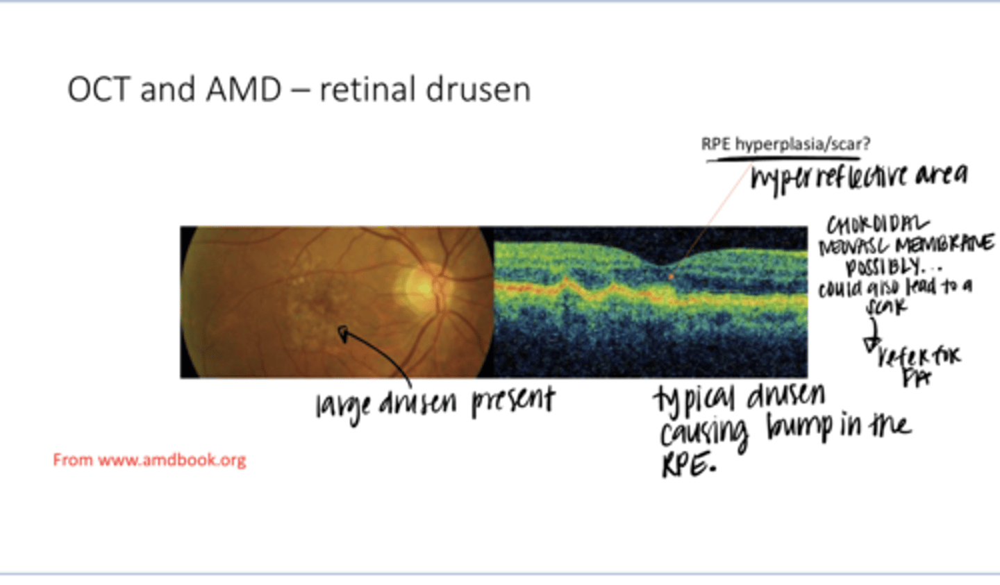
OCT of AMD -- Retinal Drusen Present (Pic)
OCT of AMD -- Retinal Drusen Present (Pic)

If you see a scar in the macula of an AMD patient, what should you do about it?
REFER -- could be d/t choroidal neovasc

What are the bumps in the RPE?
Drusen

What can be seen on an OCT of AMD?
-hyperreflectivity (Scarring or choroidal neovasc membrane)
-Serous detachment (dark)

If a drusen is clear inside, what is this?
drusenoid detachment -- RPE and Bruchs have seperated from each other
Is a drusenoid detachment a bad prognostic sign?
Yes
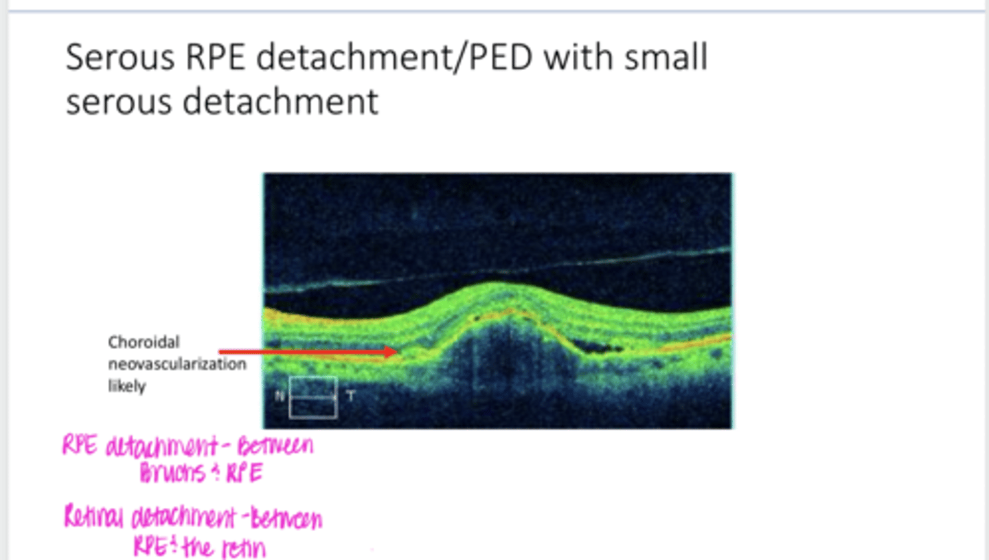
Where is a serous RPE detachment?
Between Bruchs and RPE

Where is a serous retinal detachment?
Between the retina and RPE
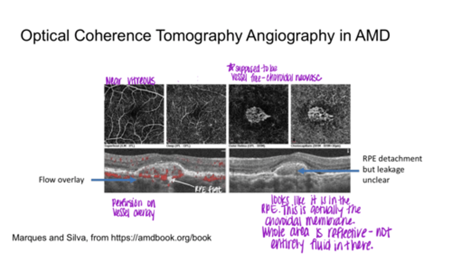
What are you looking for on OCT-A with AMD?
vessels in the outer retina
What is this photo of? How do you know this?
This is actually the choroidal membrane. The whole area is reflective meaning it is not entirely fluid in there.
Why would you do AMD genetic testing?
shows the long-term risk, and might encourage "at risk" people to modify their lifestyle
What are the genes that are responsible for an increased risk of AMD?
-CFH Y402H
-C3
-ARMS2/HTRA1
What did Awh et al do?
Reaxamined AREDS2 data
What was the conclusion of Awh et al AREDS data study?
concluded that individuals with 2 high-risk CFH alleles and no ARMS2 risk alleles had an increased risk of vision loss when taking an AREDS supplement
Should genetic testing be performed on all AMD patients before prescribing the supplement?
Yes
Does the American Academy of Ophthalmology recommend routine genetic testing in AMD?
No -- but need to keep watching this
Should everyone with intermediate/severe AMD get an AREDS supplement?
Yes
What does FAF look for?
Lipofuscin
Lipofuscin is a dominant source of _______
fluorophores
When/Where does lipofuscin accumulate?
Accumulates in RPE cytoplasm as a byproduct of incomplete degradation of photoreceptor outer segments
Does OPTOS have the capability to do FAF?
Yes
Dark areas of FAF (hypoautofluorescence) = ?
RPE missing/degenerated; cannot accumulate lipofuscin
When will you get dark areas on FAF?
Window Defects
Bright areas of FAF (hyperautofluorescence) = ?
areas of lipofuscin
Bright areas on FAF indicate what about the RPE?
poorly functioning RPE

Fundus Autofluorescence (Pic)
Fundus Autofluorescence (Pic)

Drusen will (Hyper/Hypo)Autofluoresce on FAF
Hyper

Window Defects will (Hyper/Hypo)Autofluoresce on FAF
Hypo
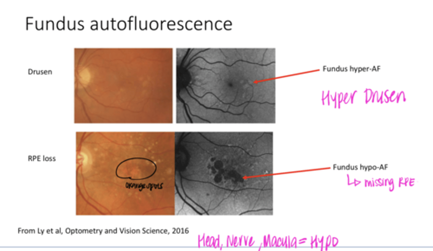
What is orange color on fundus photo mean?
RPE window defect w/ effacement (missing retina on top)

Can large loss on FAF change your F/U and management schedule?
Yes -- and sometimes pushes patients from early to mild/moderate AMD

AMD Differentials and Fundus Autofluorescence -- Possible Stargardts Disease (FAF)
AMD Differentials and Fundus Autofluorescence -- Possible Stargardts Disease (FAF)

What are the hyperautofluorescent areas depicting? (see pic)
areas of lipofuscin
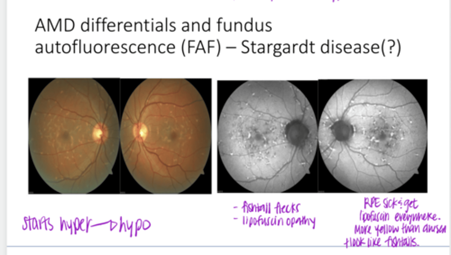
Is this drusen? How do you tell the difference between drusen and lipofuscin? (see pic)
No, this has fishtail appearance and is more yellow than drusen. This is lipofuscin

Will the hyperautofluorescent areas eventually become hypoautofluorescent? (see pic)
Yes
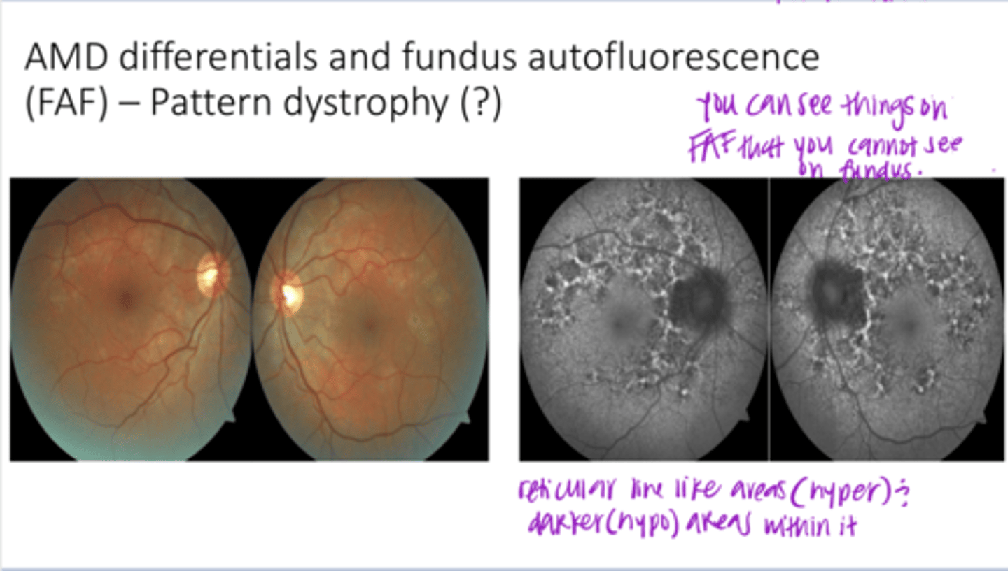
AMD Differentials and Fundus Autofluorescence -- Pattern Dystrophy or Inflammatory Disease?? (FAF)
**reticular (line like) areas of lipofuscin (hyper) and within are darker areas (hypo)
AMD Differentials and Fundus Autofluorescence -- Pattern Dystrophy or Inflammatory Disease?? (FAF)

Is the macula spared in this pic?
Yes

Is this AMD?
No -- does not affect the macula

Fundus Autofluorescence -- Serous Fluid (Pic)
Fundus Autofluorescence -- Serous Fluid (Pic)
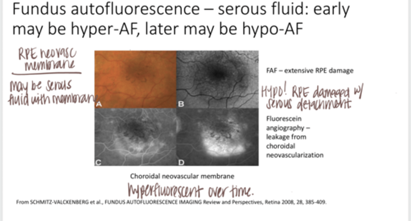
When a RPE neovascular membrane is present, can there be serous fluid?
Yes

With choroidal neovasc, will there be leakage on FA?
Yes -- more over time as seen in pic

Auto-Fluorescence -- Hemorrhaging (Pic)
Auto-Fluorescence -- Hemorrhaging (Pic)
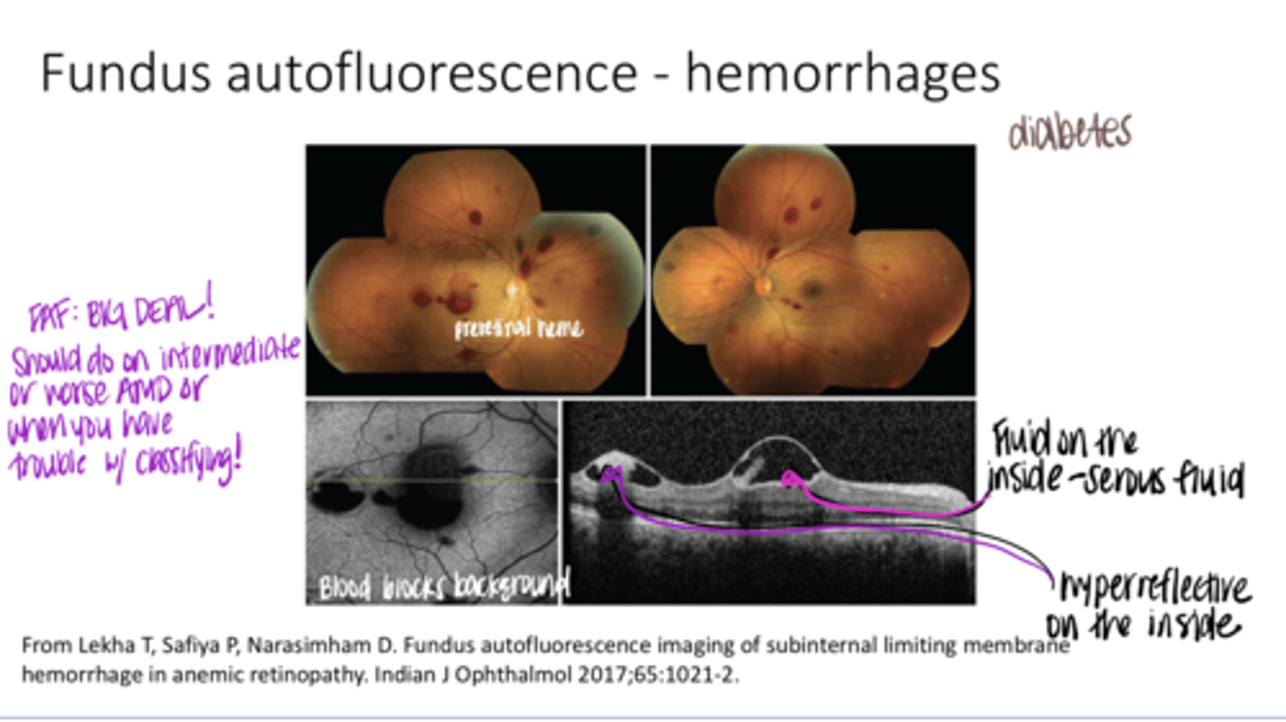
What is going on in this FAF picture?
Blood is blocking the background of the photo
You should do an FAF on every patient with what levels of AMD?
any AMD at intermediate stage or worse
What are the three macular (carotenoid) pigments in the macula?
lutein, zeaxanthin, meso-zeaxanthin
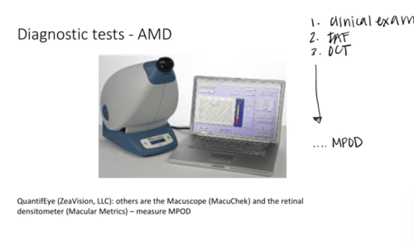
How are the three macular pigments measured?
macular pigment optical density (MPOD)

Commercially available devices that assess macular pigments make use of what?
heterochromatic flicker photometry

Are there studies that correlate vision to MPOD?
Yes -- a thicker MPOD will correlate to LESS glare

Are there studies that suggest that AMD risk will increase with a lower MPOD score?
Yes -- fewer anti-oxidants and less blockage of UV light which catalyzes oxidative reactions
What are the ALSTAR study results?
subjects with abnormal dark adaptation prior to developing AMD were 2x as likely to develop AMD at 3 years
Will you prescribe supplements based on a decreased dark adaptation period?
No
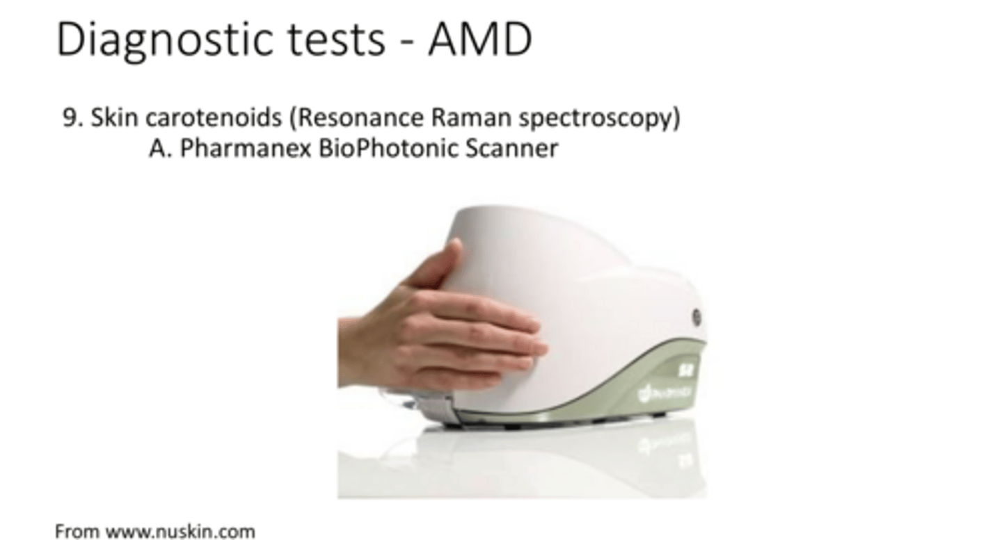
Measuring Skin Carotenoids for Diagnostic Marker of AMD (Pic)
**correlates w/ levels of carotenoids in the macula
Measuring Skin Carotenoids for Diagnostic Marker of AMD (Pic)