Anatomy and Physiology Chapter 9 Joints
1/84
There's no tags or description
Looks like no tags are added yet.
Name | Mastery | Learn | Test | Matching | Spaced | Call with Kai |
|---|
No analytics yet
Send a link to your students to track their progress
85 Terms
arthrology
study of joints
fibrous joints
dense regular connective tissue
cartilaginous
cartilage being located at the joint
Synovial
type of membrane located at this joint, cavity at the articulation
diarthrosis
freely movable joints
example: synovial joints
amphiarthrosis
slightly moveable joints
example: syndesmosis and symphysis
synarthrosis
Immovable joints/doesn't move
example: sutures, synostosis (adults)
sutures
skull only
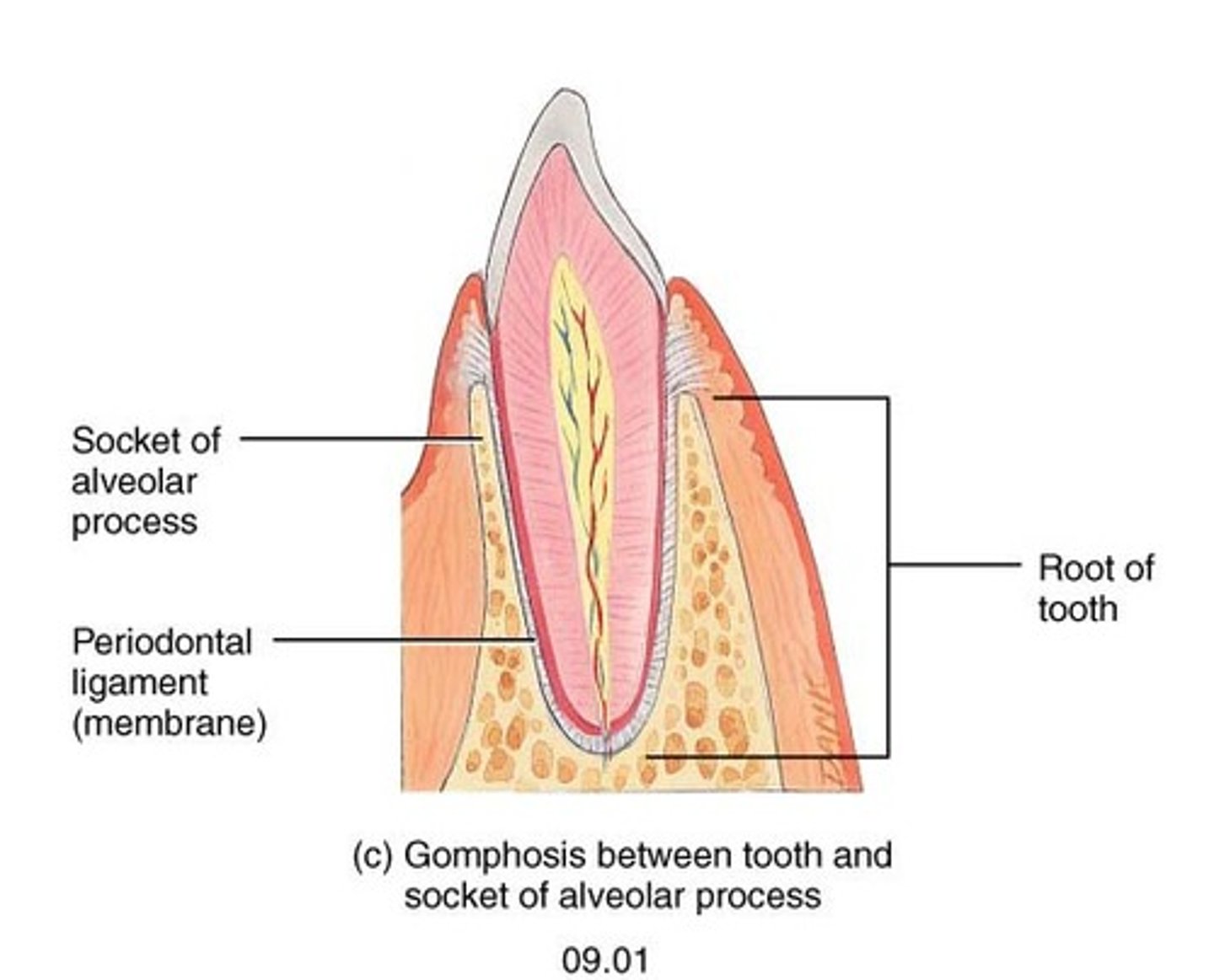
gomphoses
anchor teeth into sockets
syndesmoses
interosseous membrane.
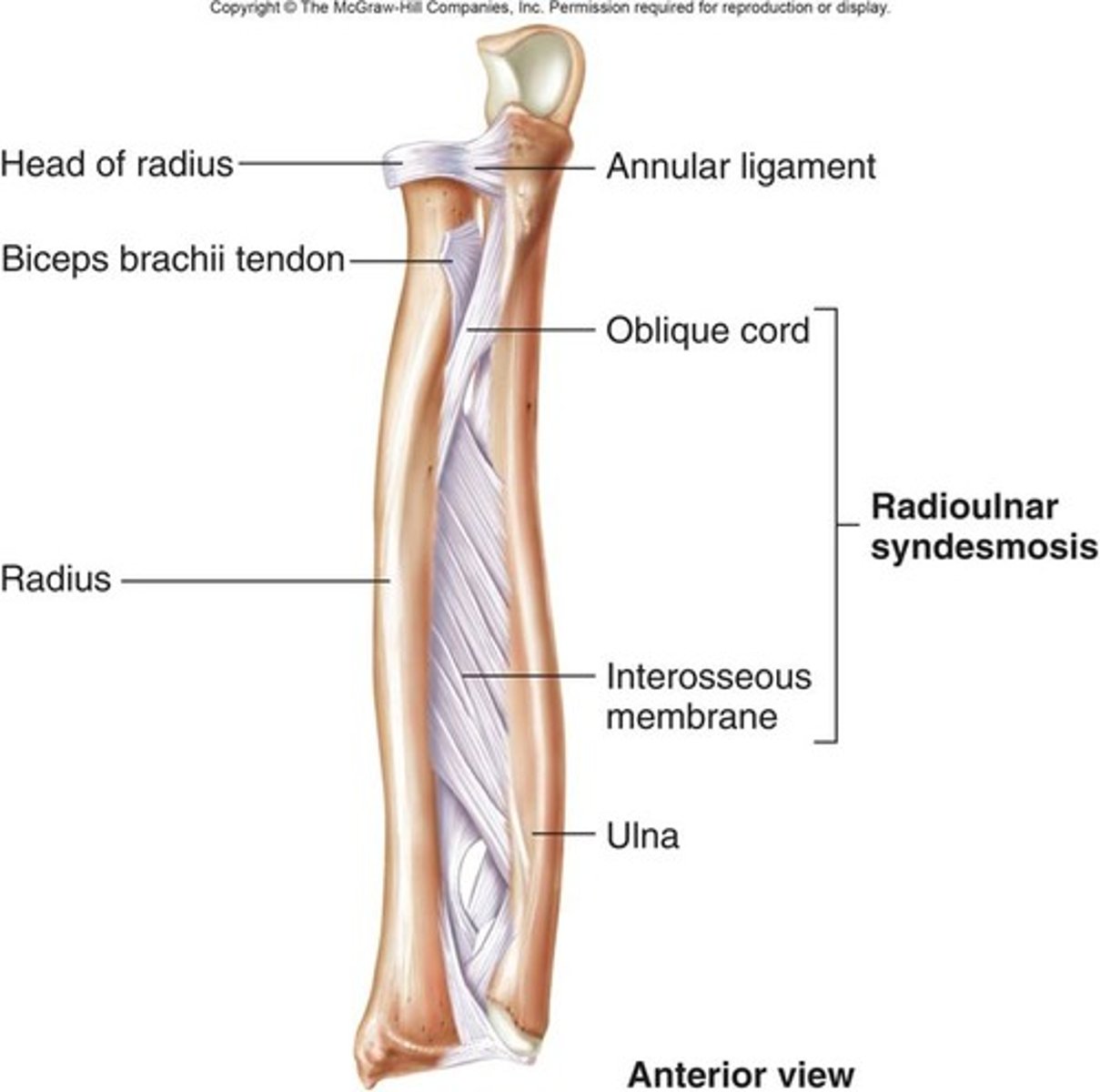
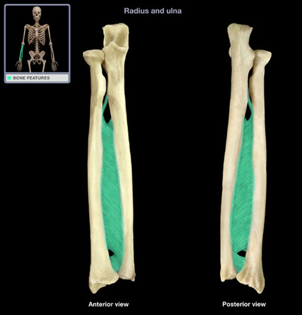
interosseous membrane
sheet of collagen fibers that attach the shafts of two bones. between radius and ulna
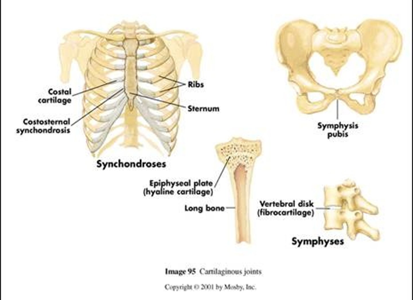
cartilaginous joints
two bones bound together by cartilage.
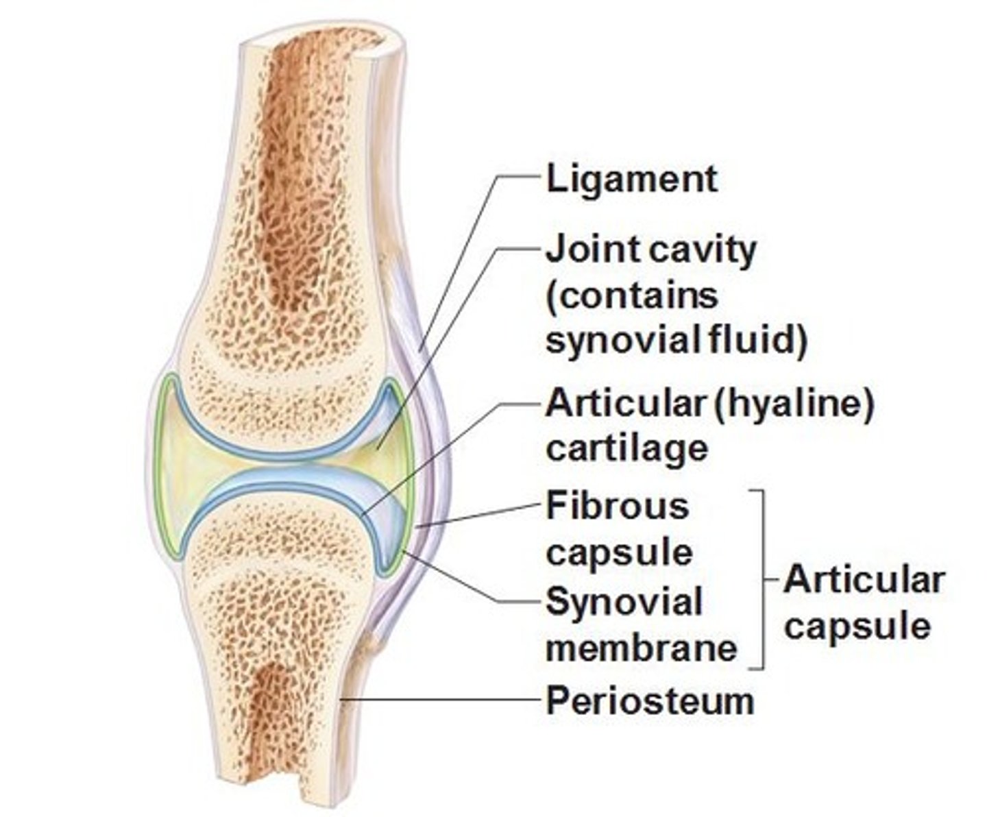
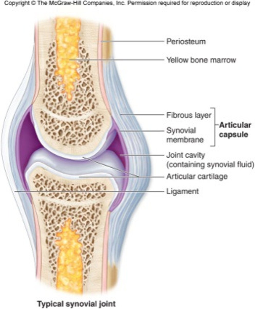
synovial joint anatomy
the relationship between mobility and stability joints
stable(yet limited mobility)
suture
interosseous membrane
intervertebral joints
knee joint
glenohumeral joint
mobile(yet unstable)
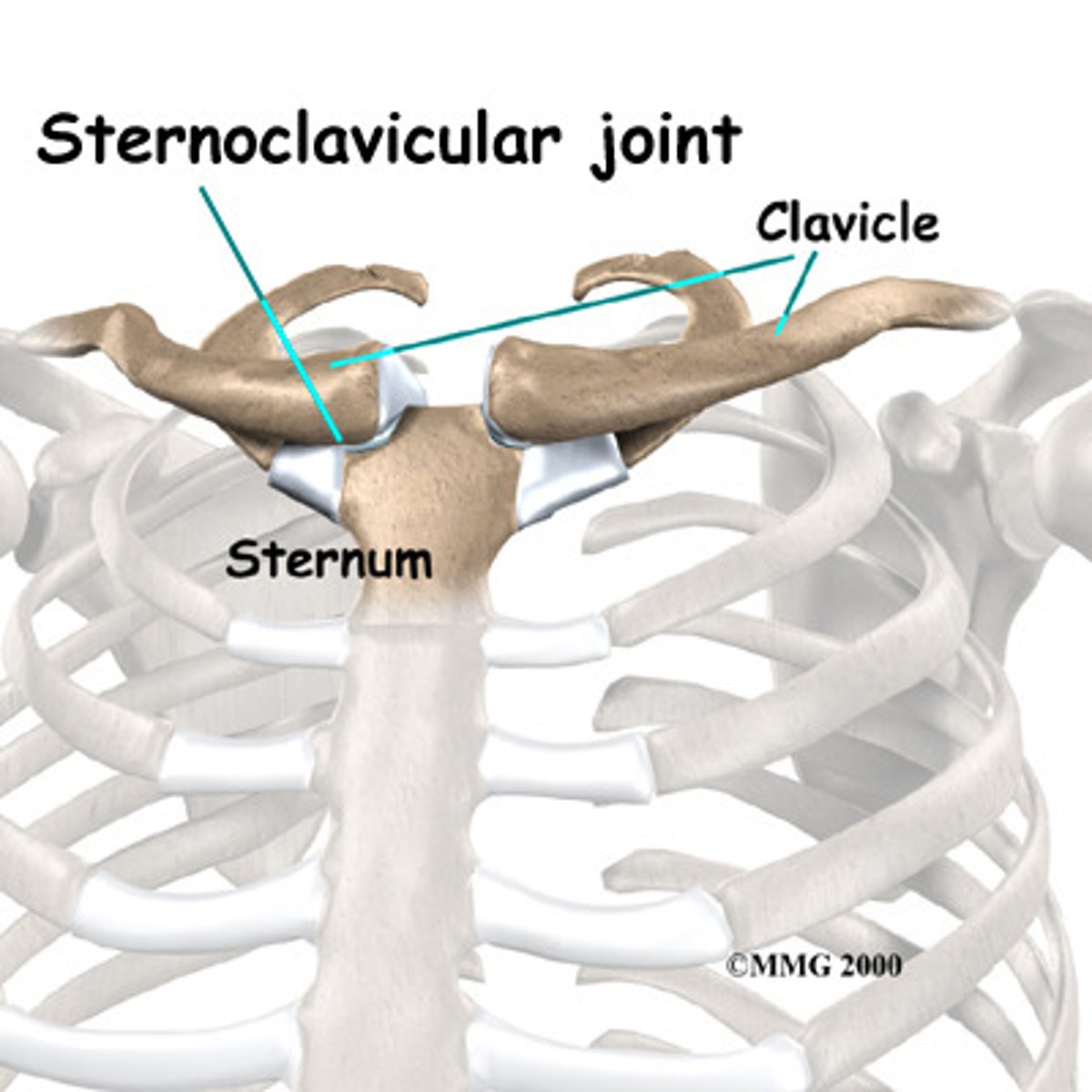
Sternoclavicular joint
synovial joint, diarthrosis, 1st rib and sternum, sternocostal
types of synovial joints
ball and socket: multiaxial joints, shoulders and hips
hinge joints: monaxial, elbow, knee, interphalangeal joints
saddle joint: biaxial
pivot: monaxial
gliding: ampharthroses
condyloid: biaxial
flexion
decreases the angle of a joint. (bending elbow or wrist)
extension
straightens a joint and returns a body part to the anatomical position
hyperextension
extension of a joint beyond 180 degrees or over the anatomical position
dorsiflexion
raising of the toes as when you swing the foot forward to take a step (heel strike)
plantar flexion
extension of the foot so that the toes point downward as in standing on tiptoe
abduction
movement of a part away from the midsagittal line (midline)
Example: raising arm to the side, spreading finger
adduction
movement towards the midsagittal line
when does supination and pronation occur?
in the forearm and foot
supination
rotation of forearm so that the palm faces forward
pronation
rotation of forearm so the palm faces to the rear
elevation
movement that raises a bone vertically. mandibles are elevated during biting and clavicles during a shrug
depression
lowering the mandible or the shoulders
protraction
movement of a bone anteriorly (forward) on a horizontal plane. thrusting the jaw forward, shoulders or pelvis forward
retraction
movement of a bone posteriorly
what occurs during lateral and medial excursion?
Side to side grinding movements occurring during chewing
Lateral excursion
sideways movement to right or left
Medial excursion
Movement back to the midline
Opposition
Movement of the thumb to approach or touch the fingertips that allows you to grasp an object and hold on to it
Reposition
Movement back to the anatomical position
Inversion
Movement in which the soles are turned medially
Eversion
Turning the soles to face laterally
Synostosis
a joint formed by fusion of two bones and the boundary between them disappears
Synchondrosis
cartilage bridge between two articulating bones (immovable)
Gomphosis
dense connective tissue (fibrous joints)
Example: periodontal ligaments
Symphysis:
is a cartilaginous joint in which the ends of the articulating bones are covered with hyaline cartilage, but a broad, flat disc of fibrocartilage connect to the bones (slightly movable)
Synovial membrane releases:
synovial fluid into the joint cavity
Functions of synovial fluid:
a. lubrication
b. shock absorption
c. provide nutrients for the tissue in the joint
d. protect articular cartilage (because it contains phagoytes)
Characteristics of articular cartilage
a. hyaline cartilage
b. no perichondrium
c. surfaces are normal slick and smooth
d. the matrix contains more water than other cartilage
When articular cartilage is damaged (arthritis)
a. the cartilage matrix is to break down
b. the surface is rough
c. more friction occur
d. normal synovial joint function is unable to continue
Bursae
a. sac-like structures filled with a small amount of fluid that is similar to synovial fluid
Tendon sheaths
tube-like bursae
Bursea are situated to:
reduce friction in some joints
Bursea are found in:
the tendon sheaths, beneath the skin that covers vone, and most of synovial joints (shoulder, knee, elbow joints)
The stronger and more stable a joint...
the less mobility it has
The weaker and less stable a joint...
the more R.O.M. (range of motion) it has
Dislocation occurs when:
the articulating surfaces of a synovial joint are foced out of position
Types of movements that may be permitted at a synovial joint:
a. linear motion (gliding)
b. angular motions (flexion, extension, adduction, abduction, and circumduction)
c. rotation
d. other special movements (eversion, inversion, protraction, retraction, depression, elevation, opposition)
Examples of gliding joints include:
inter-carpals
inter-tarsals
vertebrae with ribs
clavicle with sternum
ilium with sacrum
Gliding join is:
biaxial or multiaxial
Hinge joint is:
angular, monoaxial
Condylar joint is:
angular, biaxial
Examples of condylar joints
a. radiocarpal joint
b. medacarpophalangeal joints 2-5
c. metatarsophalangeal joint
Pivot joint is:
rotaion, monoaxial
Examples of pivot joints
a. Atlanto axial joint
b. proximal radio-ulnar joints
Ball and socket joint is:
angular, circumduction, rotation, tri-axial (multi-axial)
Example of a hinge joints
a. The articulation between tibia and talus bone (ankle joint)
b. elbow joint
c. interphalangeal joint
Examples of ball and socket joints:
a. shoulder joint
b. hip joint
Examples of saddle joint
a. the joint between carpal (trapezium bone) and metacarpal I (thumb)
The joint that permits the greatest ROM (Range of motion) is the:
shoulder joint
The should joint is mainly stabilized by:
a group of muscles (rotator cuff muscles) that move the humerus bone and reinforce the joint capsule
Baseball pitchers are the greatest risk of developing:
rotator cuff injury
The most common athletic knee injuries involve:
menisci
In the elbow joint the largest and strongest joint is the:
articulation between the humerus and the ulnar bone (humero-ulnar joint)
The elbow joint is mainly stabilized by:
a strong and thick articular capsule
Functions of the intervertebral discs:
a. act as shock absorber
b. prevent bone-to-bone contact
c. contribute the height of the individual
d. allow the movements associated with flexion, extention and rotation of the spine
A hernia disc is cause by:
protrusion of the nucleus pulposus of the intervertebral disc
The role of the menisci (medial and lateral menisci) is to:
act as cushions and conform to the shape of the articular cartilage
The menisci are responsible for:
channeling the flow of synovial fluid in the knee joint
Knee joint is a:
modified hinge joint that consists of three articulations within a single cavity
Compared to the should joint, the complete dislocation of the knee joint is rare because:
the knee joint is stabilized by 7 major ligaments
Compared with that of the shoulder, the articular capsule of the hip is very:
dense and strong
Compare with that of the hip, the articular capsule of the knee is:
weak
Five ligaments of the hip joint include;
1. iliofemoral
2. pubofemotal
3. ischiofemoral
4. ligament teres (ligaments of the head of femur)
5. transverse ligament of the acetabulum
Seven ligaments of the knee joint include:
1. patellar ligament
2. popliteal ligament (2)
3. ACL
4. PLC
5. Tibial collateral
6. Fibular collateral
The ACL and PCL:
a. limit the anterior and posterior movement of the tibia
b. maintain the alignment of the femoral and tibial condyles
The structure that assists the bursae in reducing friction between the patella and other tissues are:
FAT PADS
When the articular cartilage is damaged:
a. the matrix begins to break down
b. the expose articular surface become rough
c. the friction of the joint increases
d. normal synovial joint functions is unable to continue
Rheumatism is a general term for:
pain and stiffness affecting the musculoskeletal system
Arthritis affects the:
synovial joints