cytoskeleton 2D and 2E cell motility
1/32
There's no tags or description
Looks like no tags are added yet.
Name | Mastery | Learn | Test | Matching | Spaced | Call with Kai |
|---|
No analytics yet
Send a link to your students to track their progress
33 Terms
Cytoskeleton
The cytoskeleton is a network of protein fibres that organises the structures and activities of the cell, forming an internal framework that supports the cell and coordinates movement of structures within it.
Cytosol
The cytosol is the aqueous fluid inside the cell surrounding organelles where many metabolic reactions occur.
Cytoplasm
The cytoplasm consists of the cytosol and cytoskeleton. Therefore cytoplasm = cytosol + cytoskeleton.
Functions of the cytoskeleton
The cytoskeleton maintains cell shape, protects and stabilises cellular structures, enables cell motion via cilia and flagella, transports vesicles and organelles within the cell, and forms structures such as the mitotic spindle during cell division.
Three types of cytoskeletal fibres
The cytoskeleton contains microfilaments (~7 nm, actin), intermediate filaments (~10 nm, keratin or vimentin), and microtubules (~25 nm, tubulin). All form through polymerisation of protein subunits.
Microfilaments structure
Microfilaments are the thinnest cytoskeletal fibres (~7 nm) composed of actin. G-actin (globular actin) monomers polymerise to form F-actin (filamentous actin), and two F-actin strands twist together into a double helix.
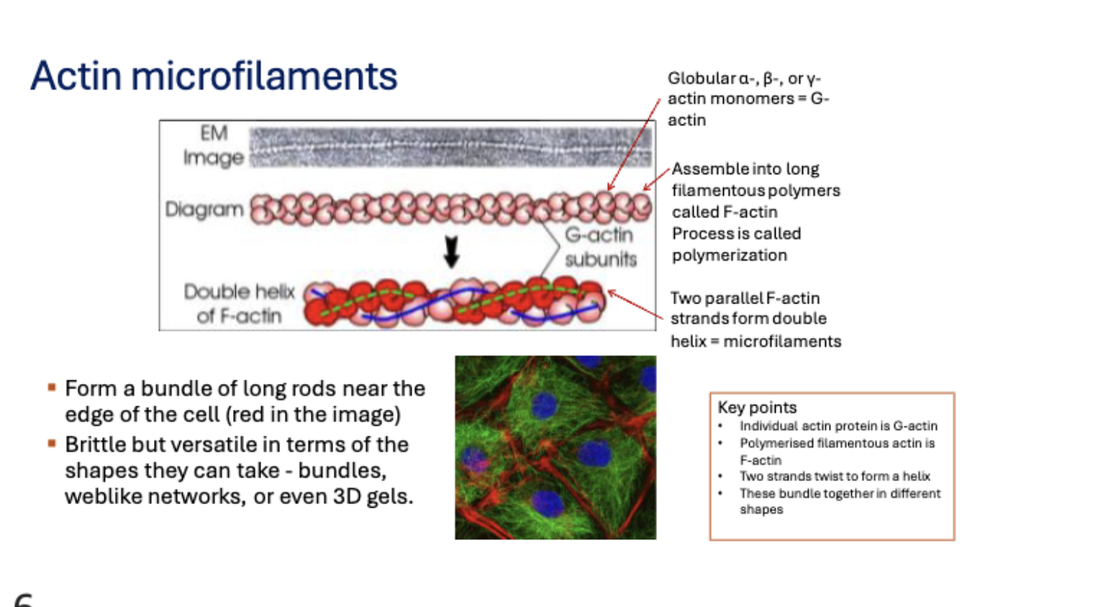
Microfilament organisation
Microfilaments can form bundles, web-like networks, and three-dimensional gels.
Functions of microfilaments
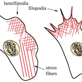
Microfilaments contract or lengthen to change cell shape, enable cell movement, form lamellipodia in migrating cells, allow intracellular movement of components, and are essential for cytokinesis.
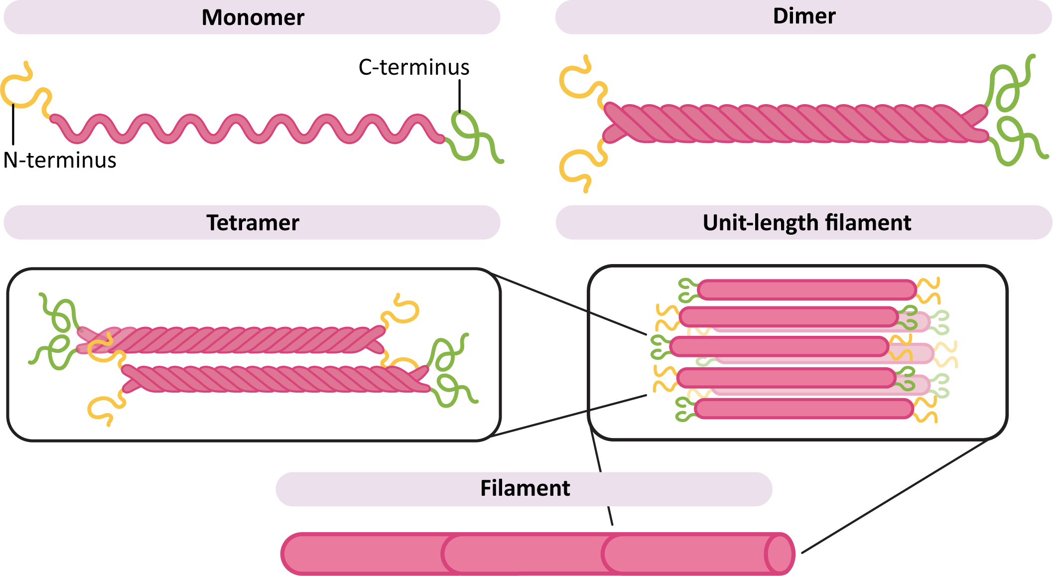
Intermediate filament structure
Intermediate filaments have a diameter of about 10 nm and are composed of proteins such as keratin and vimentin. Assembly occurs through monomers → dimers → tetramers → protofibrils → unit length filament → intermediate filaments.
Functions of intermediate filaments
Intermediate filaments provide mechanical strength, resist mechanical stress, maintain structural integrity and help position organelles within the cell.
Examples of intermediate filaments in the body
Intermediate filaments form structural components in hair, nails, the outer layer of skin, muscle cells, nerve cells and heart tissue.
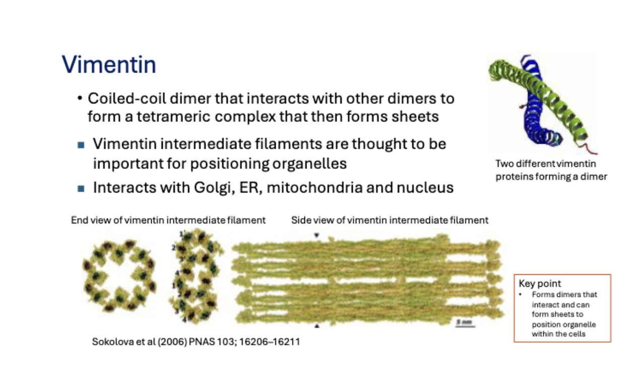
Vimentin filaments
Coiled-coil dimer that interacts with other dimers to form a tetrameric complex that then forms sheets
Vimentin intermediate filaments help position organelles and interact with the Golgi apparatus, endoplasmic reticulum, mitochondria and nucleus.
Microtubule structure
Microtubules are hollow cylindrical fibres about 25 nm in diameter composed of tubulin. Tubulin forms heterodimers of α-tubulin and β-tubulin that assemble into 13 protofilaments forming a hollow tube.
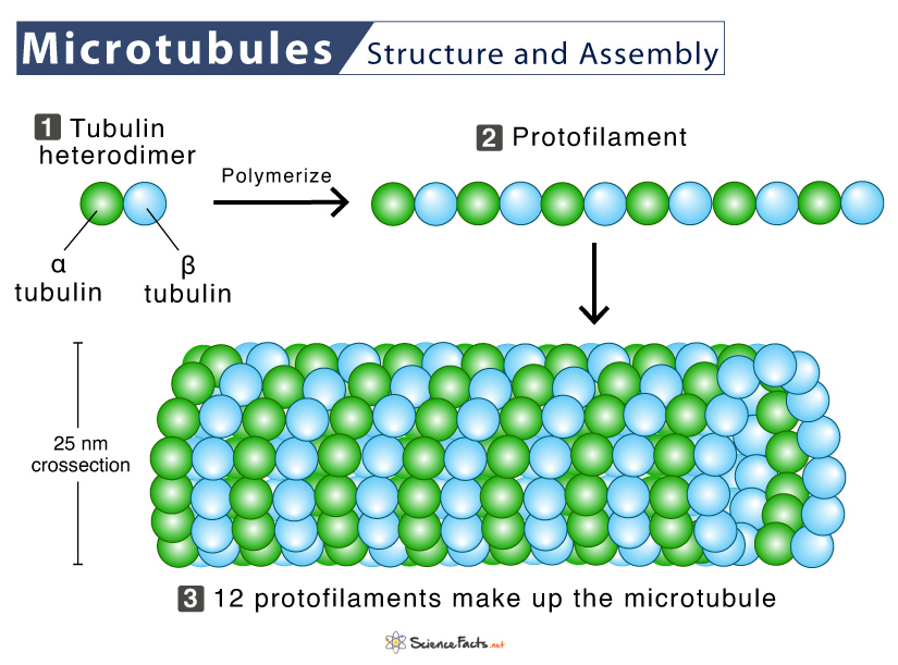
Microtubule polarity
Microtubules have two ends called the plus end and minus end. This polarity enables directional transport of vesicles and motor proteins.
Functions of microtubules
Microtubules provide structural support, form the mitotic spindle during cell division, act as tracks for vesicle transport, organise the ER and Golgi and form the core structure of cilia and flagella.
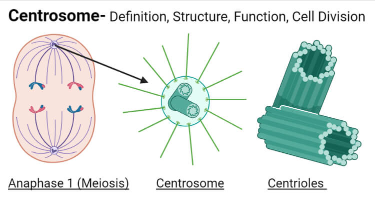
Centrosome
The centrosome is the microtubule organising centre (MTOC) of the cell and organises the microtubule network.
Centrioles structure
Centrioles are hollow cylindrical structures composed of nine triplets of microtubules and occur as a pair arranged at right angles within the centrosome.
Cells containing centrioles
Centrioles are found in animal cells, fungi and algae but not in plant cells.
Motor proteins
Motor proteins are enzymes that move along cytoskeletal fibres using ATP as an energy source, mainly travelling along microtubules to transport cellular cargo.
Why motor proteins require ATP
Motor proteins convert chemical energy from ATP into mechanical movement, allowing repeated attachment, movement and detachment along cytoskeletal filaments.
Kinesin
Kinesin is a motor protein that moves toward the plus end of microtubules, typically transporting vesicles, organelles and proteins from the centrosome toward the cell membrane.
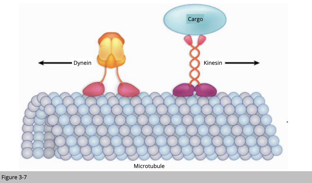
Dynein
Dynein is a motor protein that moves toward the minus end of microtubules, transporting vesicles toward the cell centre and generating movement in cilia and flagella.

Motor protein walking mechanism
Motor proteins bind to microtubules, hydrolyse ATP, undergo a conformational change and move forward approximately 8 nm per step while carrying cargo attached via receptors.
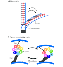
Dynein sliding mechanism
In cilia and flagella dynein attaches between adjacent microtubule doublets and uses ATP to pull one microtubule past another. Because the microtubules are anchored and cross-linked, sliding results in bending rather than separation.
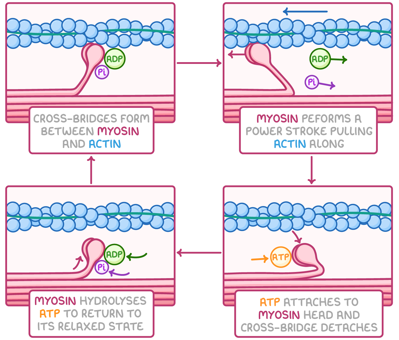
Myosin and actin sliding
In muscle cells myosin motor proteins attach to actin microfilaments and use ATP to generate a power stroke that pulls actin filaments past each other, producing muscle contraction.
Polymerisation
Polymerisation is the addition of protein subunits to a cytoskeletal filament, causing it to grow.
Depolymerisation
Depolymerisation is the removal of protein subunits from a filament, causing the filament to shrink and allowing cells to retract or change shape.
Cilia and flagella
Cilia and flagella are plasma-membrane-covered organelles built from microtubules and powered by motor proteins that enable cell movement.
Axoneme
The internal structure of cilia and flagella is called the axoneme and has a characteristic 9+2 arrangement consisting of nine outer microtubule doublets and two central microtubules.
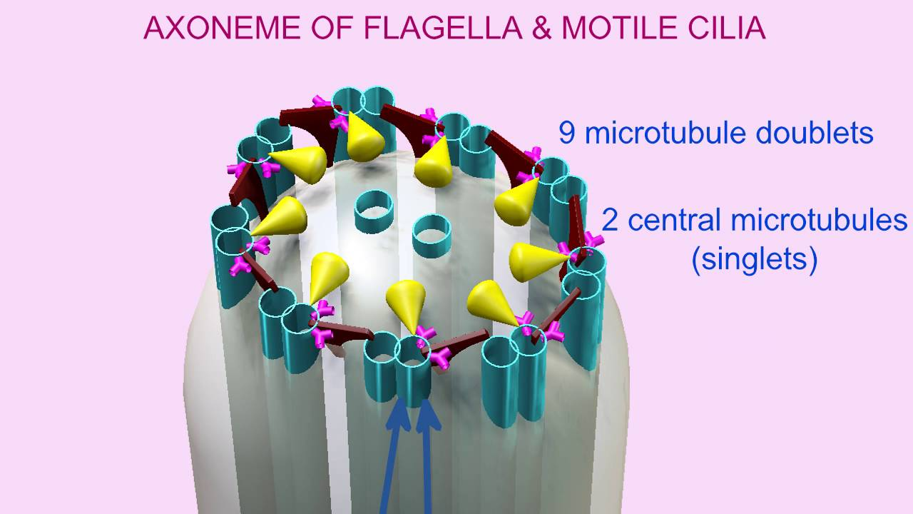
Dynein arms in cilia and flagella
Dynein arms attach to outer microtubule doublets and interact with neighbouring microtubules to generate sliding forces that produce movement.
Requirements for bending of cilia and flagella
Bending requires dynein motor proteins, microtubule sliding, ATP energy and anchorage of microtubules at the base, which converts sliding into bending motion.
Amoeboid movement
Amoeboid movement is a crawling cell movement used by amoeba and some immune cells where actin polymerises at the front forming a pseudopodium while myosin contraction at the rear pushes cytoplasm forward.
Cytoplasmic streaming
Cytoplasmic streaming (cyclosis) is the movement of cytosol and organelles through the cell driven by microfilaments, helping distribute nutrients, molecules and organelles especially in large plant cells.