INB 446L - Lab Exam 1
1/299
There's no tags or description
Looks like no tags are added yet.
Name | Mastery | Learn | Test | Matching | Spaced | Call with Kai |
|---|
No analytics yet
Send a link to your students to track their progress
300 Terms
Which plane divides the body into a front and back?
Frontal/Coronal
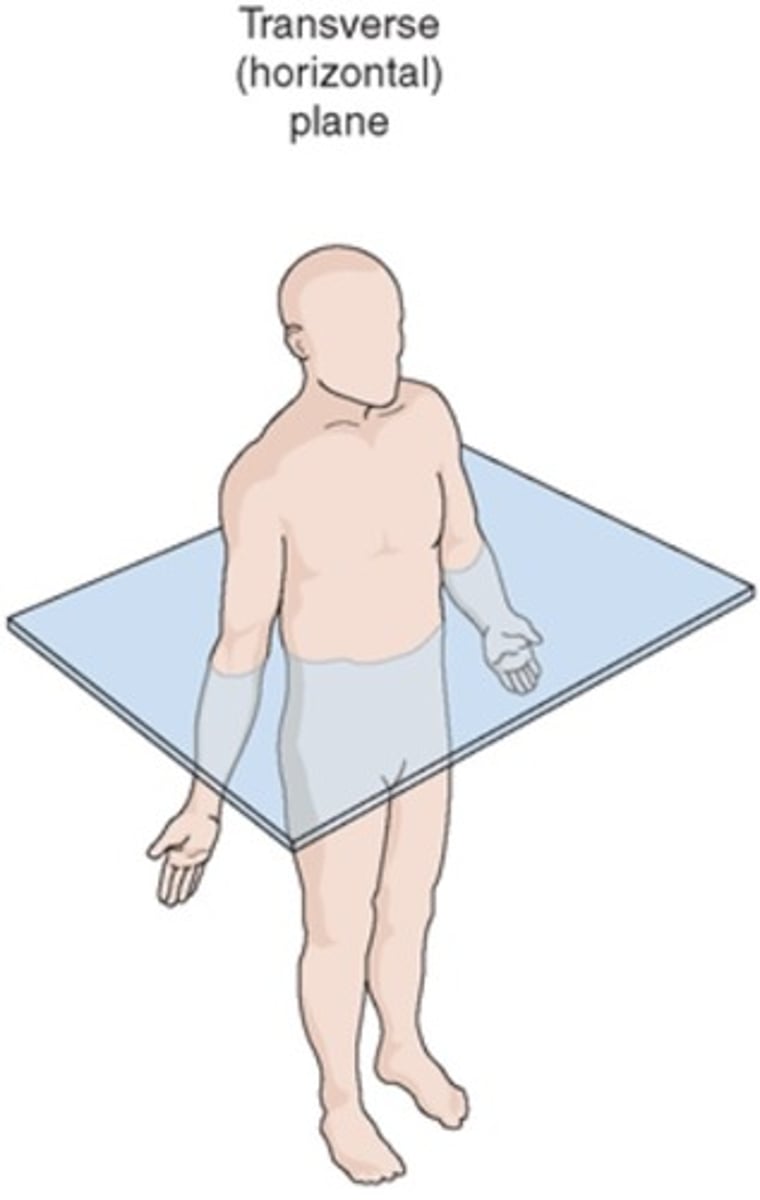
Which plane divides the body into a top and bottom?
Axial/Transverse
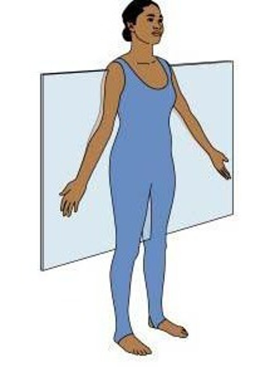
Which plane divides the body vertically into equal left and right sides?
Midsagittal
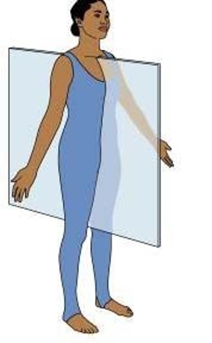
Which plane dives the body vertically into left and right sides?
Sagittal

Superficial
Closer to the surface of the body
Deep
Farther from the body surface, more internal
Medial
Closer to the midline of the body
Lateral
Away from the midline of the body
Ventral/Anterior
toward the front (belly) of the body
Dorsal/Posterior
toward the back (spine) of the body
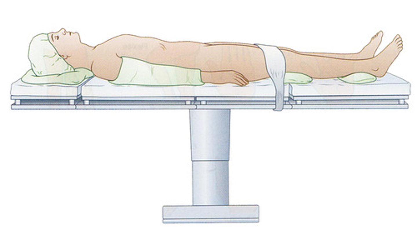
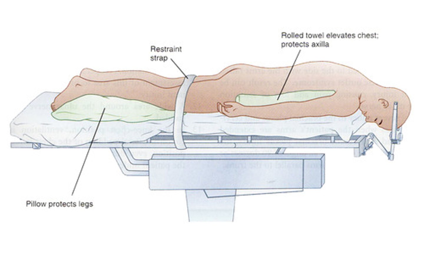
Supine
lying face up
Prone
lying face down
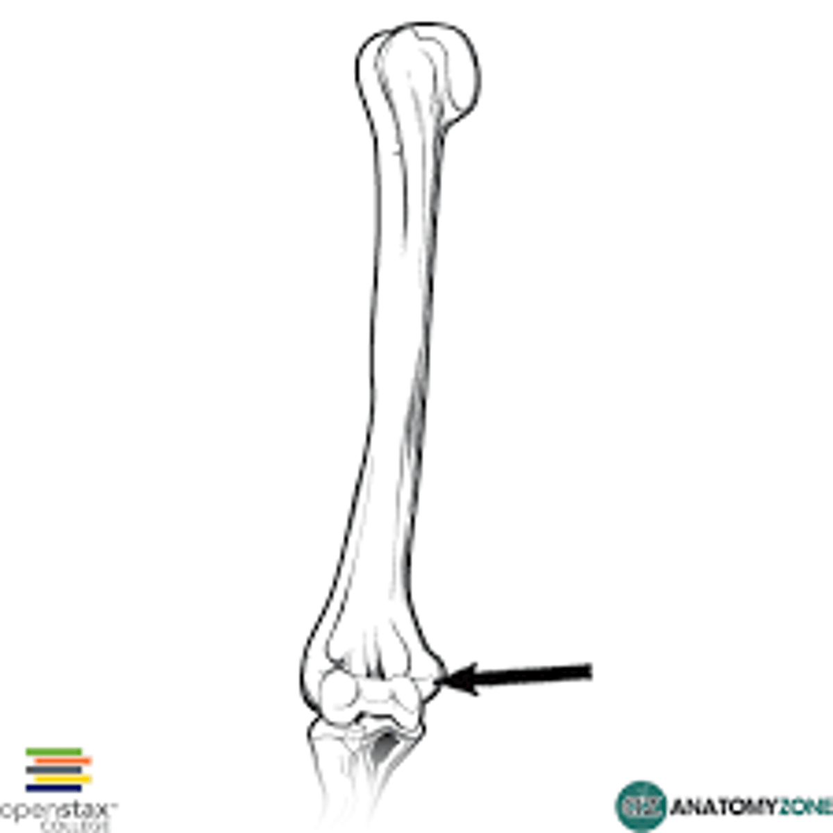
Proximal
Closer to the point of attachment
Distal
away from the point of attachment
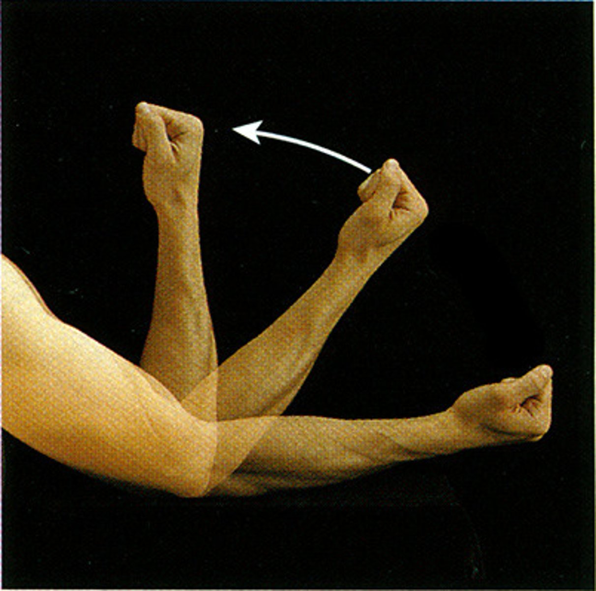
Flexion
bending a joint/decreasing the angle
Extension
Straightening of a joint/increasing the angle
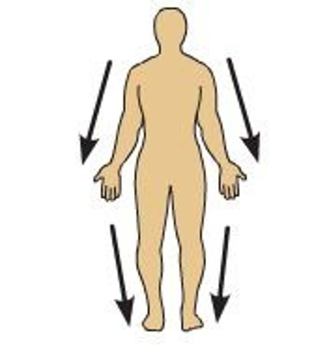
Adduction
Movement toward the midline of the body
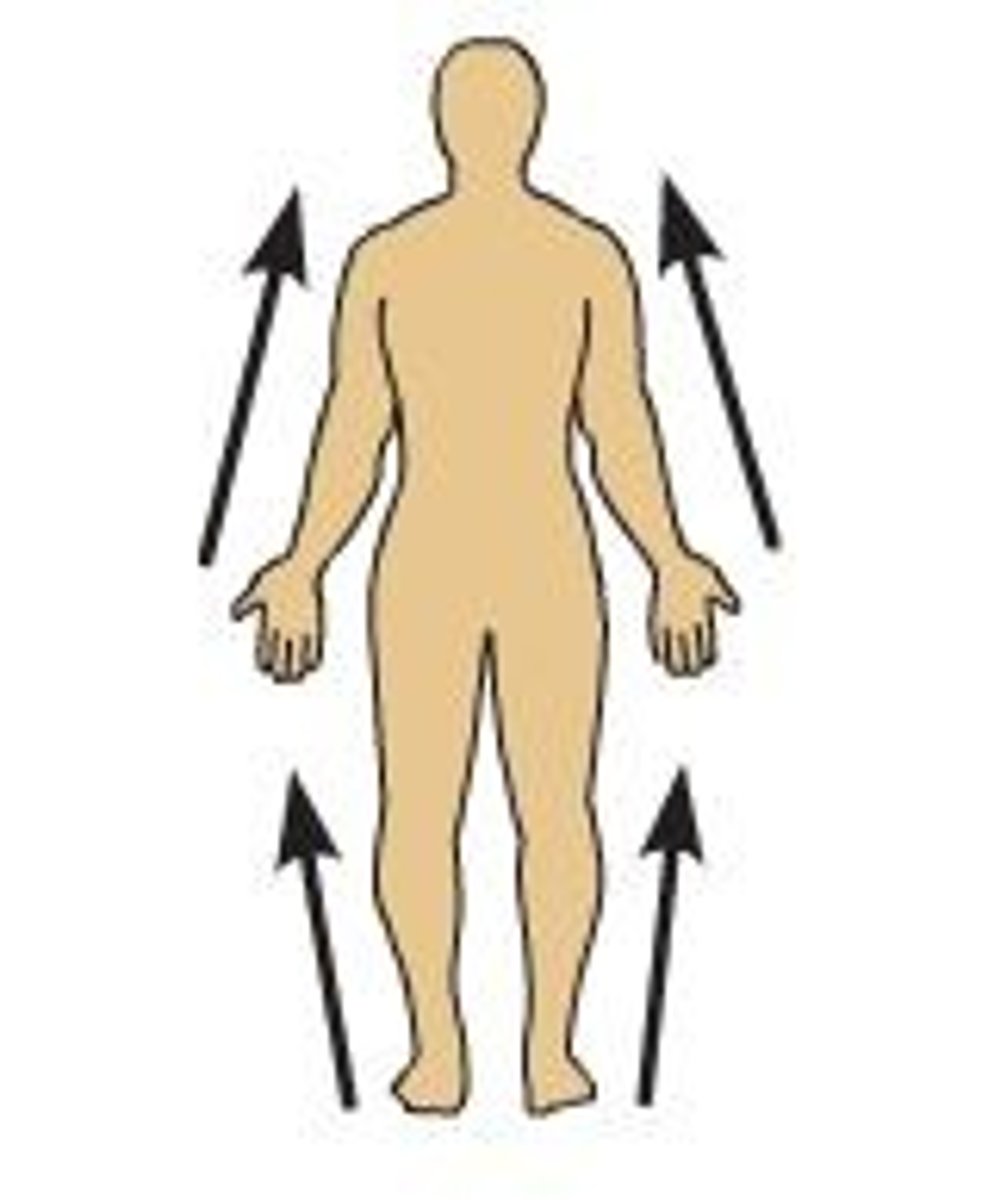
Abduction
Movement away from the midline of the body
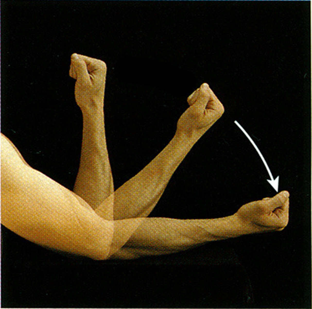
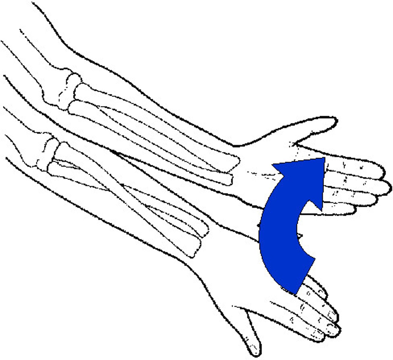
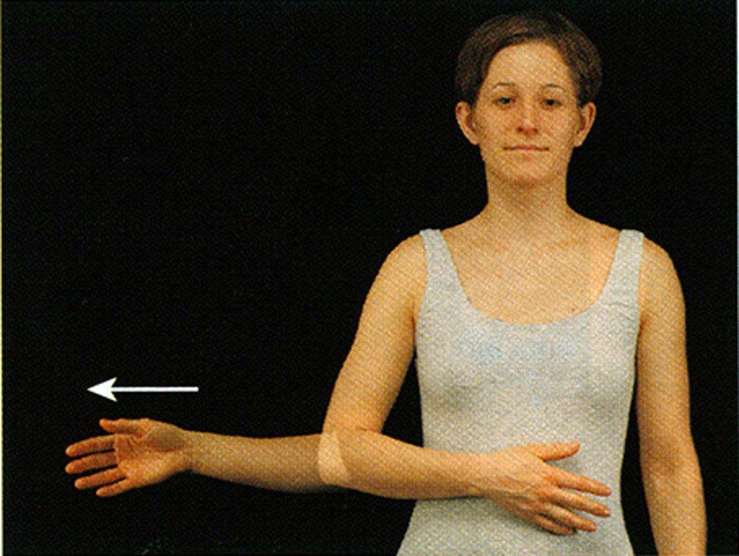
Pronation (forearm)
turning the palm downward
Supination (forearm)
turning the palm upward
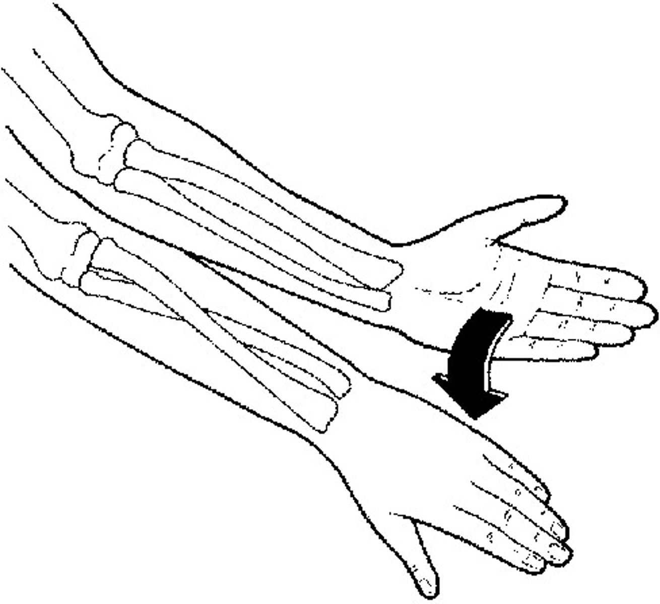
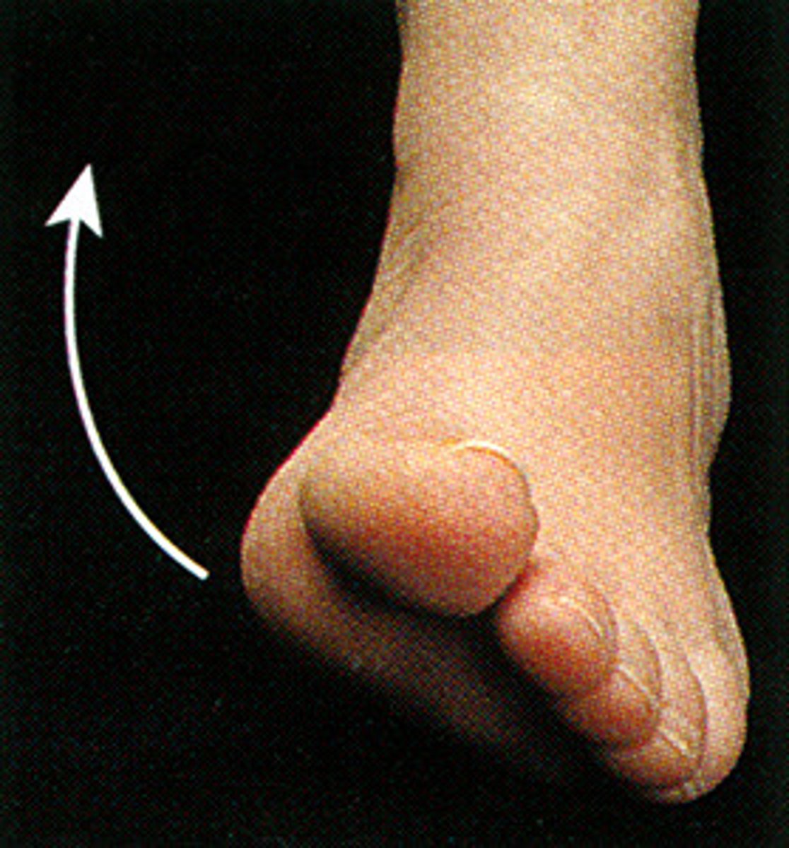
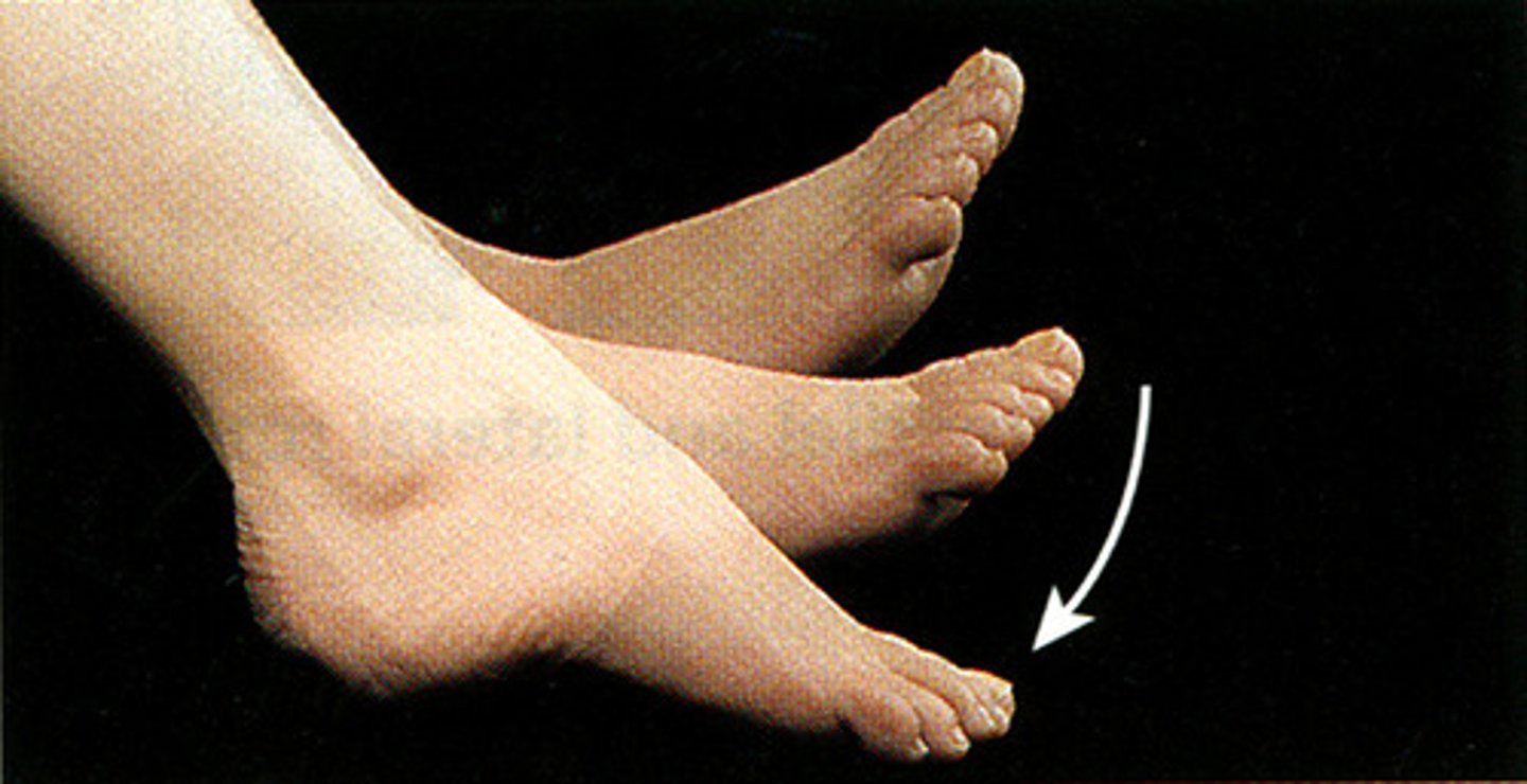
Inversion (ankle)
Turning the sole of the foot inward or medially
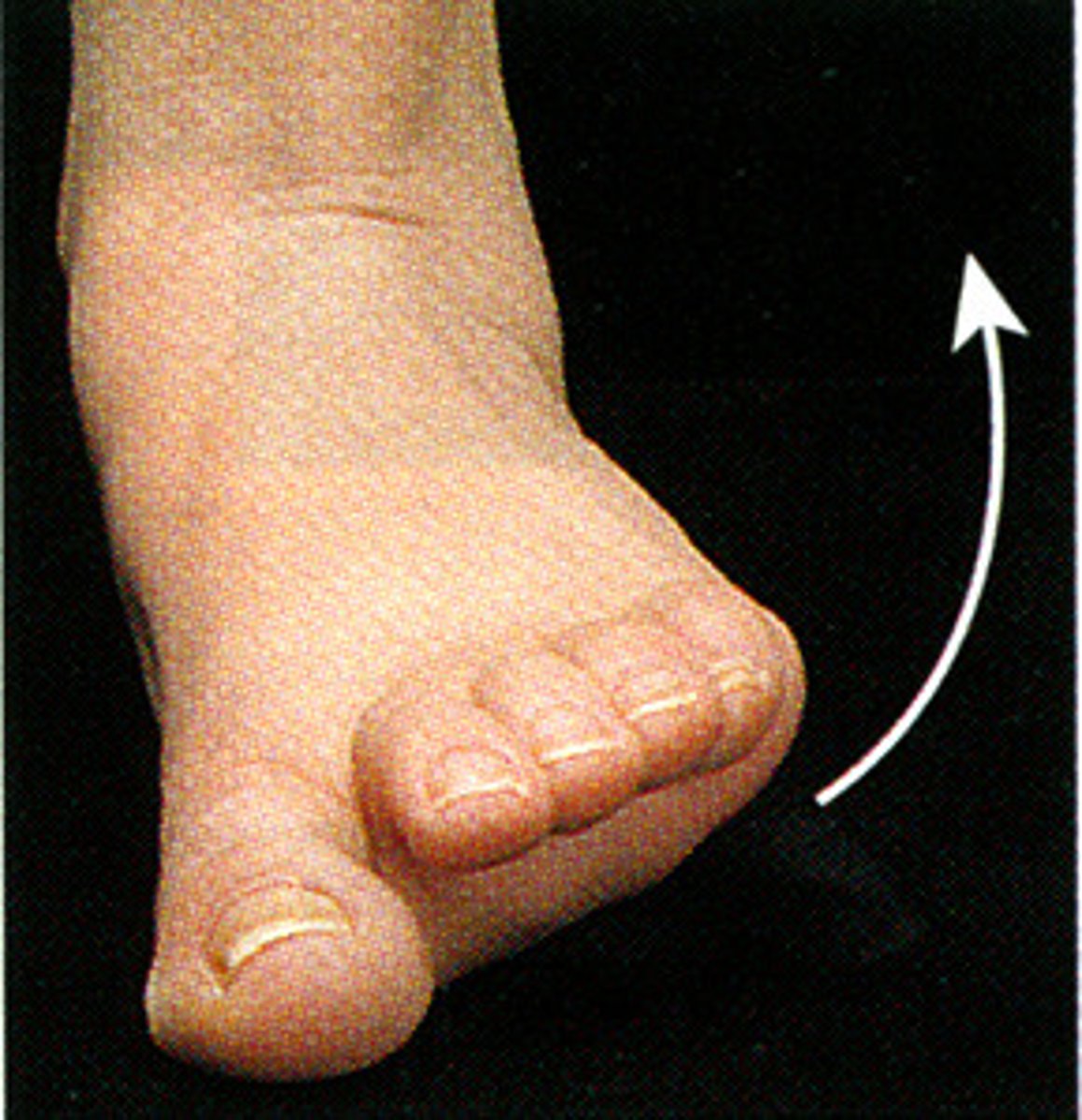
Eversion (ankle)
turning the sole of the foot outward or laterally
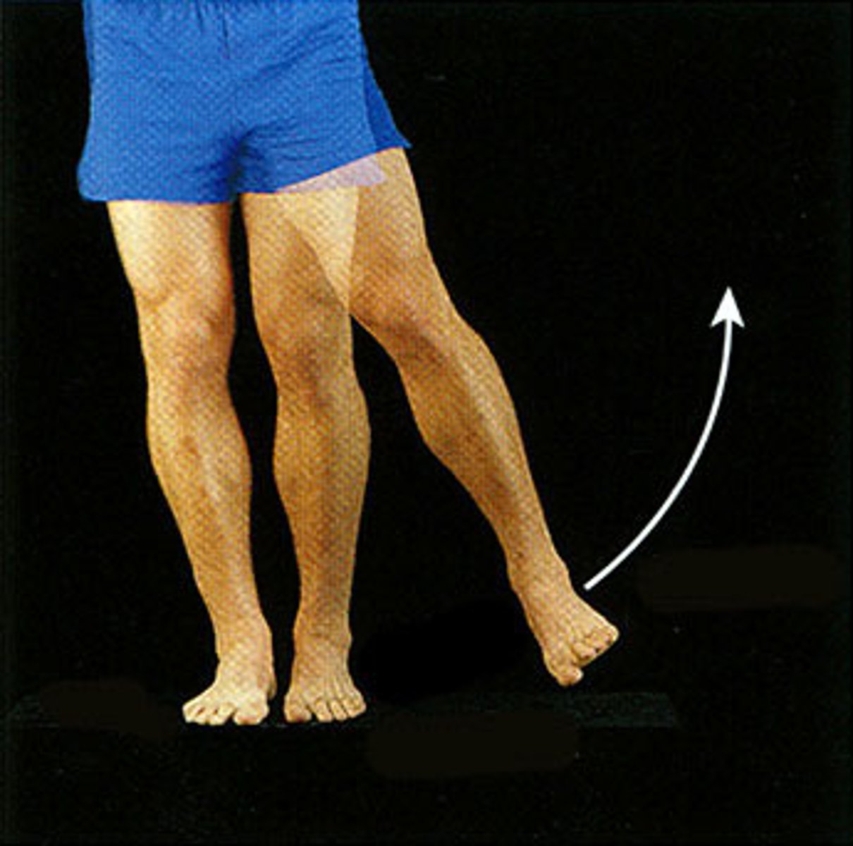
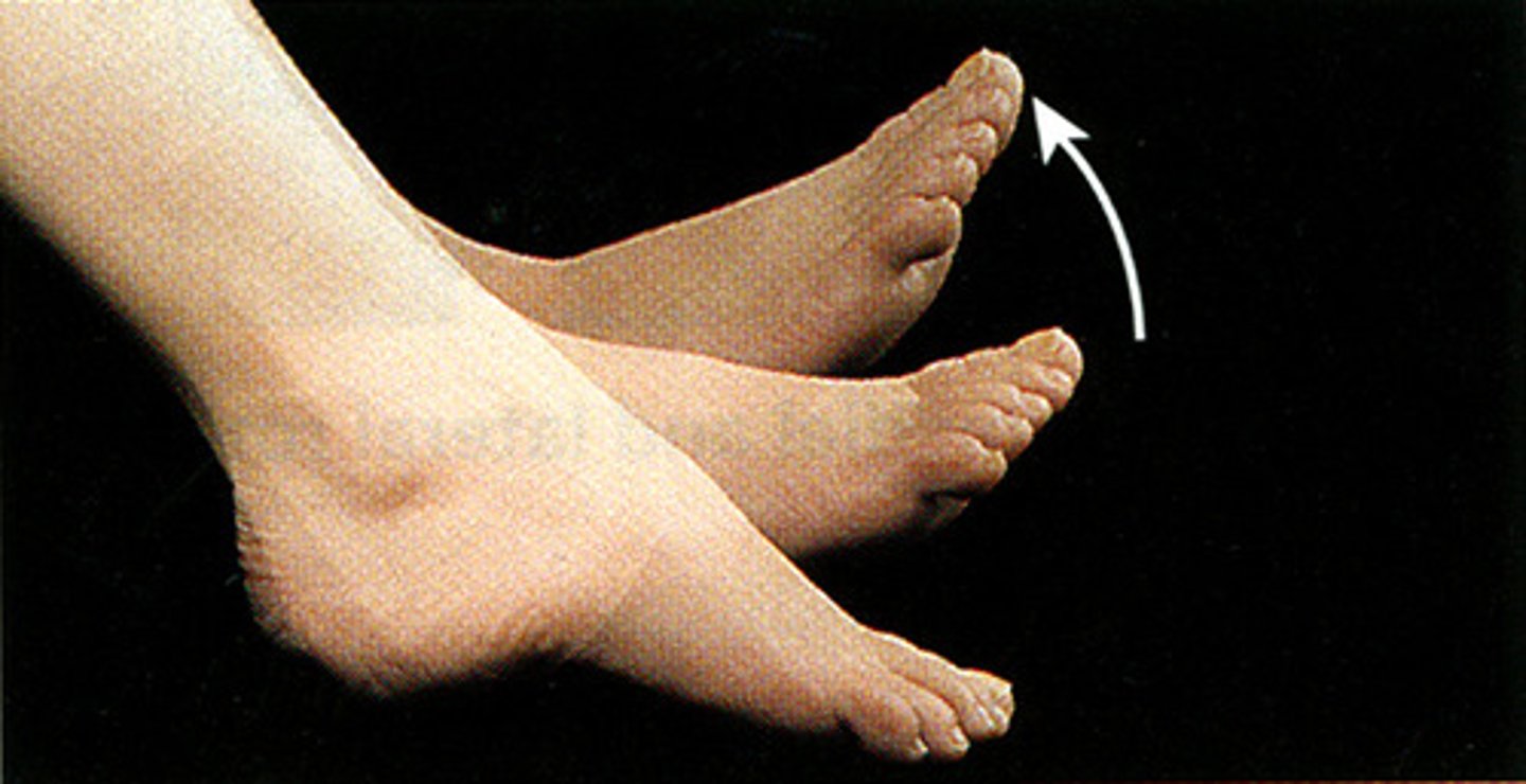
Dorsiflexion (ankle)
Lifting the foot up (towards the shin)
Plantarflexion (ankle)
pointing toes downward (away from the shin)
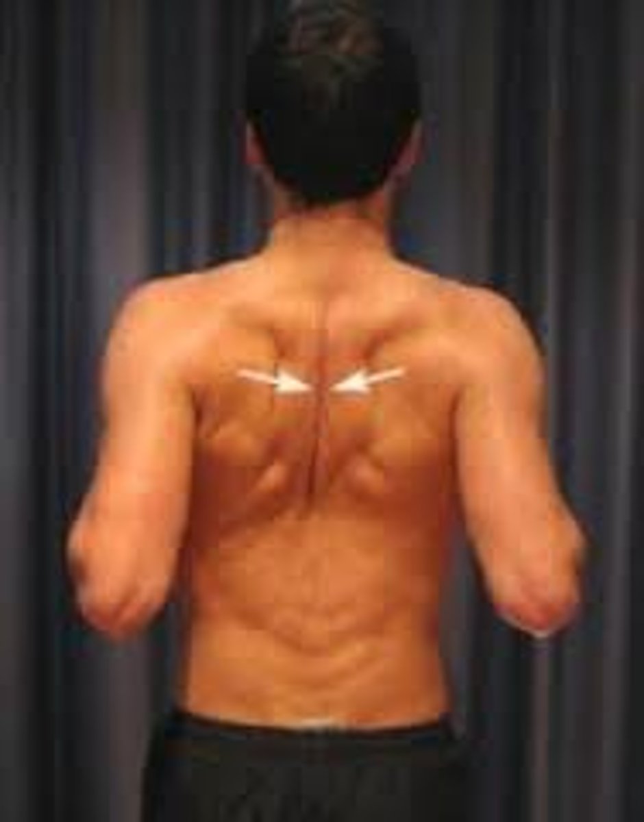
Elevation
Lifting a body part superiorly (e.g., shrugging shoulders)
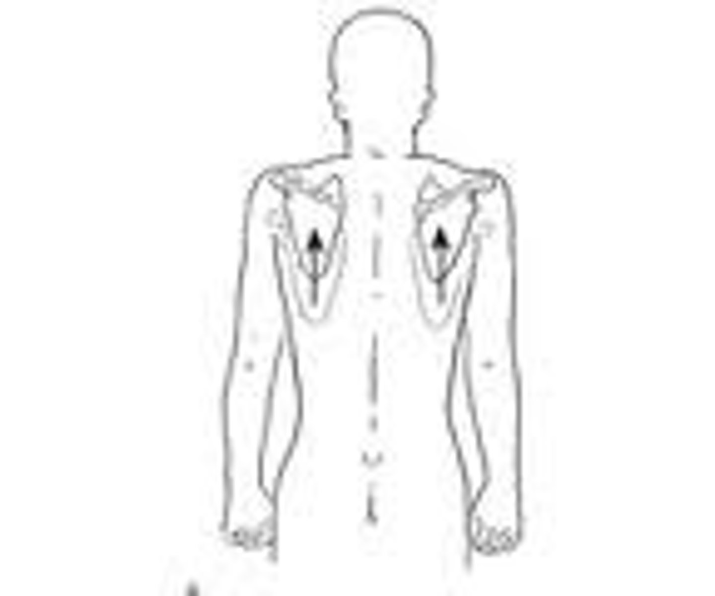
Depression
Moving a body part inferiorly (e.g., lowering shoulders)

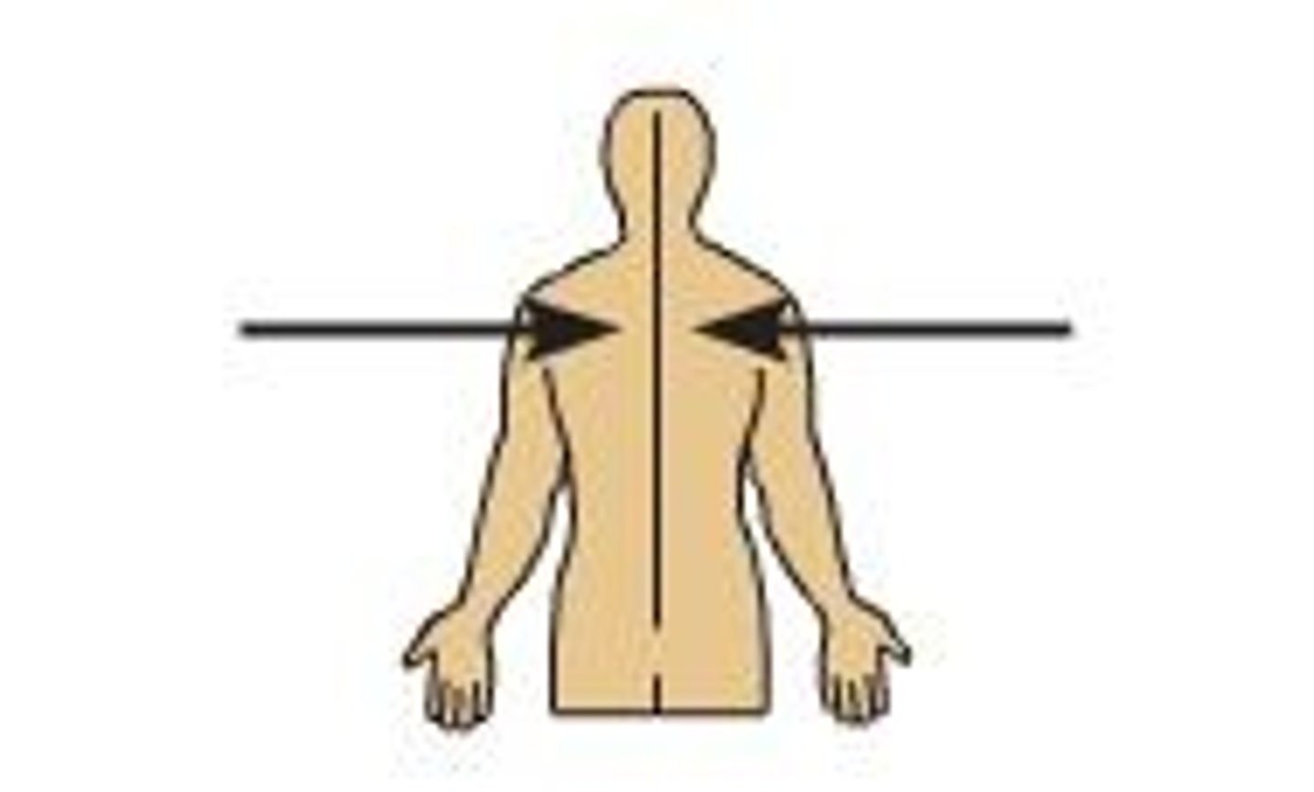
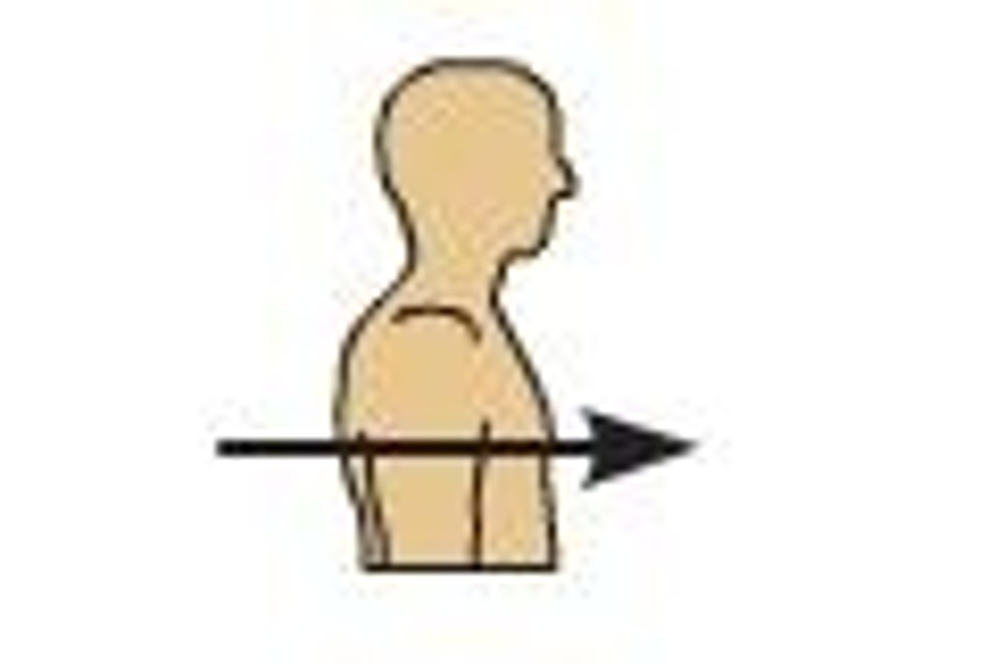
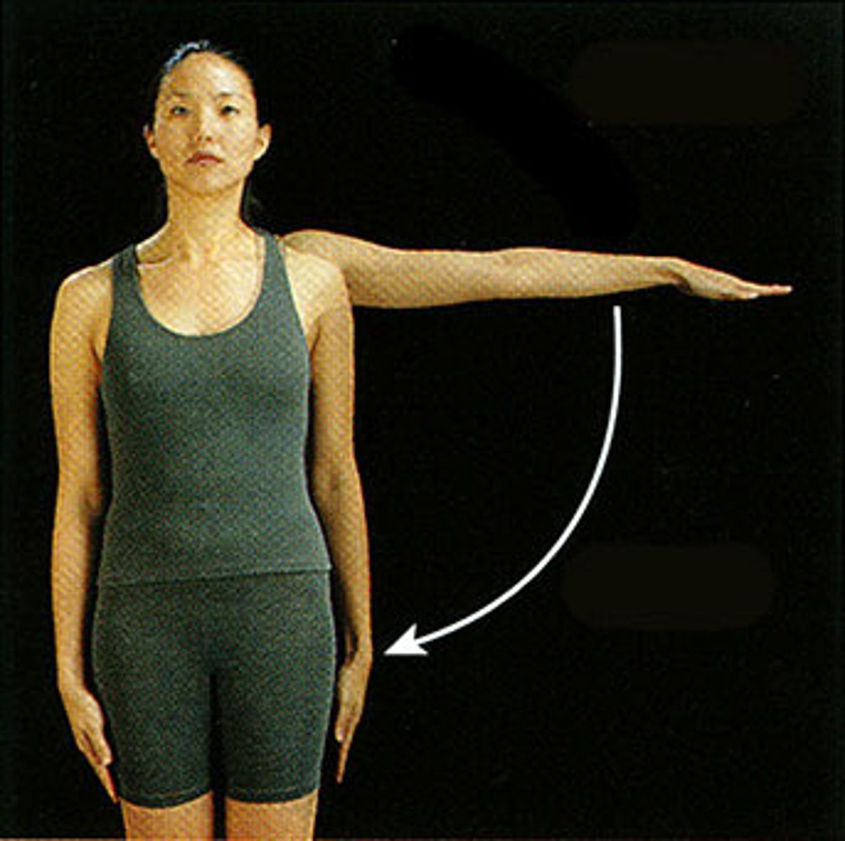
Protraction
Moving a body part forward
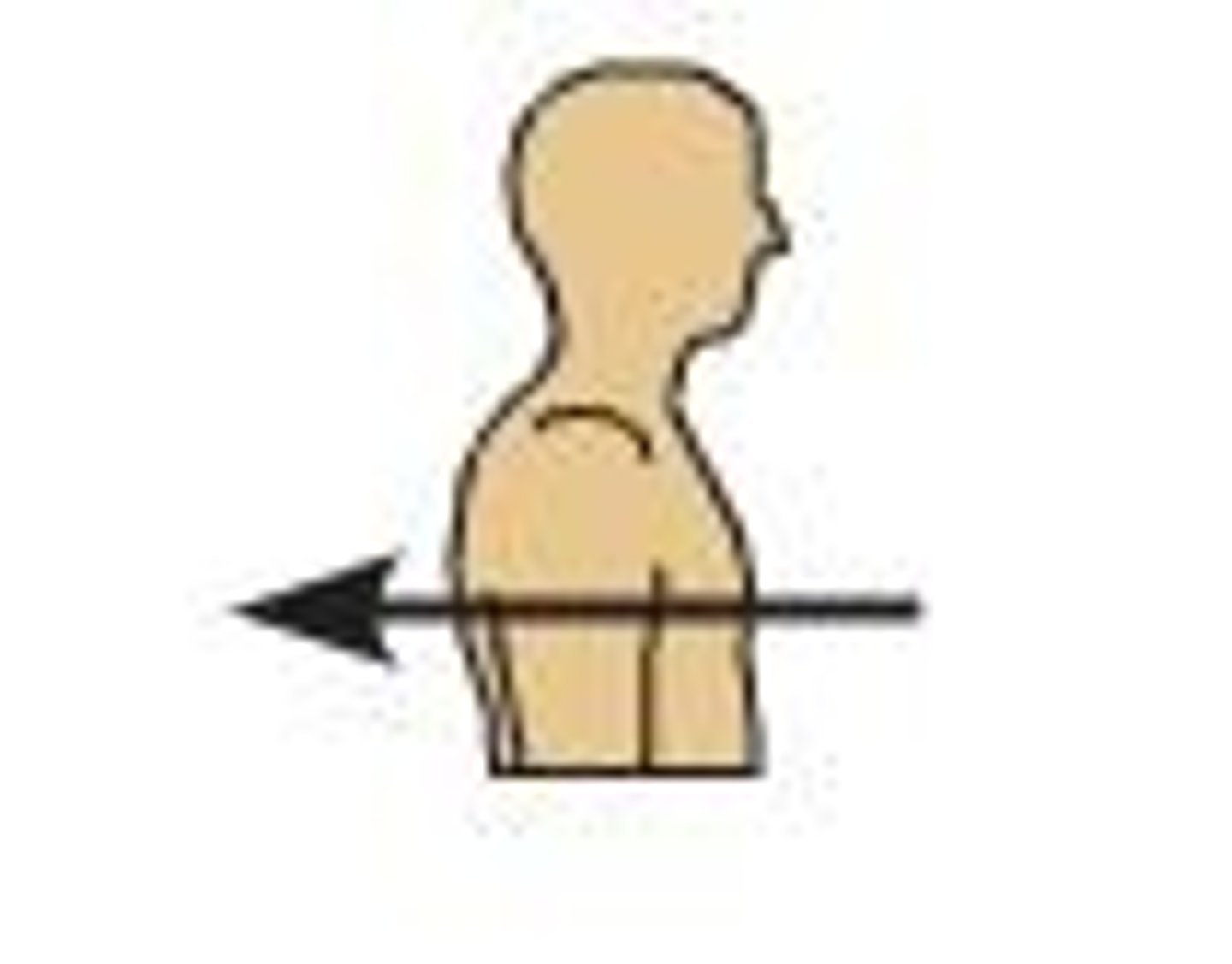
Retraction
moving a body part backward
Medial (internal) rotation
Rotating a limb toward the midline of the body

Lateral (external) rotation
Rotating a limb away from the midline of the body
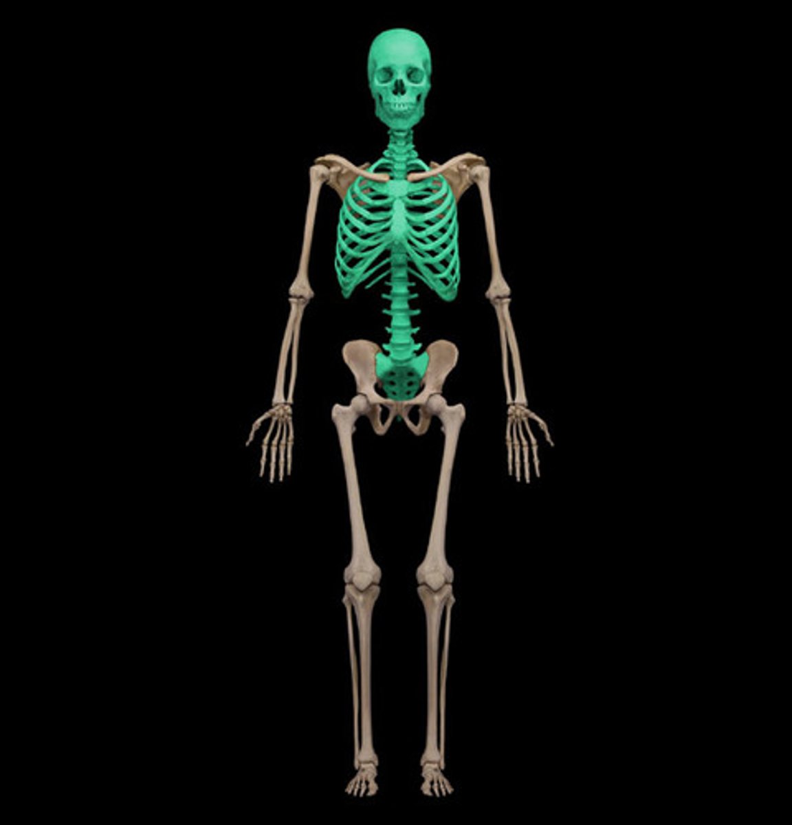
What is the main function of the axial skeleton?
To support and protect the body's vital organs, including the brain, spinal cord, and thoracic organs.

What are the bones of the axial skeleton?
skull, vertebral column, rib cage
What is the main function of the appendicular skeleton?
Facilitates movement and large range of motions
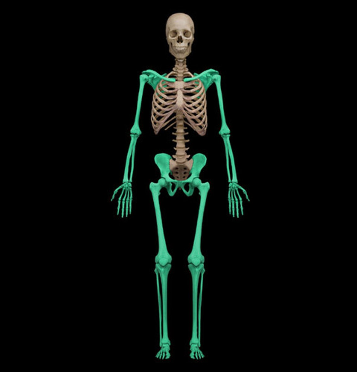
What are the bones of the appendicular skeleton?
Forearm
Hand
Thigh
Leg
Foot
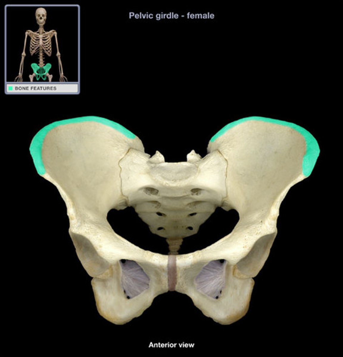
Pelvic
Clavicle
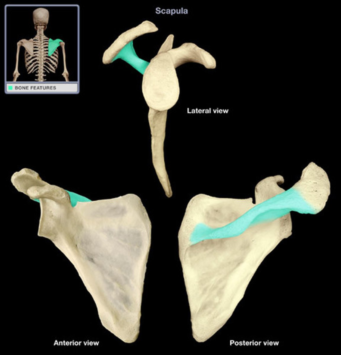
Scapula

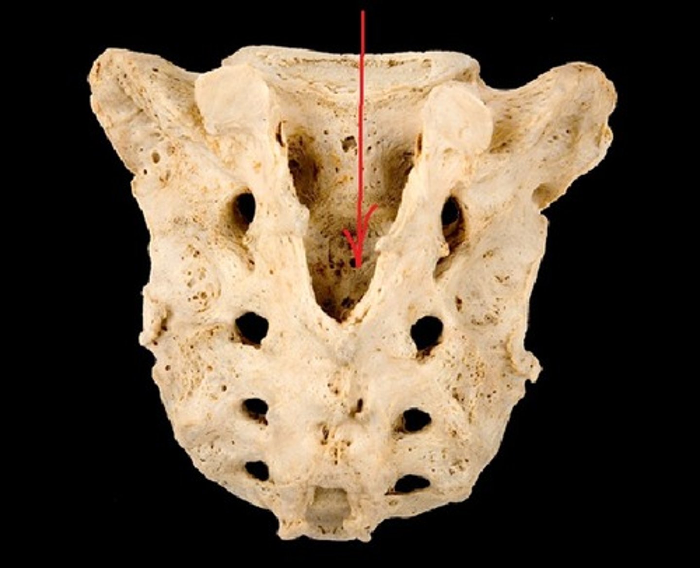
Bony Landmarks : Canal
a tunnel through a bone (e.g. sacral canal)
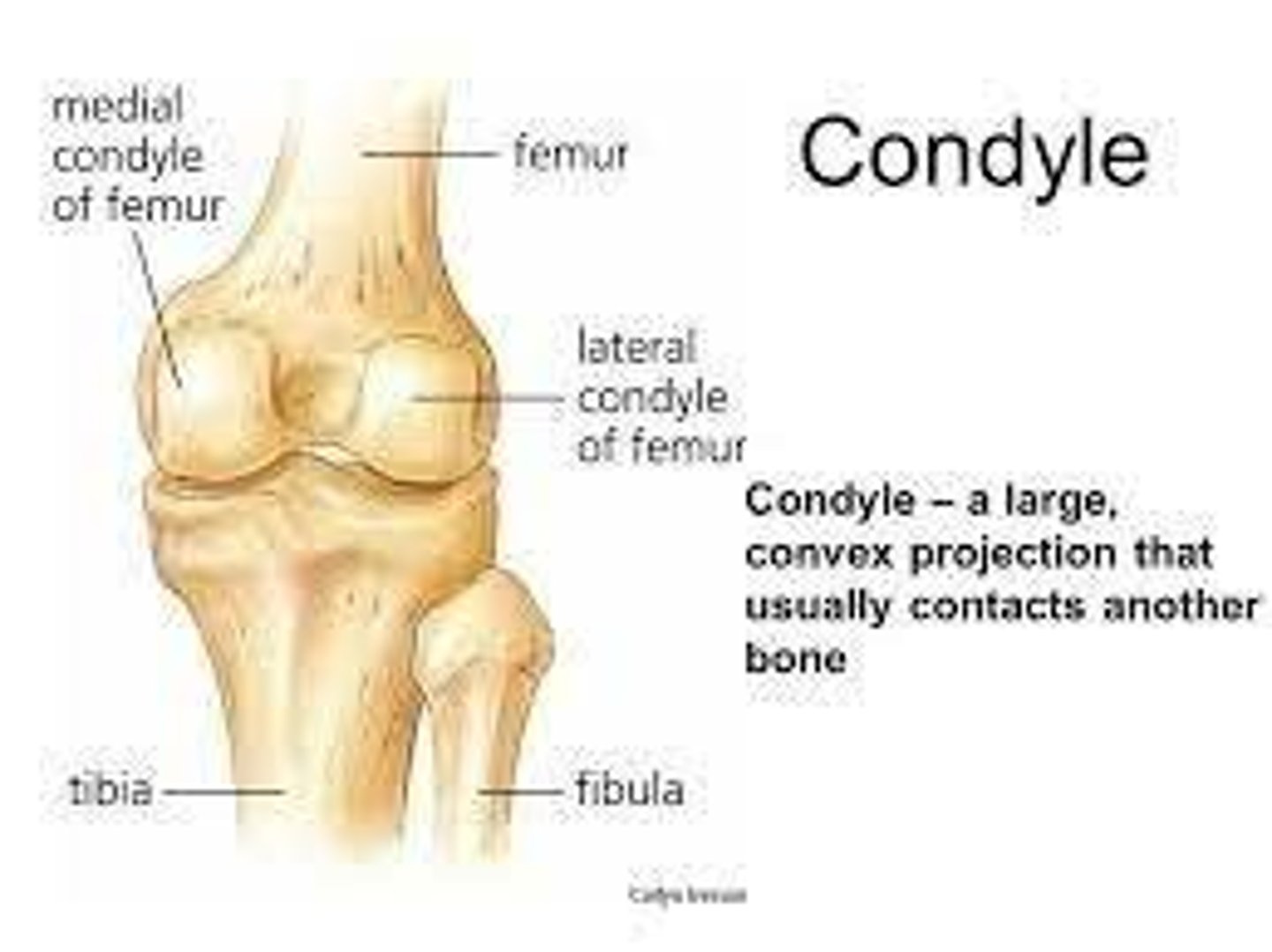
Bony Landmarks : Condyle
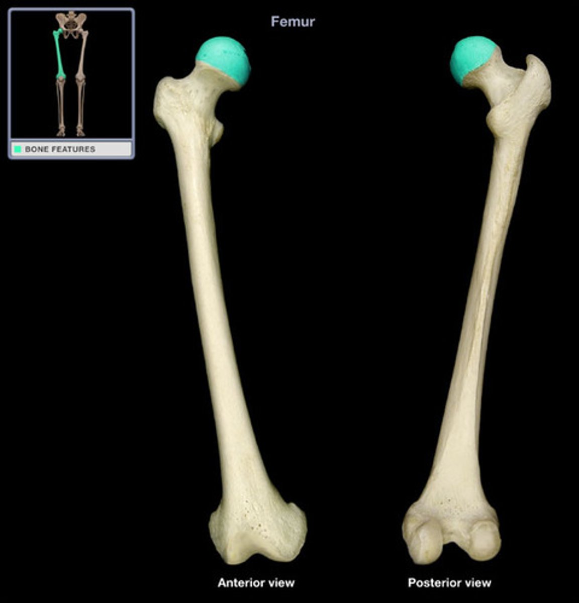
Provides structure support and absorbs most of the energy exerted by the joint (e.g. tibia or femur)
Bony Landmarks : Facet
A smooth and flat surface that helps form a gliding joint (e.g. L01 and L02 Vertebra)
Bony Landmarks : Fissure
A slit in the bone that usually houses nerves and blood vessels (e.g. maxilla)
Bony Landmarks : Foramen
A round whole where blood vessels, nerves, or ligaments pass through (e.g. occipital bone)
Bony Landmarks : Fossa
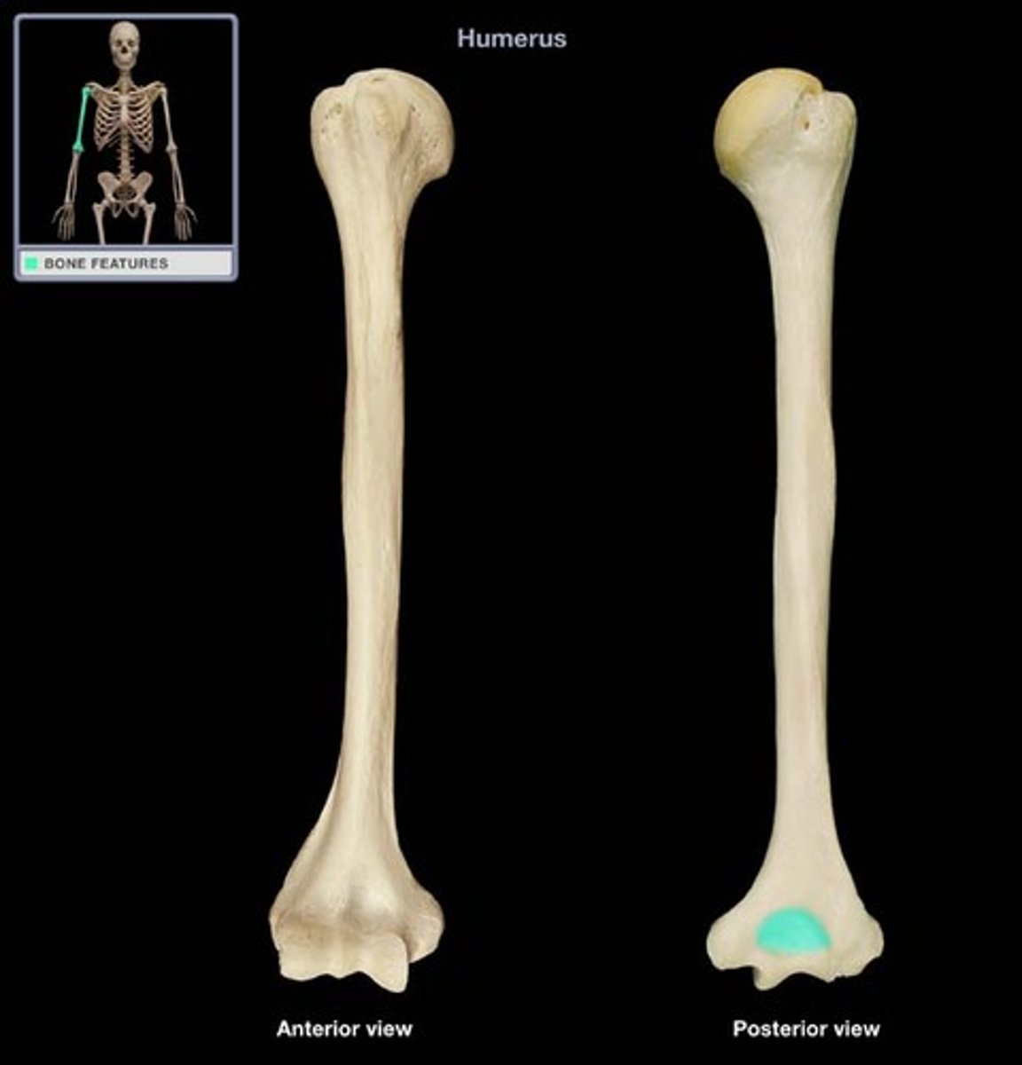
A shallow depression in a bone's surface that allows other bones to articulate with it (e.g. humerus)
Bony Landmarks : Trochanter*
A rough prominence at the upper part of the femur serving usually for the attachment of muscles (important muscle attachment site)
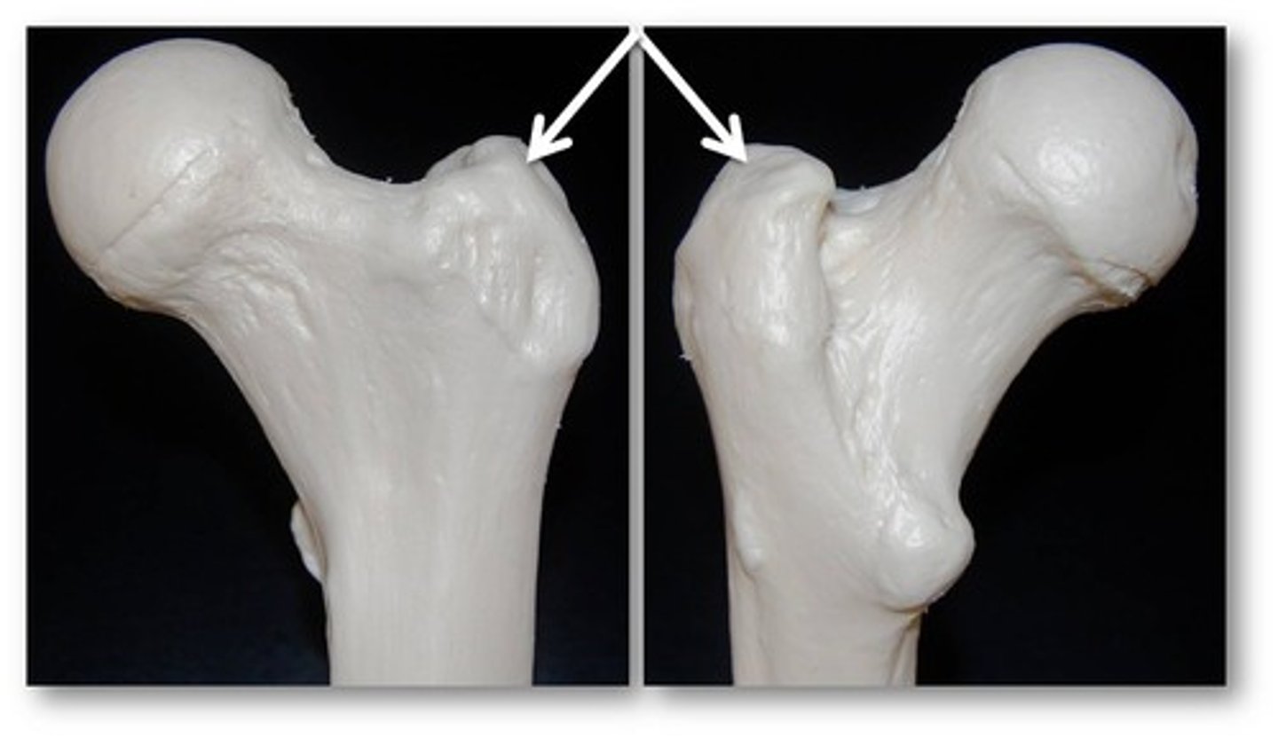
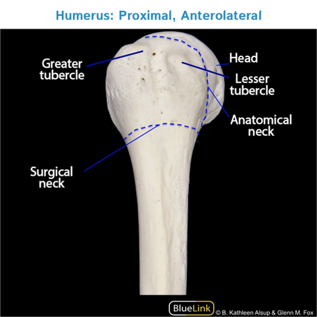
Bony Landmarks : Tubercle
A small round projection that allows connective tissues to connect to the bone (e.g. greater or less tubercle in humerus)
Bony Landmarks : Projection
An area that projects above the surface of the bone
Bony Landmarks : Head
Round surface of a joint that helps form a joint (e.g. femur)
Bony Landmarks : Epicondyle
A rounded projection that sits on top of the condyle and allows connective tissues to connect to the bone (e.g. humerus)
Bony Landmark : Spine
A more pronounced raised, sharp elevation of bone that allows connective tissues and muscles to connect to the bone (e.g. scapula)
Bony Landmark : Crest
A raised ridge projection that is part of the edge of a bone and allows connective tissues to connect to the bone (e.g. iliac crest)
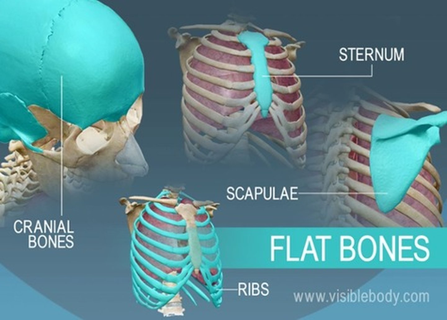
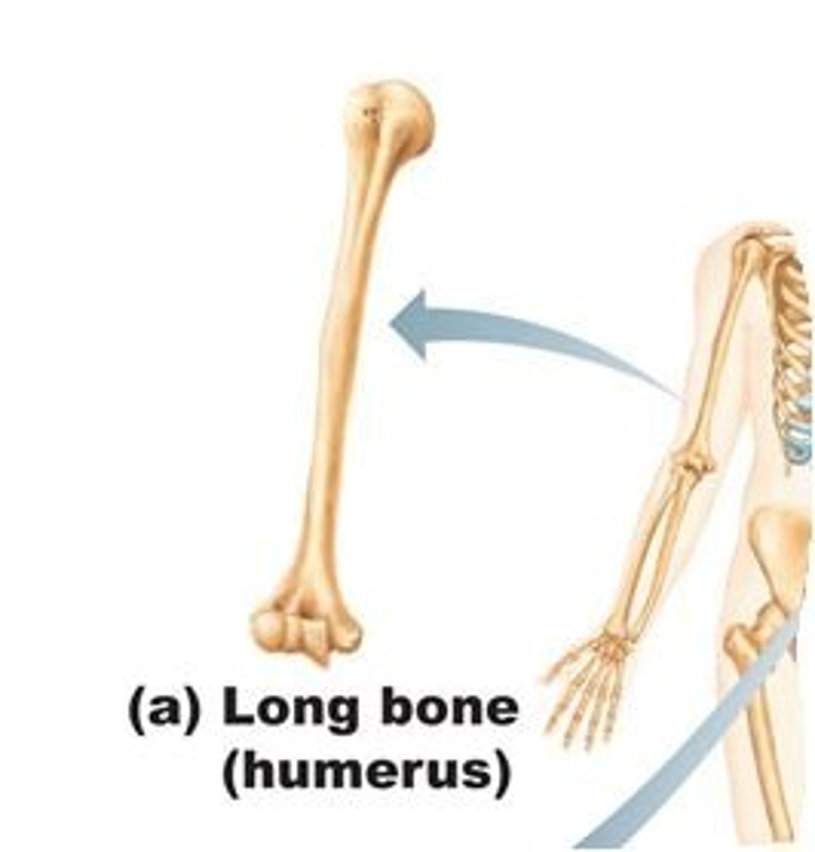
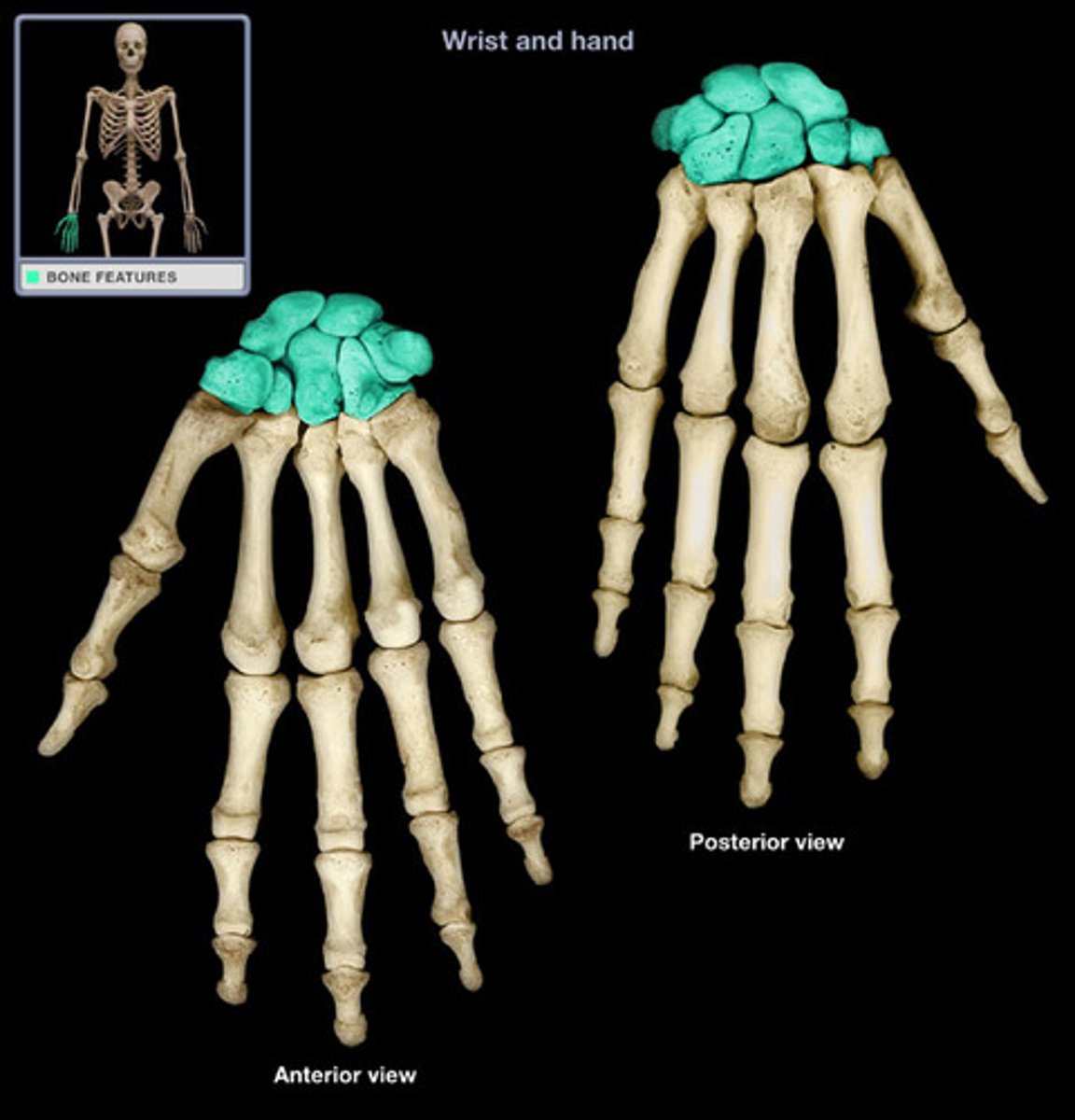
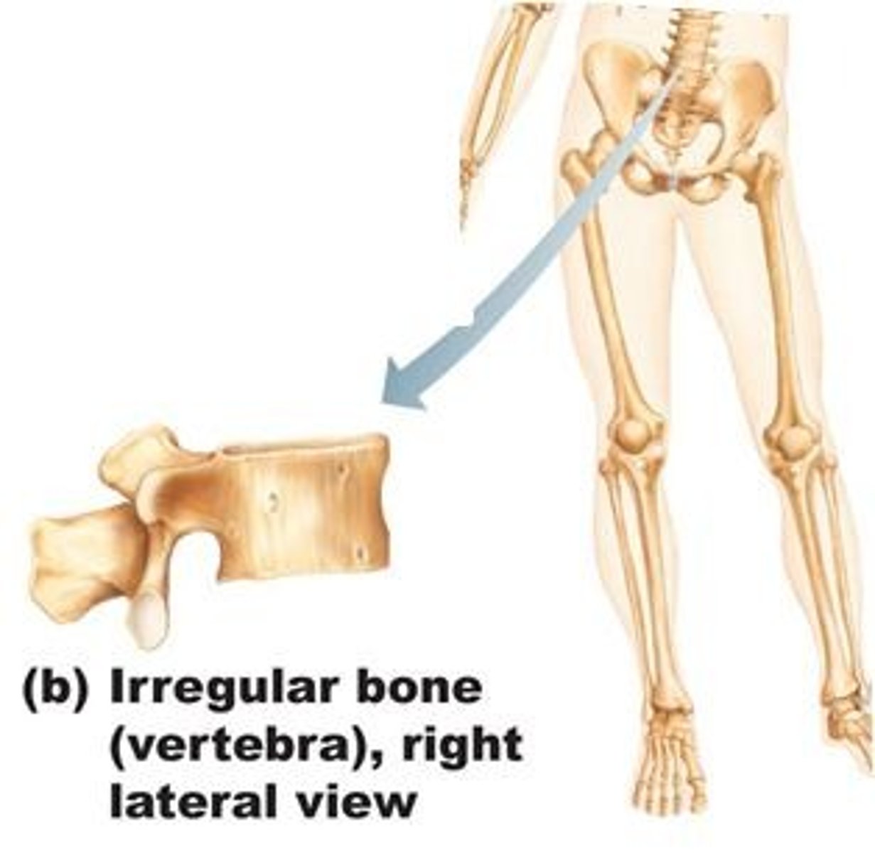
List five types of bones
Flat, irregular, short, long, sesamoid
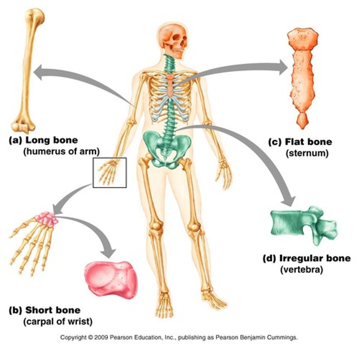
List examples of flat bones
bones of the skull, ribs, sternum, scapula
List examples of long bones
Femur, humerus
List examples of short bones
carpal bones
List examples of irregular bones
vertebrae and pelvic
List examples of sesamoid bones
patella
Which bone type reinforces tendons to protect them from wear?
Sesamoid
Which bone type protects internal organs?
flat bones
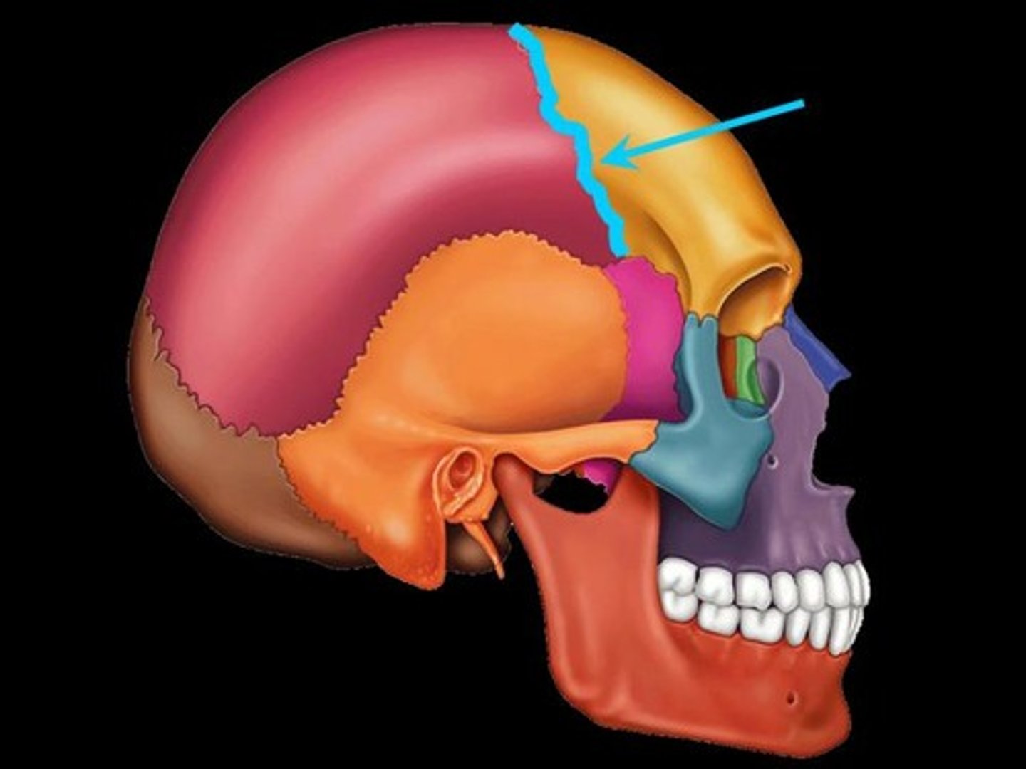
Fibrous Joints
Connected by dense connective tissue, allow little to no movement (e.g. sutures in the skull)
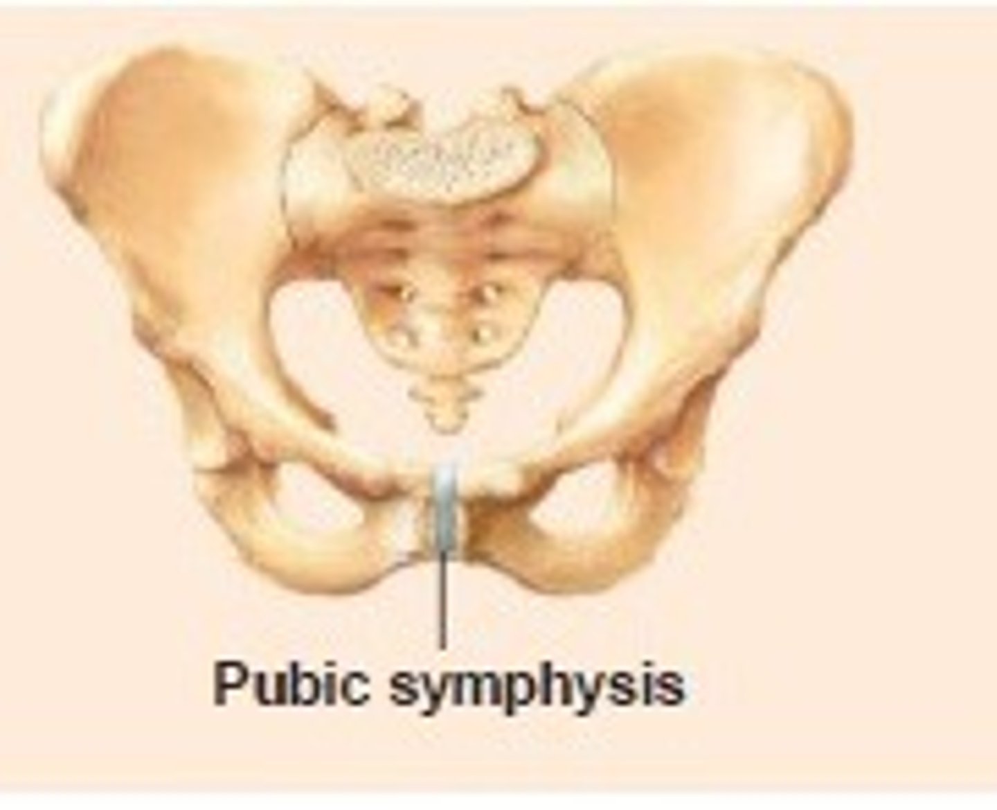
Cartilaginous Joints
Connected by cartilage, allow slight movement (e.g. pubic symphysis for child birth)
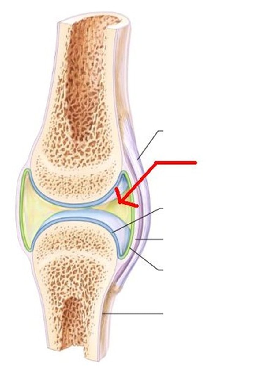
Synovial Joints
Freely movable, have a synovial cavity filled with fluid. found between bones that move against each other (e.g elbow)
List 6 types of Synovial Joints
plane (gliding), hinge, pivot, condyloid, saddle, ball and socket
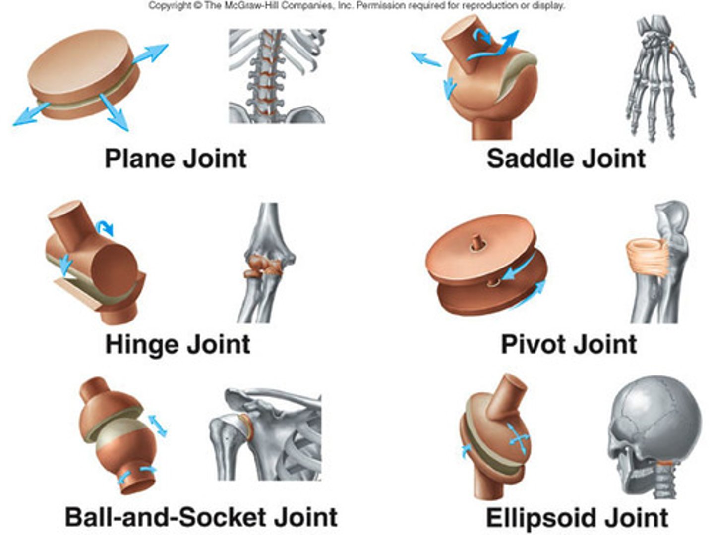
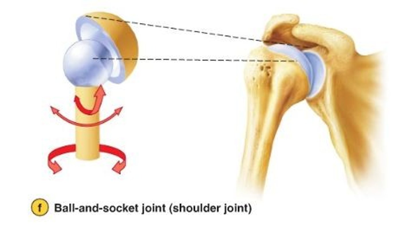
Synovial Joints : Ball and Socket
Multiaxial- Allows movement in all directions + rotation (e.g. shoulder and hip)
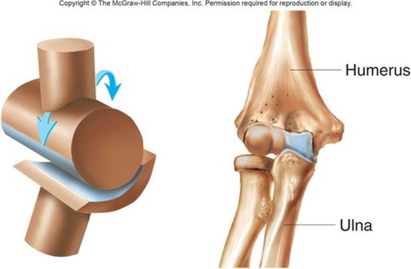
Synovial Joints: Hinge
Uniaxial- Allows movement in one plane, like a door hinge (e.g. flexion and extension of the elbow or knee)
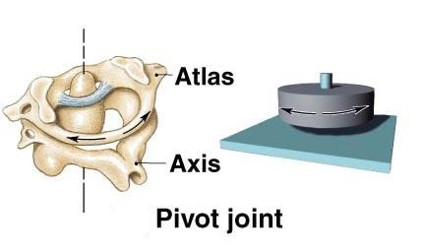
Synovial Joints: Pivot
Uniaxial- Allows rotational movement around a single axis (e.g. the joint between atlas and axis)
Synovial Joints: Saddle
Biaxial- Allows movement back and forth and side to side, but no rotation (e.g. thumb)
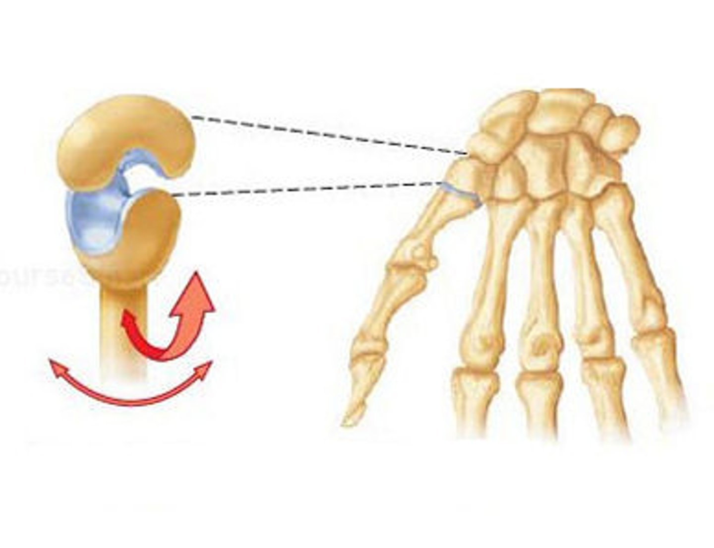
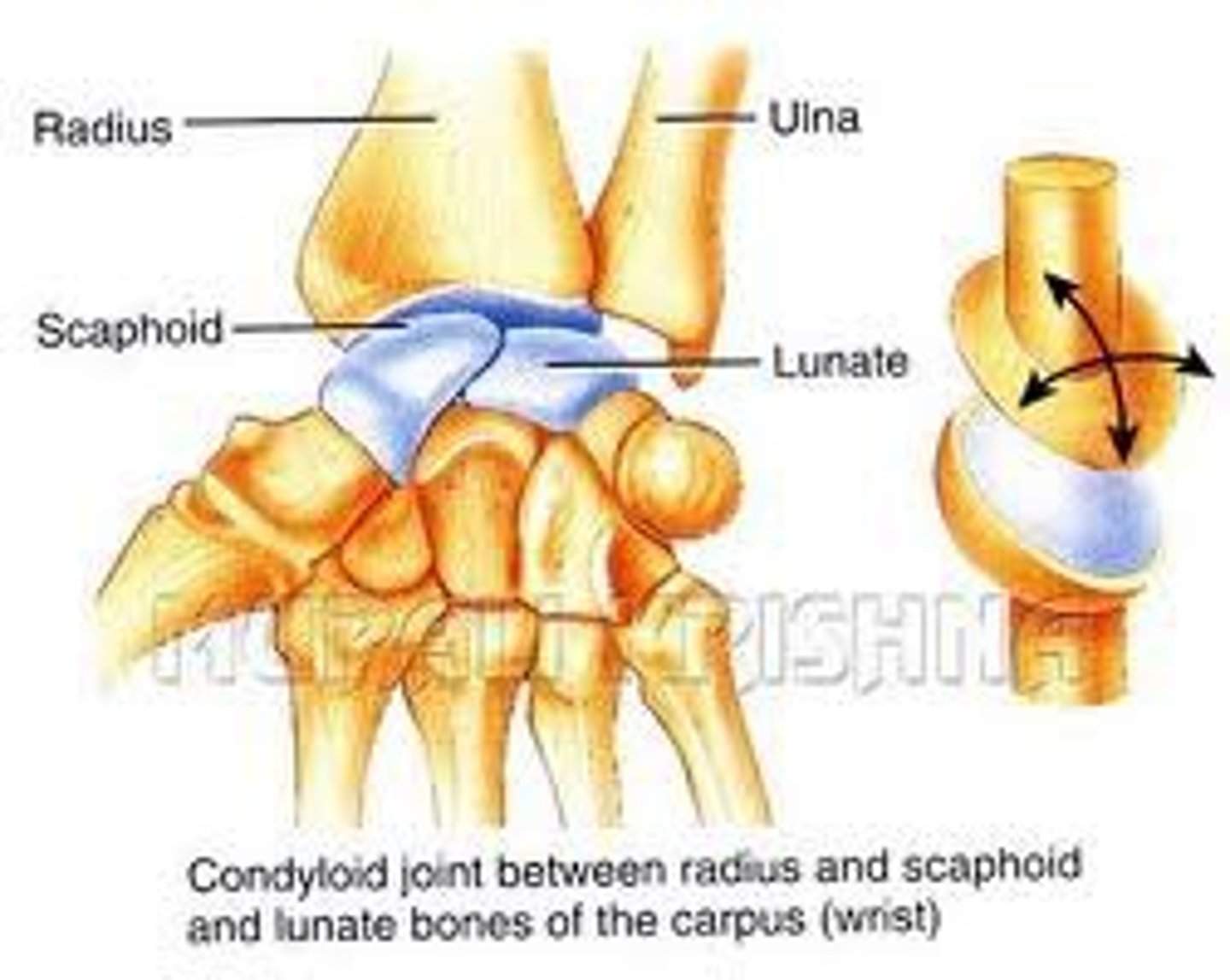
Synovial Joints: Condyloid
Biaxial- Allows movement but no rotation (e.g. wrist)
Synovial Joints: Plane (gliding)
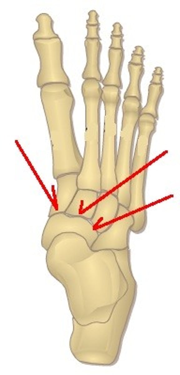
Uniaxial- Allows bones to glide past each other along the plane of the joint (e.g. Joints between tarsal bones in the foot)
What are the three uniaxial joints?
Hinge Joint, Pivot Joint, Plane Joint
What are the two biaxial joints?
Condyloid Joint, Saddle Joint
What is the ONLY multiaxial joint?
Ball and Socket Joint
What is the significance of sternal angle?
The sternal angle is the junction between the manubrium and the body of the sternum. It is a reference point for the base of the heart and where the 2nd rib attaches to the sternum. It also aligns with the T4 and T5 thoracic vertebrae
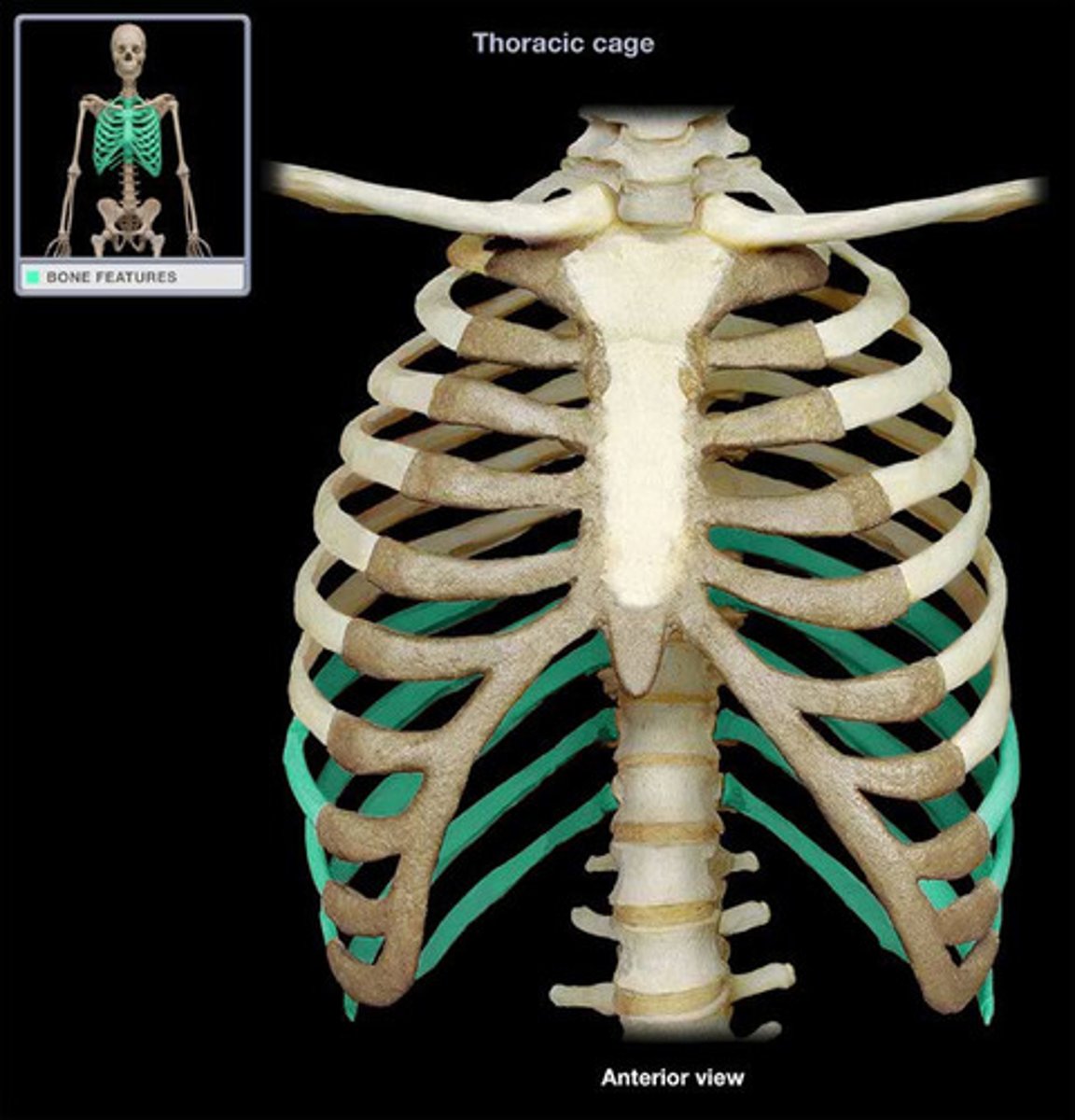
Which ribs are true ribs? Why are they classified as true ribs?
1-7 because they connect directly through the sternum
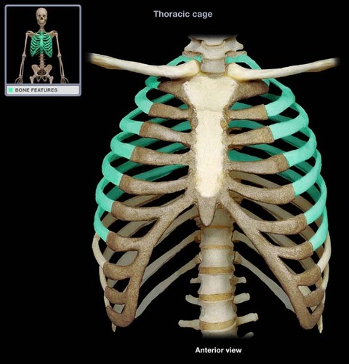
Which ribs are false ribs? Why are these classified as false ribs?
8-12 because they do not connect directly through the sternum; the cartilage for ribs 8-10 connect to the cartilage above them; ribs 11 and 12 do not connect to the sternum at all
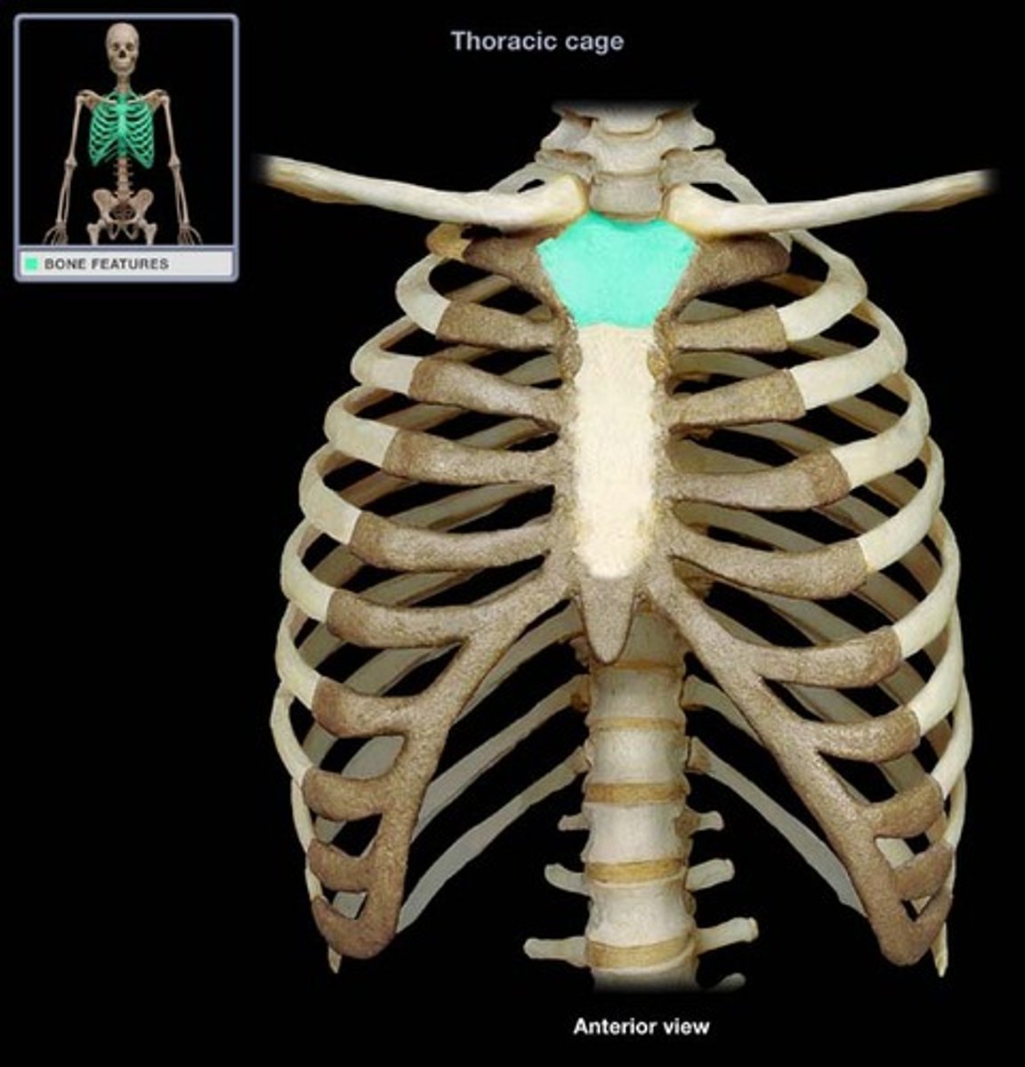
Manubrium
The broad, upper part of the sternum. It connects to the clavicles and the first two ribs, providing structural support to the upper chest and stabilizing the ribcage
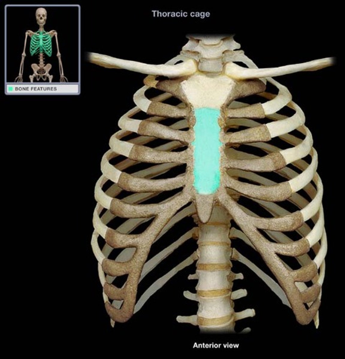
Body of The Sternum
The long, central part of the sternum, located below the manubrium. It connects to the second through seventh ribs and serves to protect the heart and lungs
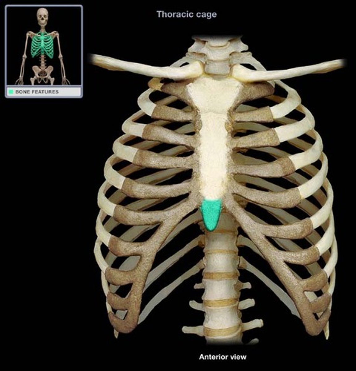
Xiphoid Process
The small, cartilaginous lower tip of the sternum. It serves as an attachment point for abdominal muscles and ligaments, and it plays a role in protecting the underlying organs, including the heart and liver.
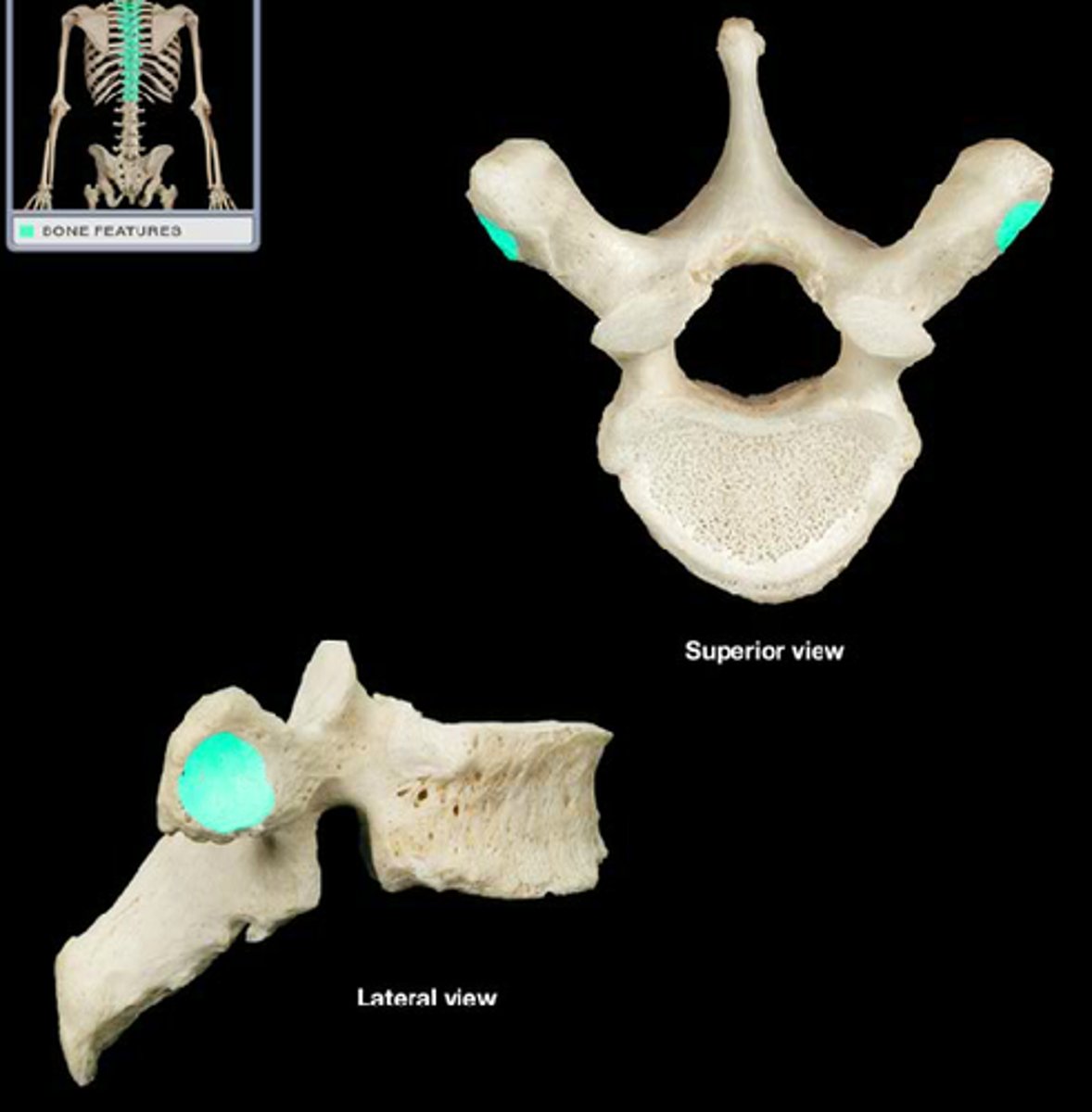
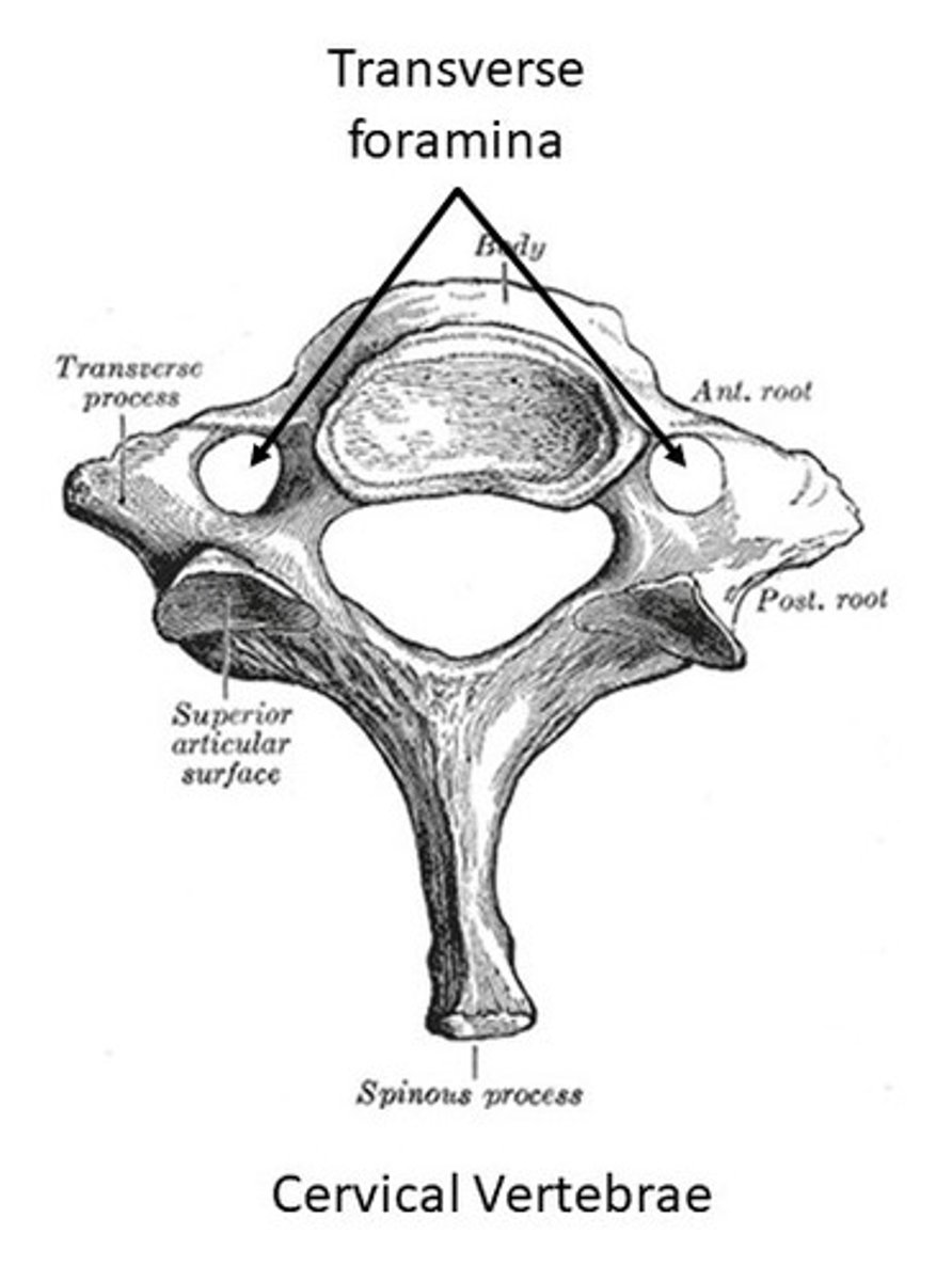
List unique features of cervical vertebrae
Small size, transverse foramina (two little holes), atlas and axis
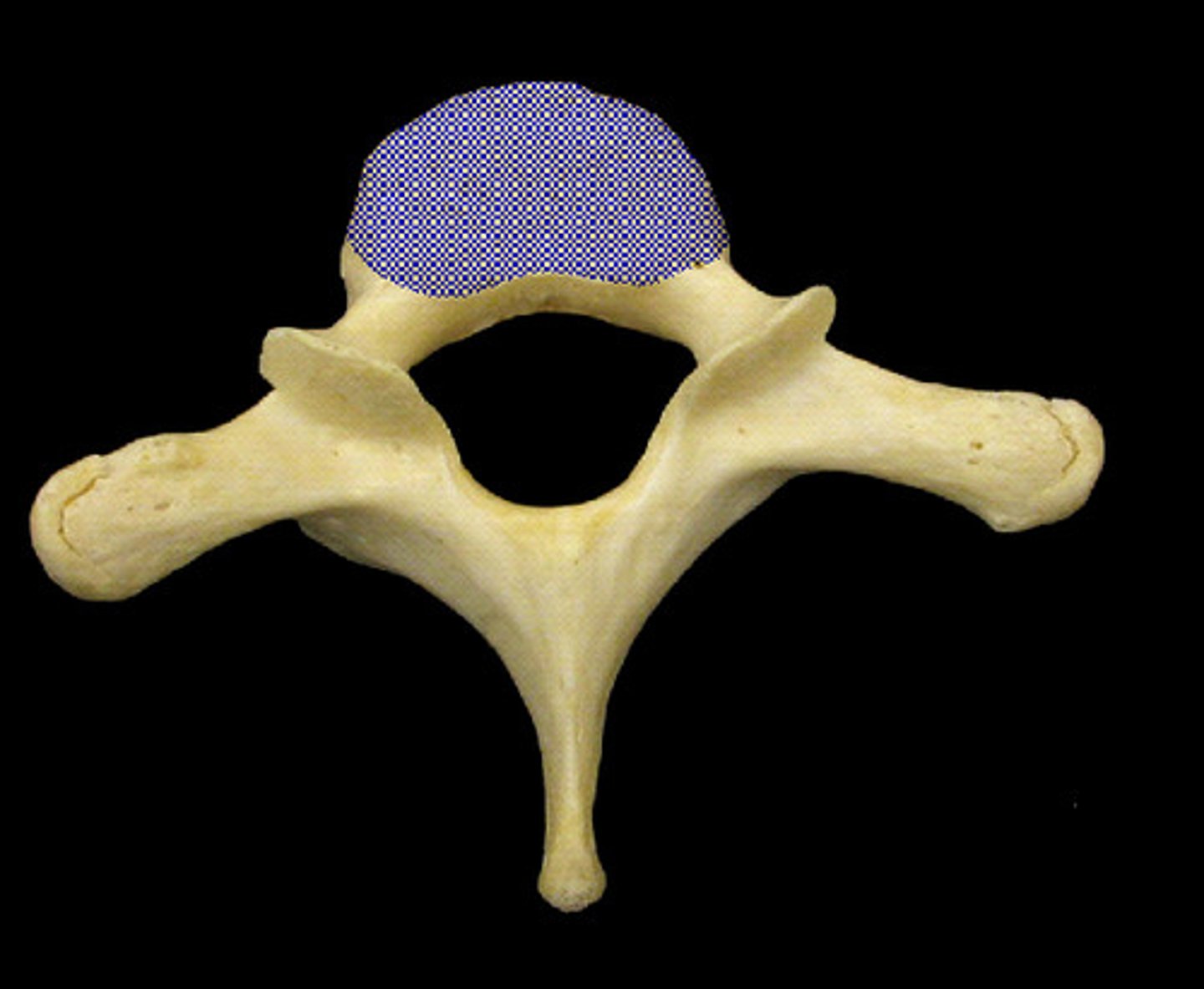
List unique features of thoracic vertebrae
Medium size (giraffe), costal facets for ribs, longer spinous processes
List unique features of lumbar vertebrae
Largest size (moose) , thick bodies, short spinous processes
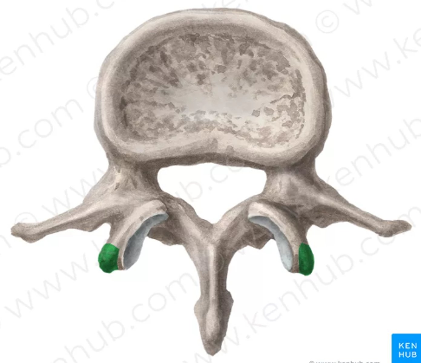
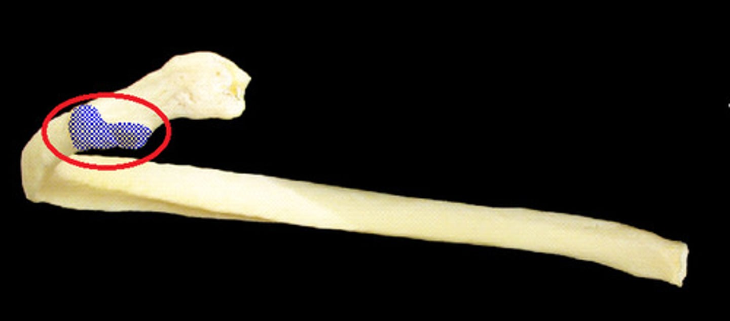
Define the Head of the Rib
rounded, posterior part of a rib that connects to the vertebral column
What does the head of the rib articulate with?
It articulates with the vertebral bodies at the costal facets, typically with two adjacent vertebrae, allowing for rib movement during breathing
Define the Neck of the Rib
the slender portion of the rib located between the head and the tubercle. It helps connect the head of the rib to the body (shaft) and provides a surface for muscle attachment
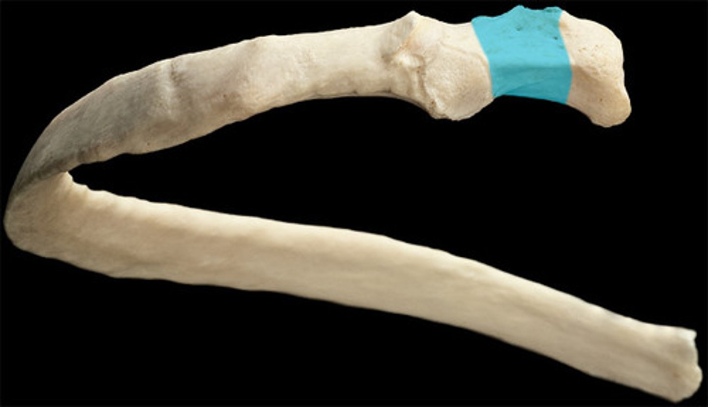
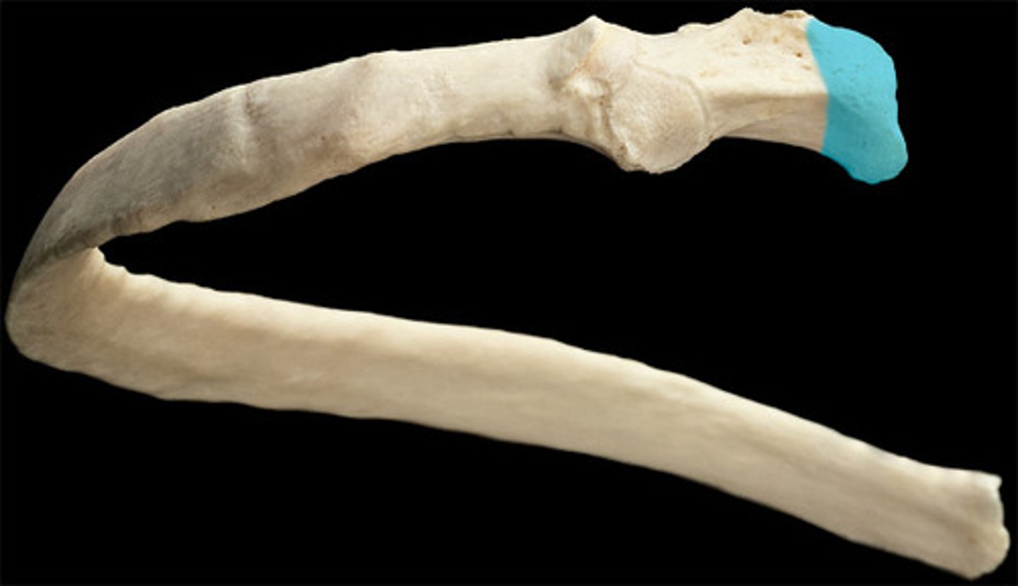
Tubercle of the Rib
small bump located on the posterior surface of the rib, near the neck
What does the tubercle of the rib articulate with?
the transverse process of the corresponding thoracic vertebra, which helps in the movement of the rib during breathing.
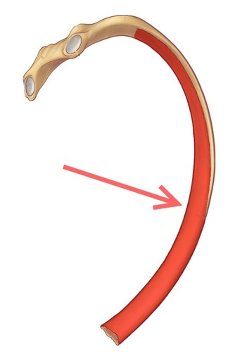
Define the Body (shaft) of the Rib
the long, curved, middle portion of the rib. It extends from the tubercle to the front of the chest and provides protection to the thoracic organs
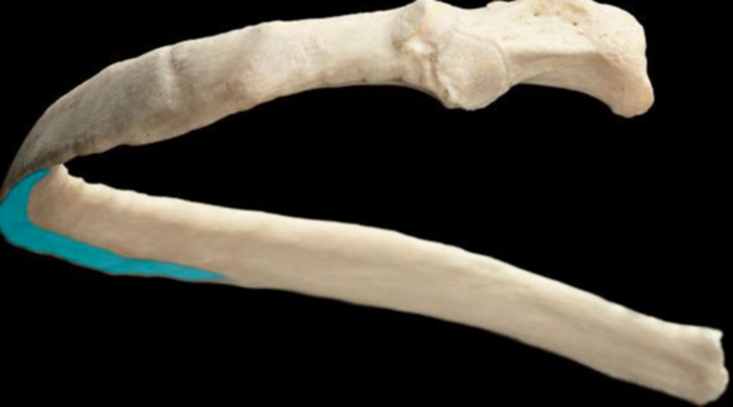
Define the Costal Groove of the Rib
a groove located along the inferior border of the rib's body (shaft)
How many cranial bones are there? Name them
8. Ethmoid, sphenoid, frontal, parietal (2), temporal (2), occipital
List paired cranial bones
parietal and temporal
Cranial Bones: Ethmoid
Between the eyes, forming part of the nasal cavity and the orbit
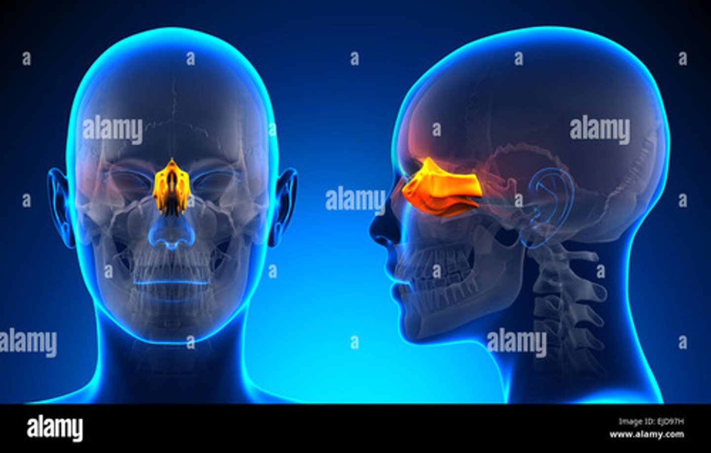
What is the main function of the ethmoid bone?
To support the structure of the nasal cavity and contribute to the formation of the nasal septum
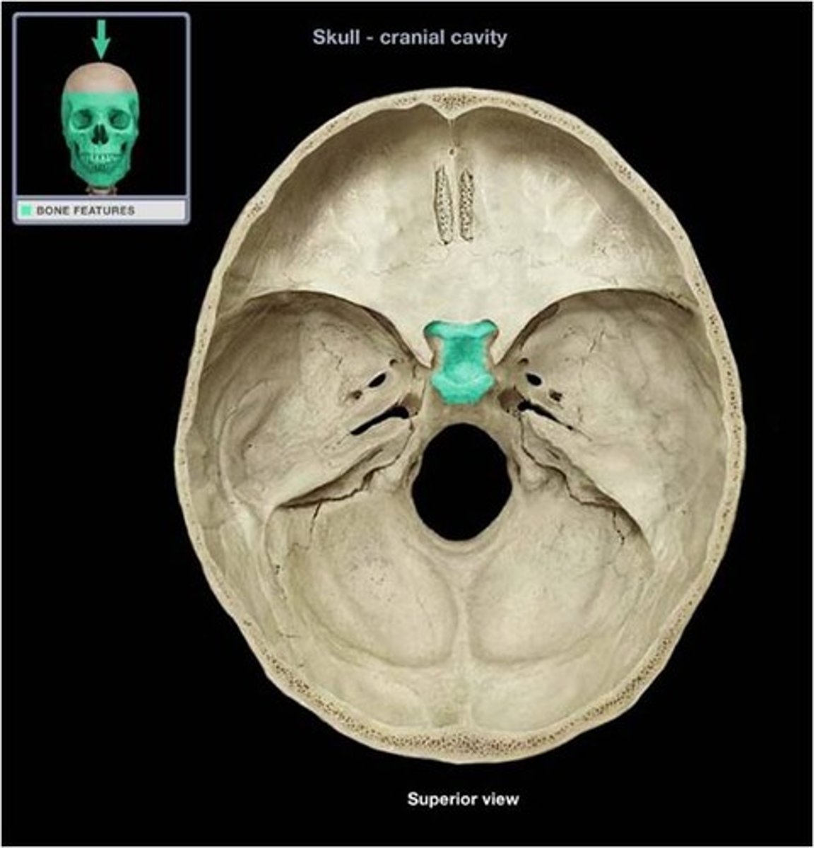
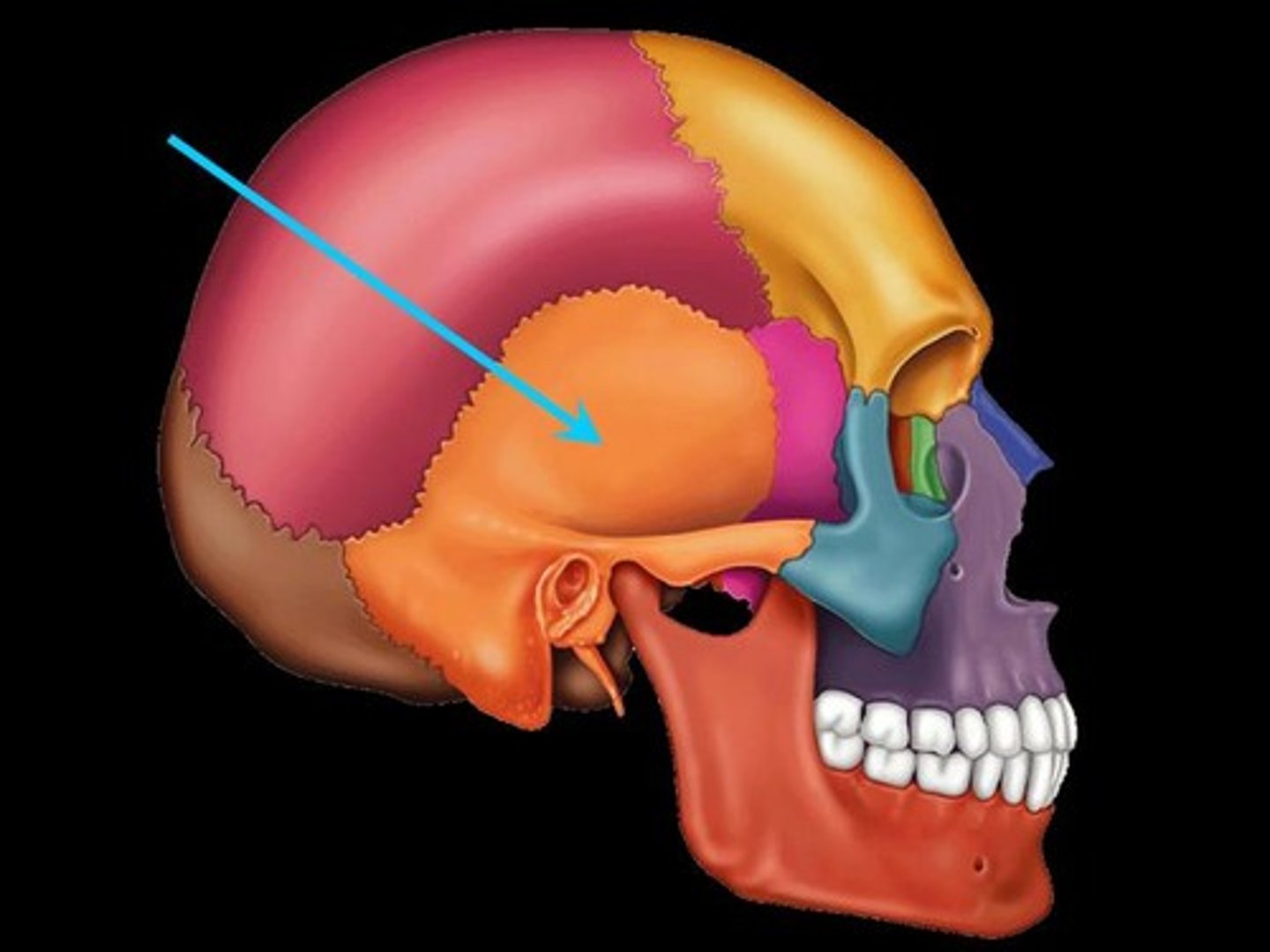
Cranial Bones : Sphenoid
a butterfly-shaped bone located at the base of the skull, in front of the temporal bones
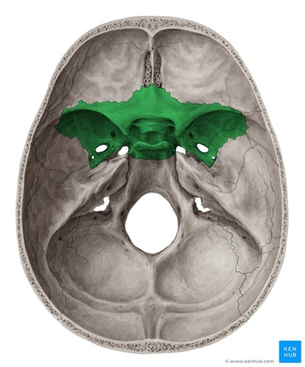
The sphenoid bone houses the...
pituitary gland found in the sella turcica (turkish saddle)
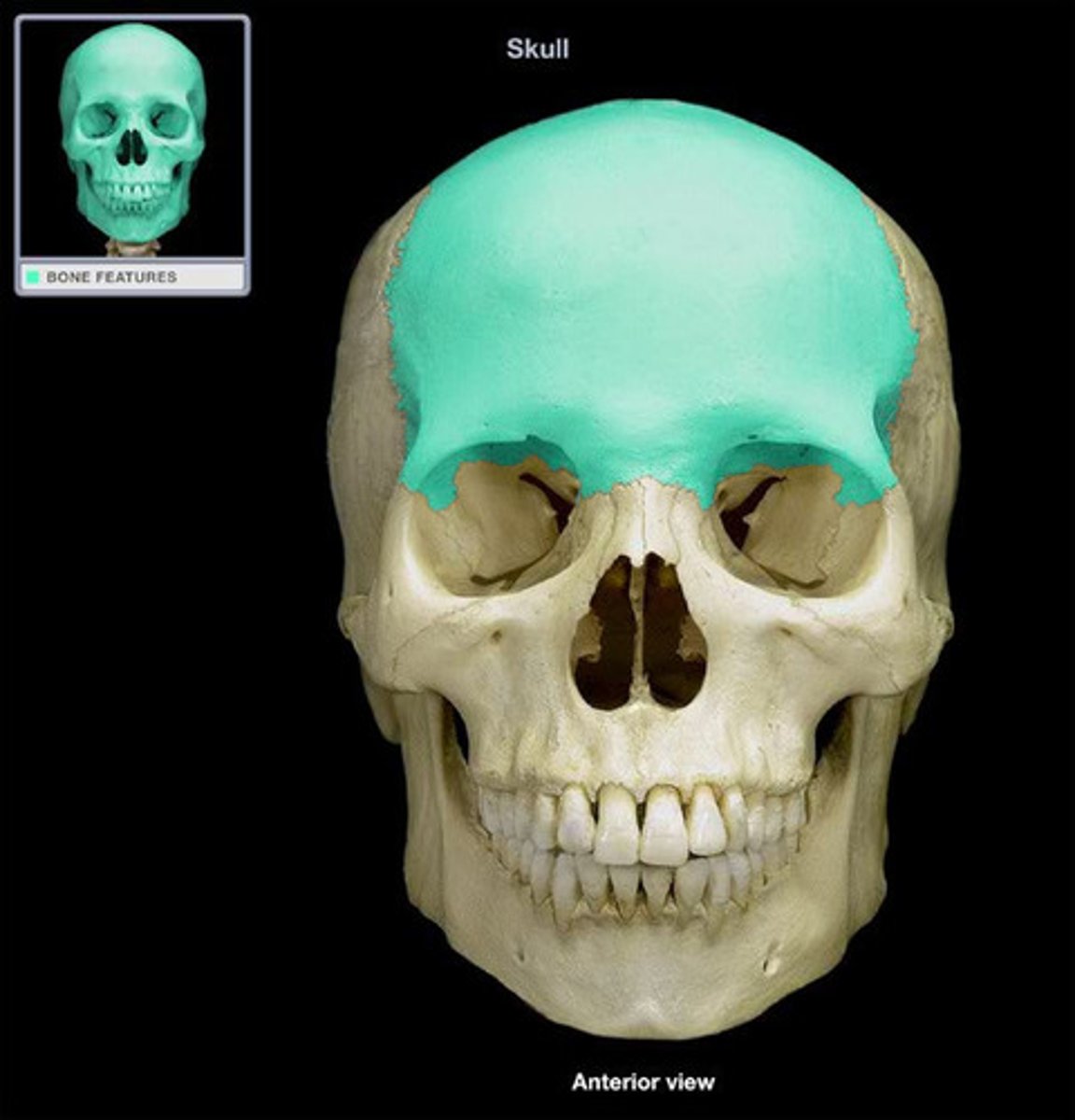
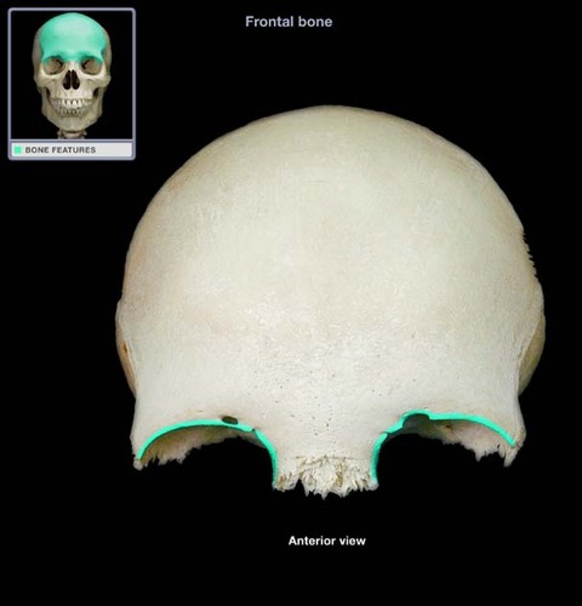
Cranial Bones : Frontal
forms the forehead & orbit
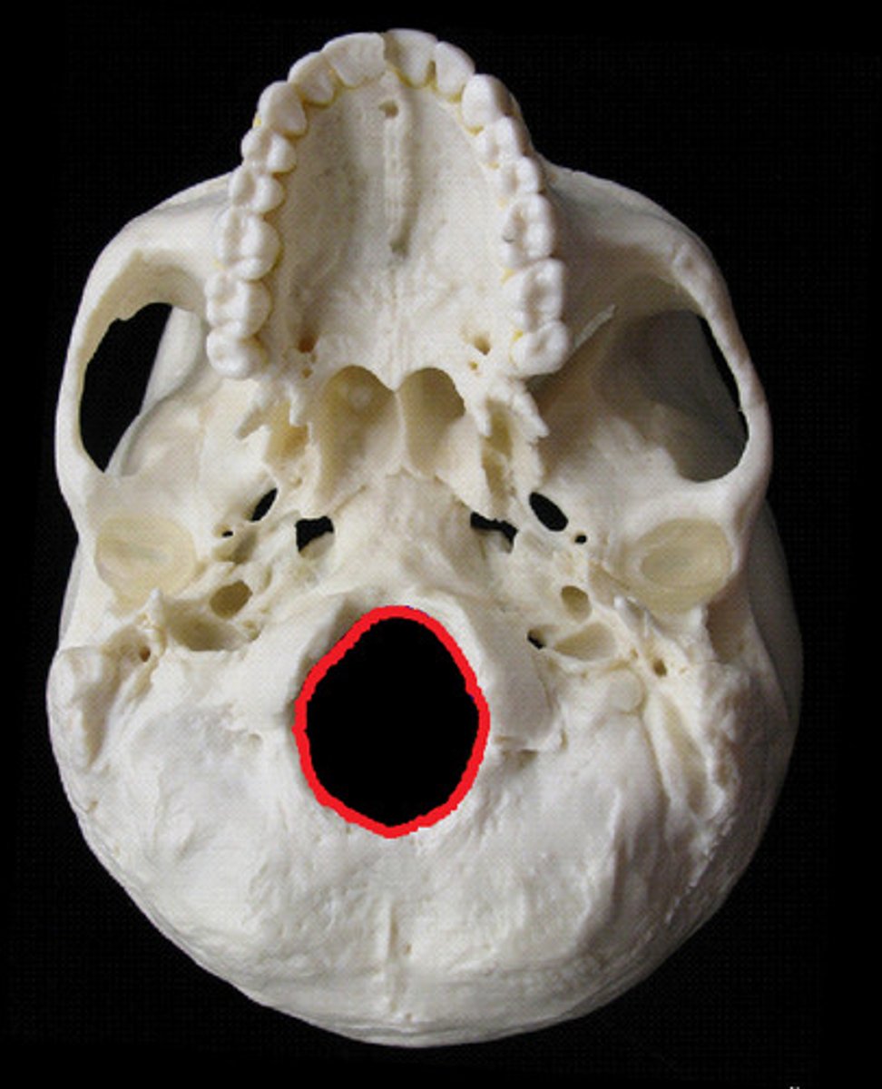
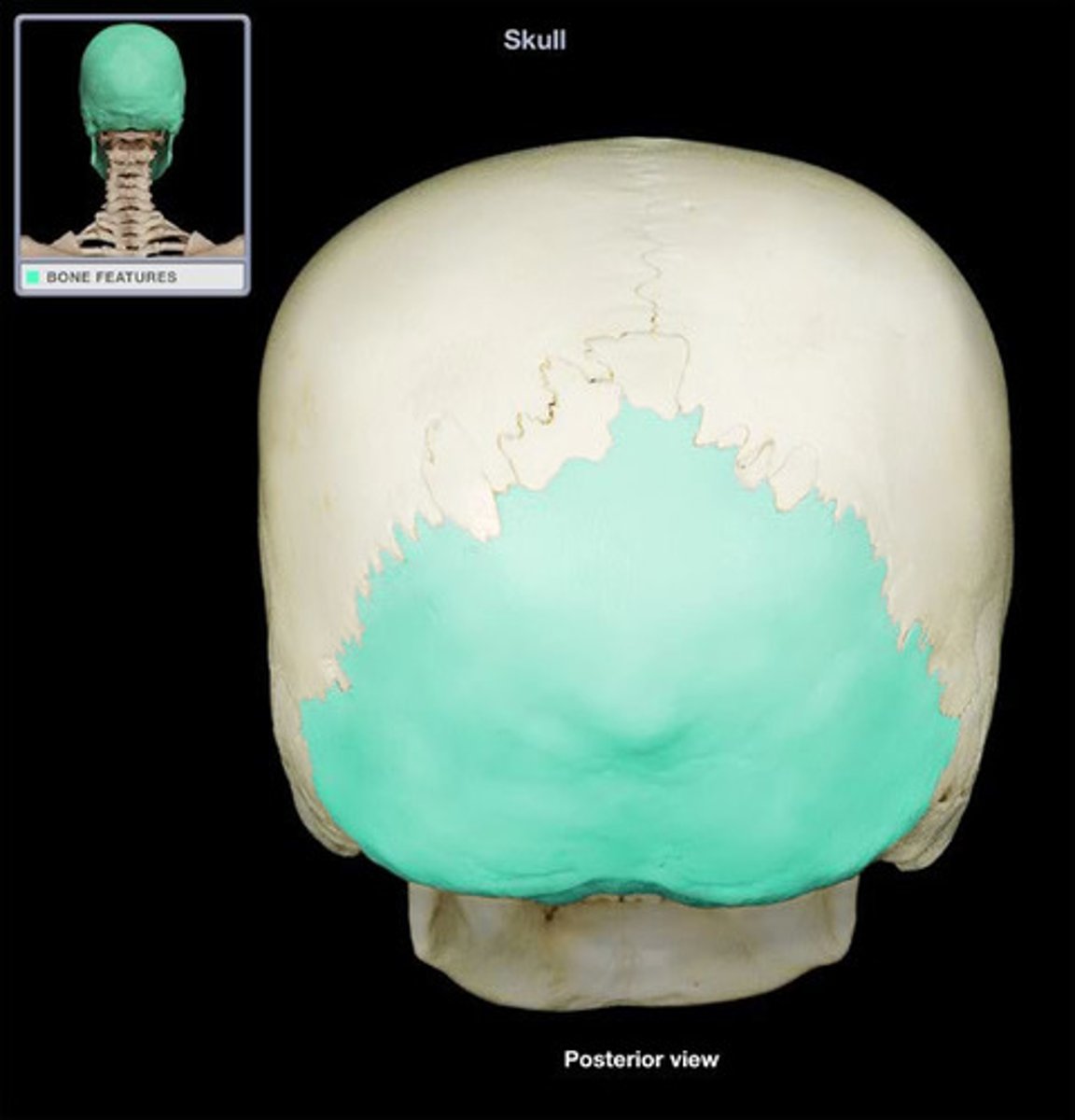
Cranial Bones : Occipital
back of head, contains the foramen magnum
Cranial Bones : Temporal
has several key features, including the external auditory meatus (ear canal), mastoid process, and zygomatic process
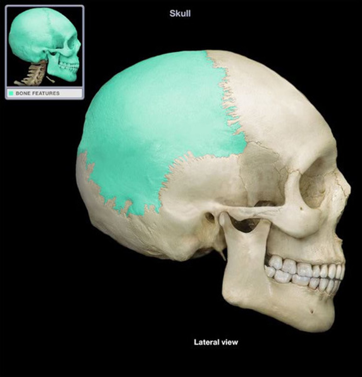
Cranial Bones : Parietal
two large, flat bones that form the top and sides of the skull. They are located on either side of the head and meet at the midline along the sagittal suture.
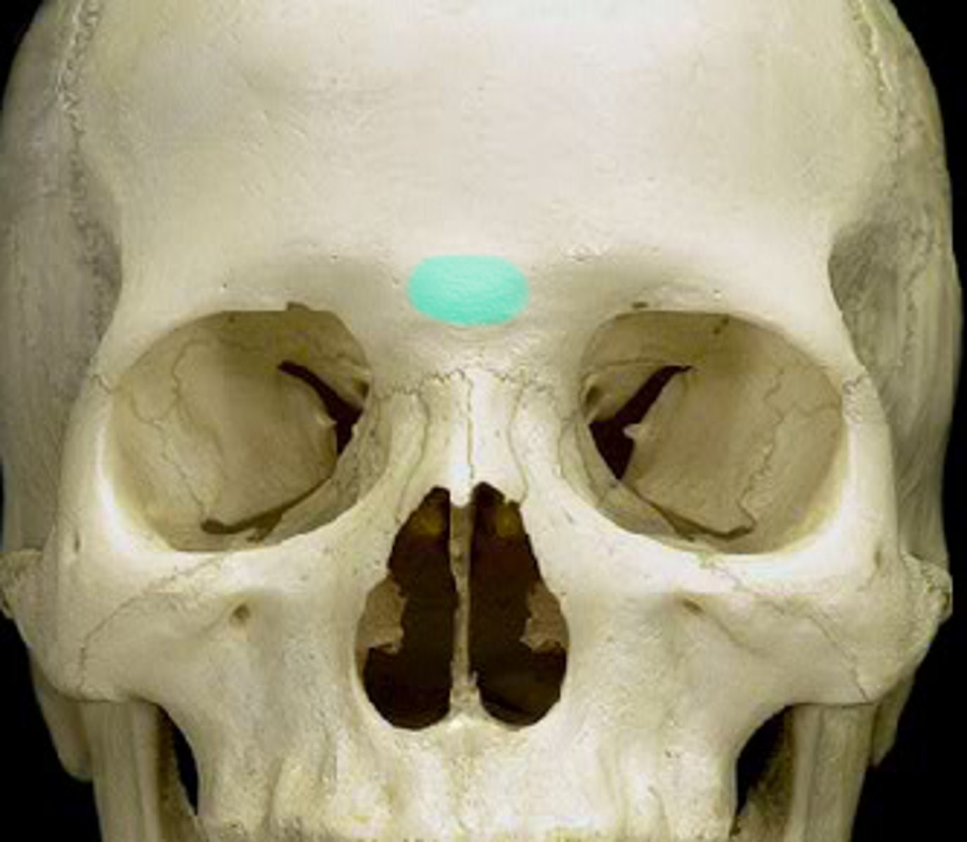
Frontal Bone : Identify the Glabella
smooth, flat area located between the eyebrows
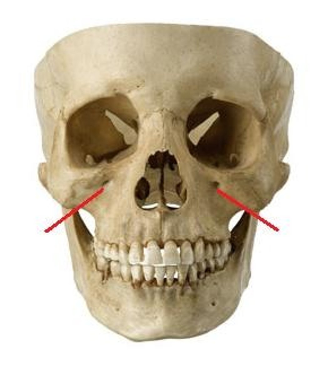
Frontal Bone: Identify the Supraorbital Margin
the thickened upper edge of the eye socket (orbit) that forms the bony ridge above the eye. Contains the supraorbital notch
Frontal Bone: Identify the Supraorbital Notch
a small indentation or opening located on the supraorbital margin of the frontal bone, just above the eye socket
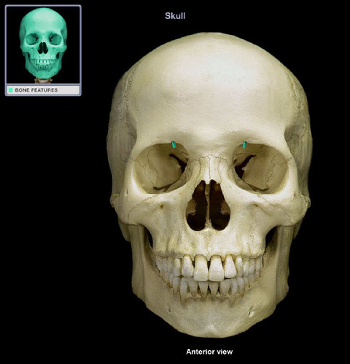
What is the significance of the supraorbital notch?
It allows for the passage of the supraorbital nerve, artery, and vein, which supply sensation and blood flow to the forehead and upper eyelid.
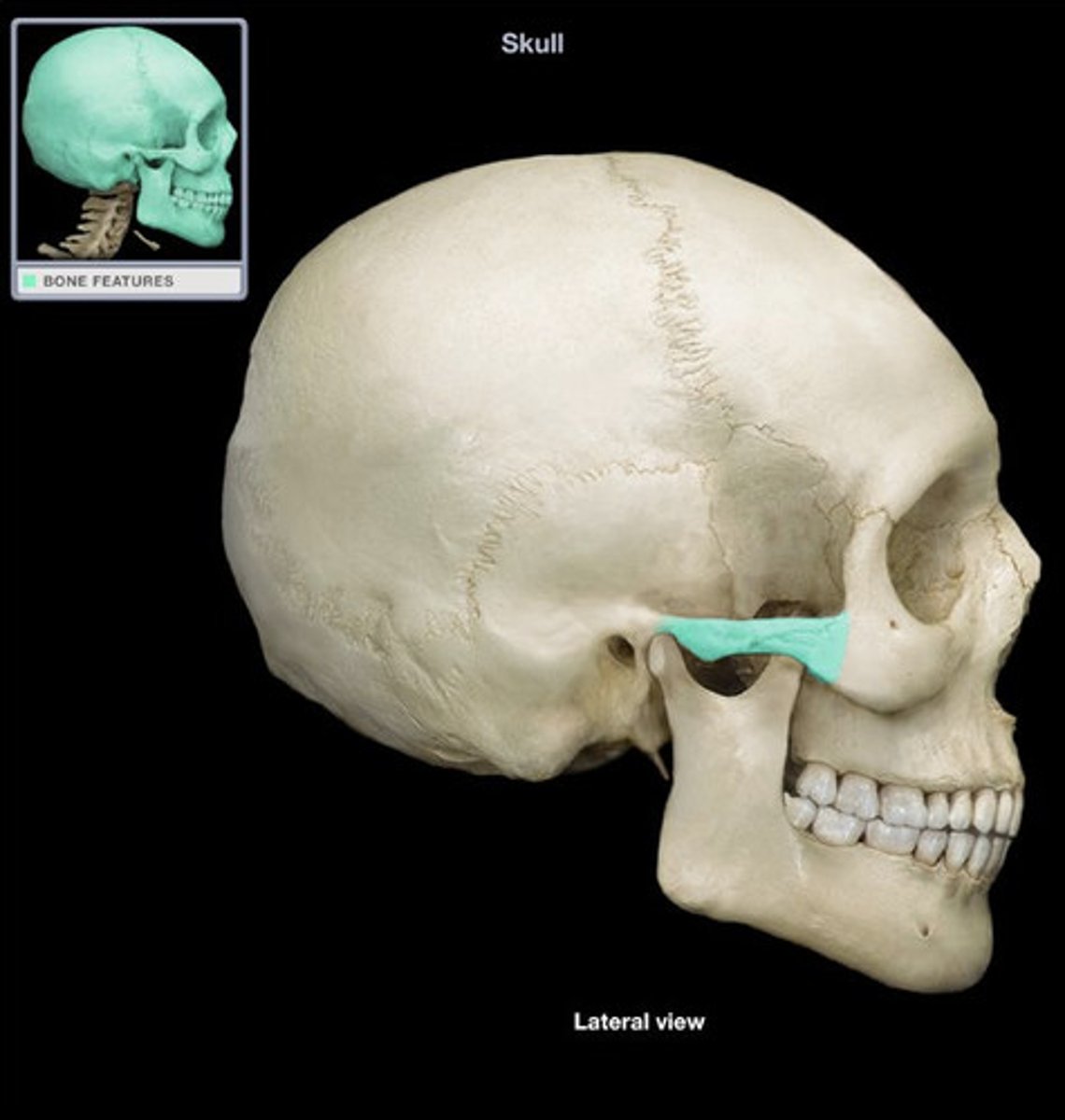
Temporal Bone : Identify the Zygomatic Process
a bony projection that extends from the temporal bone and articulates with the zygomatic bone
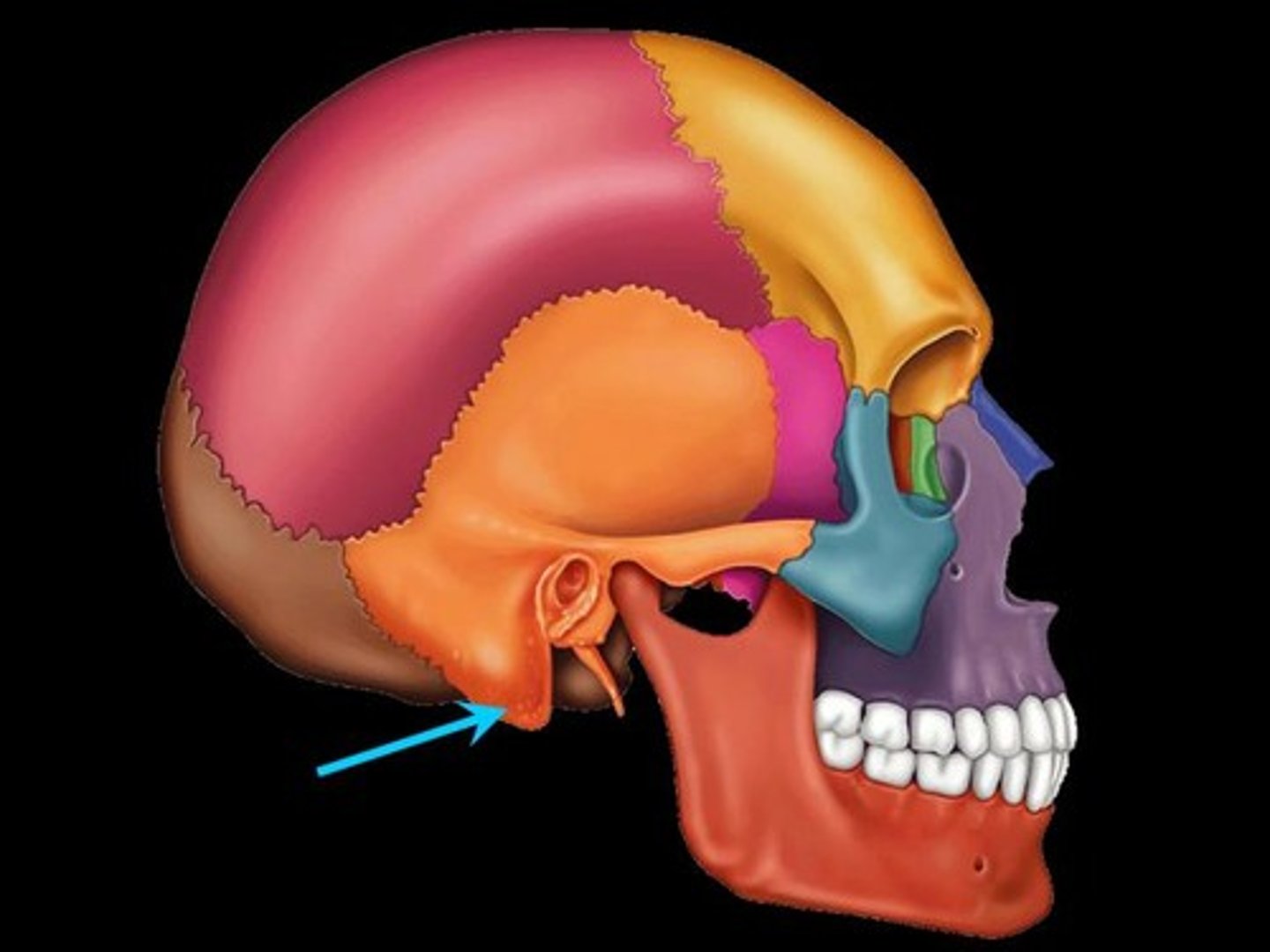
Temporal Bone : Identify the Mastoid Process
a prominent, rounded projection located on the posterior part of the temporal bone, just behind the ear