Hand Pathology
1/50
There's no tags or description
Looks like no tags are added yet.
Name | Mastery | Learn | Test | Matching | Spaced | Call with Kai |
|---|
No analytics yet
Send a link to your students to track their progress
51 Terms
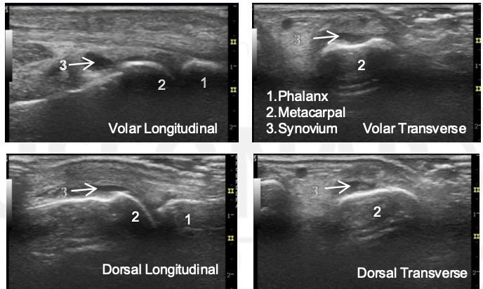
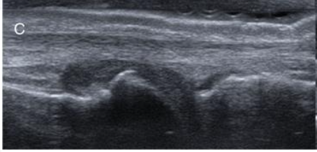
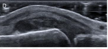
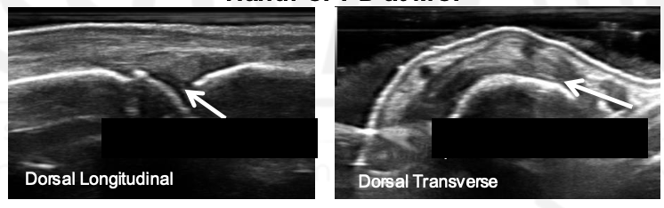
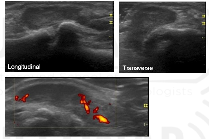
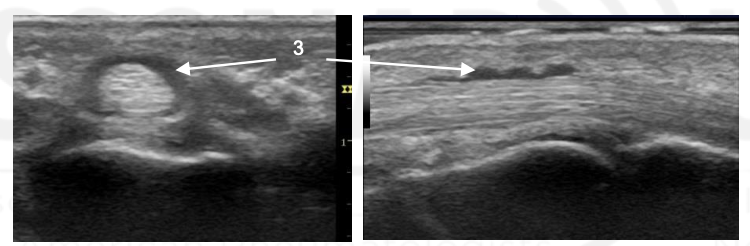
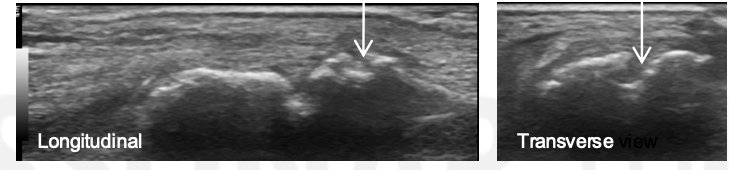
What is the arrow pointing to?
Synovial Distension
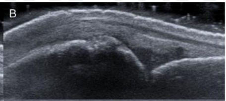
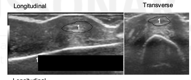
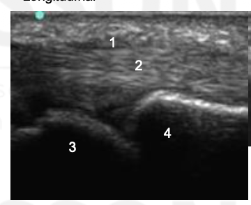
What is the abnormality?
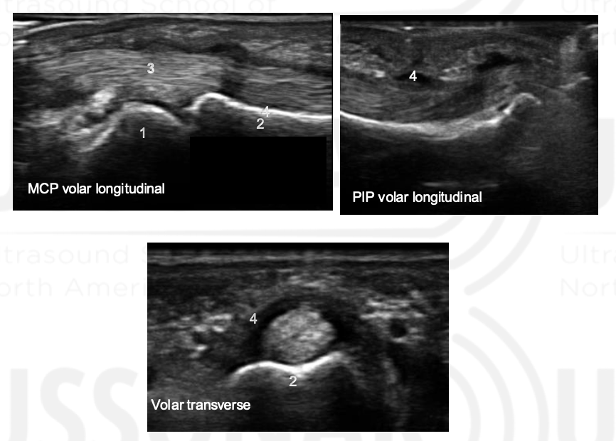
1: Synovium, 2: Flexon Tendon, 3: Proximal Phalanx, 4: Middle Phalanx
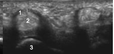
1: Synonym, 2: Flexor Tendon; 3: Osteophyte
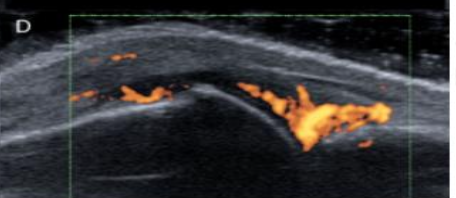
Grade The Synovial Hypertrophy
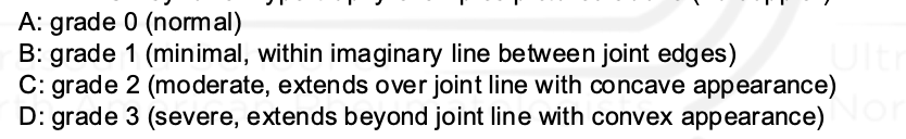
Grade this
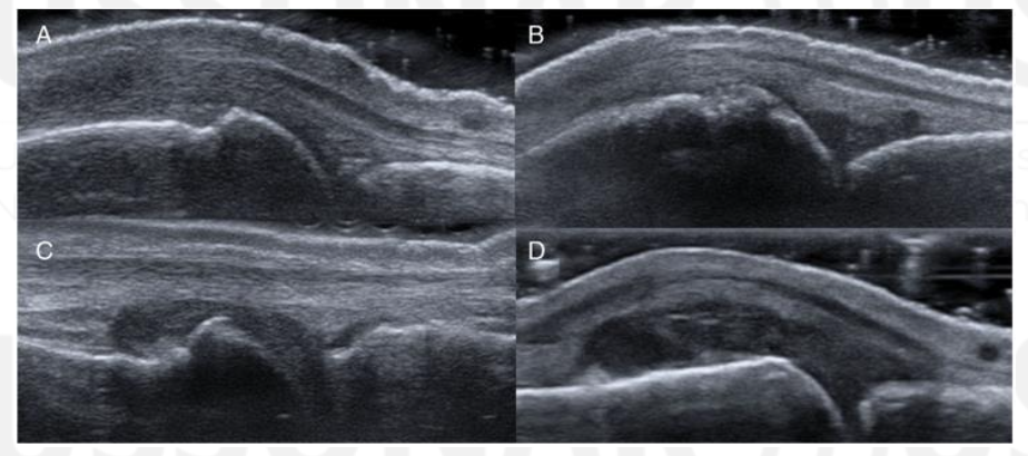
B: grade 1 (up to 3 single signals — 1 confluent + 2 single — 2 confluent)
Minimal synovial hypertrophy up to the imaginary horizontal line connecting 2 joints; Power Doppler up to 3 single signals — 1 confluent + 2 single — 2 confluent
Grade 1: minimal
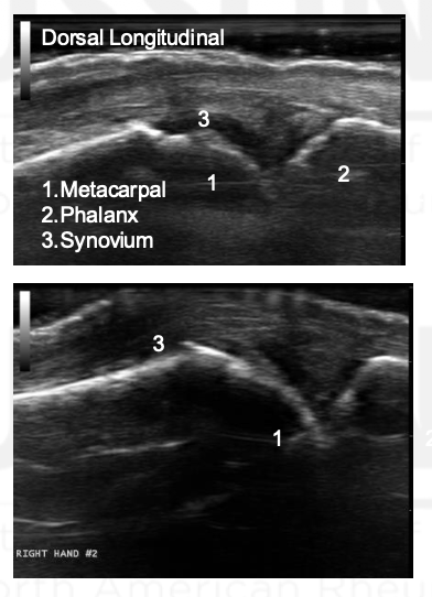
Moderate synovial hypertrophy protruding over the joint line along with concave surface; Larger than grade 1 but <50% of synovial hypertrophy covered by signals
Grade 2: moderate
Severe synovial hypertrophy producing beyond the joint line with convex surface; more than 50% of SH area covered by signals
Grade 3: Severe
Grade this doppler
Grade 2: Larger than grade 1 + <50% of SH area
Grade this doppler
Grade 3: >50% of the synovial hypertrophy is covered by signals
What grade is the synovial hypertrophy?
Grade 0
Grade this synovial hypertrophy
Grade 1: minimal within imaginary line between joint edges
Grade this synovial hypertrophy
Grade 2: moderate, extends over joint line with concave appearance
Grade this synovial hypertrophy
Grade 3: severe, extends beyond joint line with convex appearance
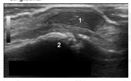
What is 1?
This is extensor calcification tendinopathy at the PIP.
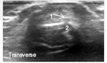
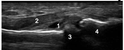
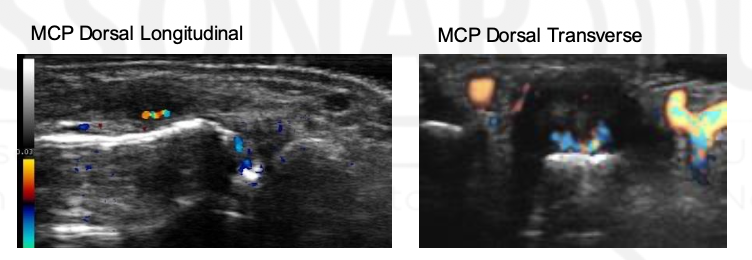
What is 1 and 2?
CPPD at the MCP
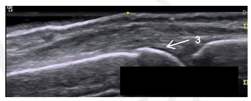
What is 3?
This is the interface sign in a normal MCP
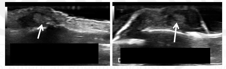
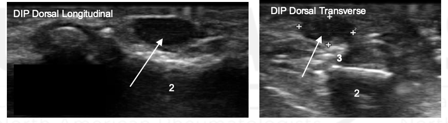
What is the arrow pointing to?
CPPD, this is the pseudo double contour sign
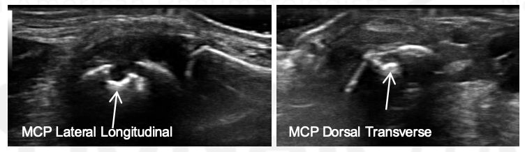
What view is this? What is the arrow?
DIP, tophus
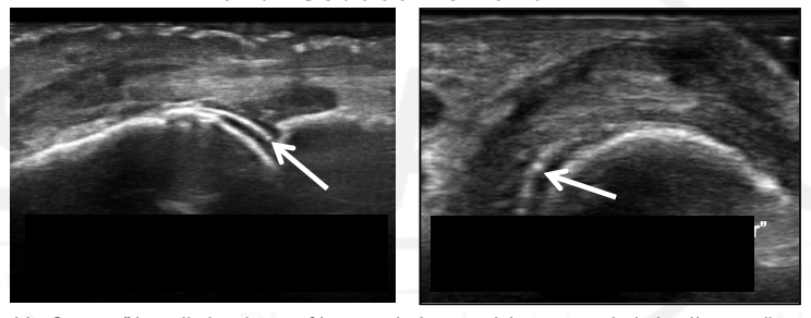
What view is this? What is the arrow?
MCP; Gout
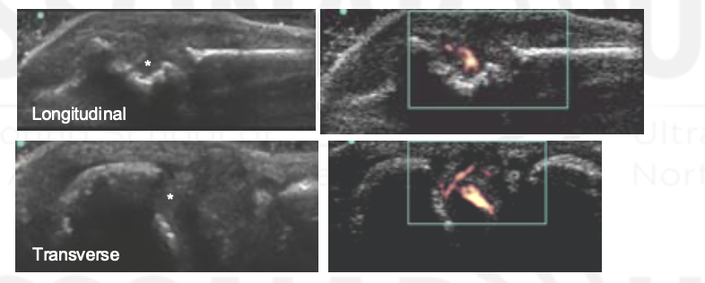
What view is this?
Extensor Tendon
Digital Vessel
Tophus
This is gouty extensor tendinopathy at the PIP.
What is this?
What is this?
Rheumatoid Nodule
Hypoechic/anechoic center
More Homogeneous than tophi
no calcifications or shadowing
hypoechoic trim
No anechoic center; homogenous and hypoechoic tissue.
What is this?
Rheumatoid Nodule
Hypoechic/anechoic center
More Homogeneous than tophi
no calcifications or shadowing
hypoechoic trim
No anechoic center; homogenous and hypoechoic tissue.
What are 1 and 2?
1 - Middle phalanx
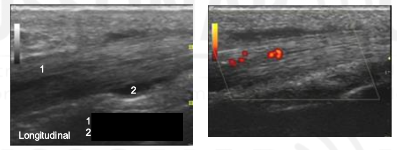
Ganglionic Cyst
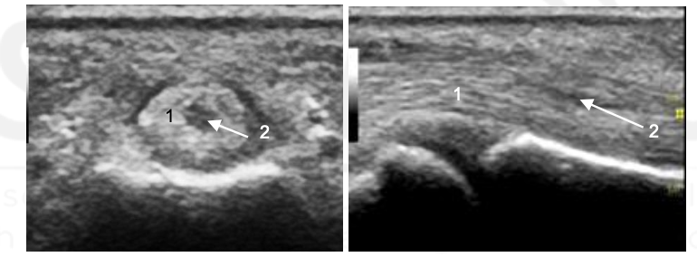
What is the arrow pointing to?
Ganglionic Cyst
How do you differentiate Ganglions, Neurofibromas/Schwannomas, and Giant Cell Tumors?
Ganglions - Anechoic + posterior acoustic enhancement + stalk from synovial cavity or tendon sheath; if nodular or doppler signal inside = cancer
Neurofibromas/Schwannomas - nerve tail + posterior acoustic enhancement
Giant Cell Tumors - Hypoechoic - posterior acoustic enhancement
What is posterior acoustic enhancement?
The region deep to a fluid collection will be relatively hypoechic as more sound waves are able to reach that tissue than the tissue to either side.
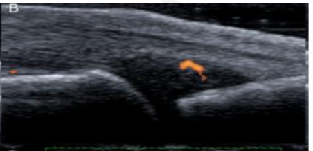
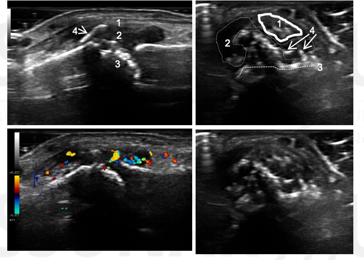
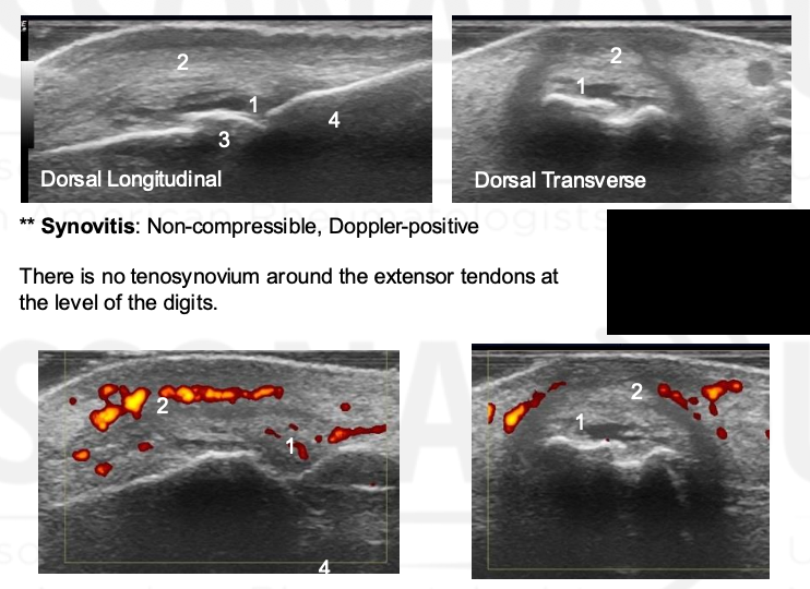
What is going on here? Name 1-4.
Extensor Tendon
Hypoechoic tendinopathic portion of tendon
Metacarpal
Phalanx
This is extensor tendinitis. Interestingly, there is no tenosynovium around the extensor tendons in teh digits, but there can still be Doppler of the tissues around the extensor tendon.
Peri-extensor tendinitis is seen here. Not common in RA. Why? Because RA is a primarily synovial disease.
How does US compare to MRI with extensor tendinitis?
US sensitivity for extensor tendonitis is 15% while it is 44% for flexor tenosynovitis.
What happened here?
Normal flexor tendon
Torn fibers of tendon
What is 3?
Tenosynovial swelling
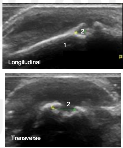
What is a normal A1 pulley thickness?
What is the thickness of trigger finger pulleys?
A1 pulley thickness: 0.24 - 0.6 mm
Trigger finger pulley thickness: 1.1 - 2.9 mm
Name 1-4
Thickened A1 pulley
Tendon thickening compared to adjacent tendon. Also note tendon is thicker distal to A1 puley. On dynamic imaging, this impinged onto the A1 pulley.
MCP
Phalanx
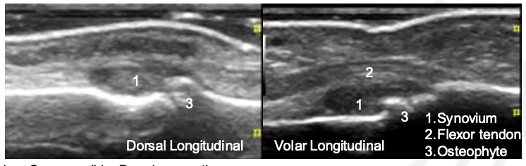
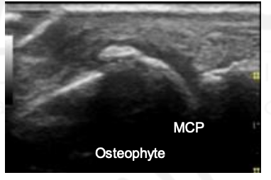
What is this?
Osteophyte
Do not be suprised to see synovial hypertrophy and effusion in OA hand joints on US exam.
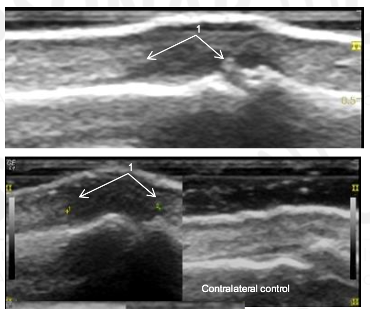
What is this?
Osteophyte
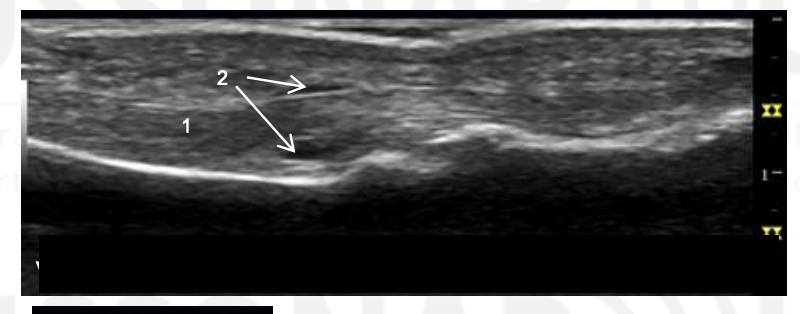
What is this?
Torn extensor tendon edges
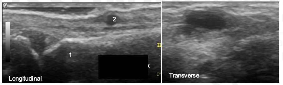
Name 1 and 2.
Tendon
Tendon sheath effusion
Note that the tenosynovial effusion is anechoic and
compressible, and on both sides of the tendon. Compare
how the deep portion of the tenosynovial fluid is abutting
the tendon and how synovial fluid from the PIP joint
would be abutting the bone.
What is this?
Flexor tenosynovial effusion at PIP
Name 1-4.
MCP
Proximal phalanx
Flexor tendon
Tendon sheath
Name 1 and 2.
Flexor tendons
Tendon sheath fluid
Tendosynovial hyperemia
Name 1-4.
What condition is this?
IP joint of the thumb
1 - Extensor tendon
Synovitis
Proximal phalanx (eroded)
New bone formation
Psoriatic arthritis patient with swelling of the IP joint of the thumb.
What is this?
Erosion
Compared to the gold standard of multidetector CT, ultrasound sensitivity for
joint erosion is 42%, specificity is 91%, accuracy is 80%.
How do you define an erosion?
What are small, moderate, large sizes?
> 1 mm
or if is the volar aspect of the MCPs or PIPs, use > 2 mm
Small erosion < 2 mm
Moderate 2 - 4 mm
Large > 4 mm
Where is the US more sensitive for erosions?
Why?
2nd and 5th MCPs due to the increased ultrasound access to the lateral and medial sides of these MCPs.
The radial side of the 2nd MCP is the MOST affected by bone erosions, but in healthy people, it can have small lesions too.
What is this?
Erosion
What is this?
Erosion
True or false:
Synovial thickness scores + Doppler pixel scores from the MCPs correlate with progression of the modified Sharp score at 1 year.
True
In patients with RA, what are the best predictors of DAS 28 in 1 year compared with inflammatory markers, clinical joint counts, and pain severity scores?
gray scale and power doppler scores
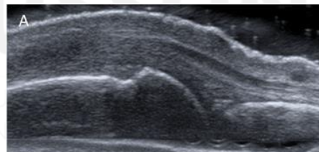
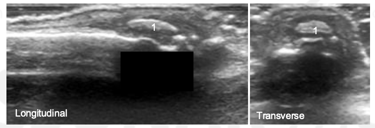
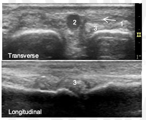
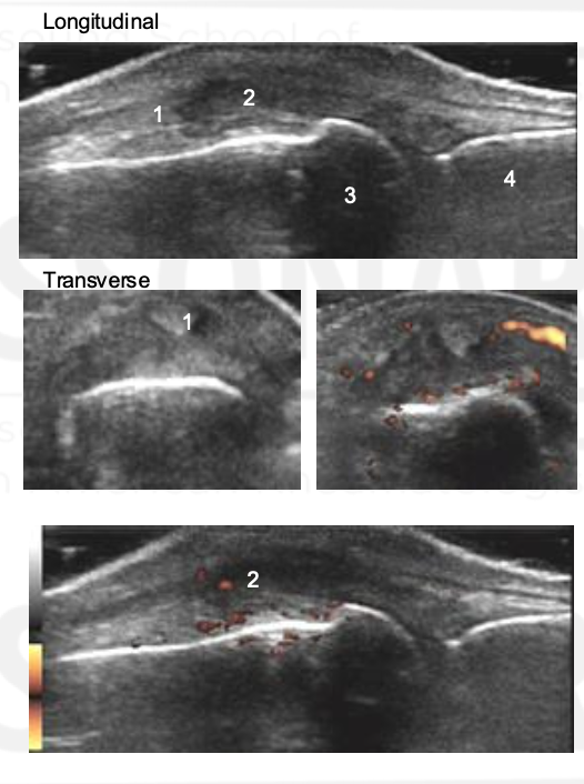
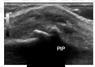
What view is this?
PIP
Synovium
Extensor Tendon
Proximal Phalanx
Middle Phalanx