Animal physiology
1/103
There's no tags or description
Looks like no tags are added yet.
Name | Mastery | Learn | Test | Matching | Spaced | Call with Kai |
|---|
No analytics yet
Send a link to your students to track their progress
104 Terms
AMOCTOBSO
Atoms
molecules
organelles
cells
tissues
organs
body
system
organism
Ephithelial TIssues
Covers body surfaces, line cavities, and forms glands.
Function: Protection, avbsorption, secretion
Connective tissues
Supports, binds and protects other tissues. Even bones, blood,adipose (fats), and cartilage
Muscle Tissue
Enables movement. Types are: skeletal, cardiac, and smooth.
Nervous Tissue
Transmits electrical signals, composes of neurons and supporting cells
Integumentary system
Eneclose/ protect internal structures
Site of many receptors
Key structures: Hair, skin, nails
Muscular systems
Enables movement (with skeletal)
Helps maintain body temperature
Key structures:
smooth, cardiac, and skeletal muscles
Skeletal systen
Enable movement (with mucular)
Supports the body
Key structures:
bones, ligaments, tendons
Nervous system
Detect & processes sensory
activate bodily responses
Structures:
Brain, spinal cord, nerves
Endocrine system
Secretes horomones
regulate bodily preocesses
Key strucutres:
pituitary gland, thyroid, pancrease, adrenal glands, testes/ ovaries
Uniary/ Renal system
Control water balance & homeostasis
remove waste from blood & excretion
key structures:
kidneys, bladder
Immune system
Protect body aganist pathogens
filter & return fluids to blood
Key structures:
Lymoh nodes, spleen, thymus
Reproductive systems
Produce sex horomones & gametes
Male = deliver gametes
female= support embryo to birth & produce milk for infant
Structures:
Testes, ovaries, uterus, mammary glands
The big 3 systems
Respiratory:
Removes carbon dioxide from the body
Delivers oxygen to blood
Circulatory system:
Delivered oxygen and nutirents to tissues
Equalizes temperature in the body
Digestive system:
Processes food for use by the body
Removes wastes from undigested food
3 parts system for homeostasis
Sensors ditect imbalance, the control center processes information and then produces and responds to restore internal body enviroment
Negative feedback
Primary method for homeostasis
The nerve cells in your skin and brain detect heat
The temperature regulatory center in the brain recieves the signal
The body triggers sweat glands
Negative = The response that reverses stimulus
Positive feedback
Amplifies responses to complete the physiological process quickly
Ie.
Baby pushes aganist cervix, streching of cervix causes nerve impulses to be sent to brain. The brain stimulates posterior pituitary to release oxytocin. The oxytocin causes smooth mucle lining of the uterus to contract.
Epiglottis
Cartilaginous flap that blocks food from entering airway when swallowing food
Its opened when breating and during resting states
Closes to cover the trachae during swallowing so food or liquid can’t enter the lungs
Larynax (voice box)
Found direcly belown the epiglottis
Home of vocal cords which fold and vibrate to producec sound/ speech
Trachea
Tube lined with cartilage rings to hold it open
Lined with cillia (which are hair-like cellular extension) that trap and expunge particulates
Main transport airqay to transport air in and out of the lungs
Lung
Comprised of multiple spongy lobes
Right is bigger than the left to accommodate because the heart on the left side.
The left and right is based on your own perspective
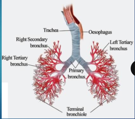
Bronchi & Bronchioli
Branched narrowings of trachea supported by cartilage rings
Terminate at alveoli
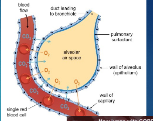
Alveoli
Where the gas exchange happens. Allows for the transprt of oxygen.
Increases absoptive surface area
The aveolar wall is 1 cell thick, where blood is on the other side (the capillaries)
Diaphragm
Responsible for inhale and exhalation
Controled by the autonomic nervous system
Relaxed when it’s curved and when flat it’s contracting
Breathing control
Controlled by medulla obolongata in the brain which searches for CO2 and pH of the blood.
pH and CO2
Detected by chemosensors in aortic and carotid bodies to prompt change in ventilation rate
More CO2 you have in your blood, the more acidic your blood is going to be.
chemosensors
Molecular/cellular sensors that detect change & produce a signal
Non-respiratory systems
Sneezing & coughing
Purges airway and obstructions/ irrtiants
Hiccups
Spasms of the diaphragm caused by sudden intake of breath
Laughing & sobbing
Emotional response influences respitaroy rhythms
Yawns
Unknown, might be linked to empathy
Gas exchange: O2 Journey
O2 dissolved in mucuos
Diffuses down into gradient into blood
Binds hemoglobin in a RBC & circulates
Hemoglobin releases it to tissues that lack O2
O2 drive cell respiration in the mitochondria
Gas exchange: CO2 Journey
Diffuses into blood & circulates to lungs
Diffuses down into gradient into aveoli
Exhale
Total lung capcity
Volume of air in the lungs after maximal inhalation
Vital capacity
Volume of air that can be exchanged by the lungs via maximal inhalation and exhalation
Residual volume
Volume of air that is always present in the lungs (20% of total lung capacity)
Tidal volume
Volume of air that is exchanged via normal breathing
Three substems of circulatory system
Pulmonary:
Move O2 (Poor blood from heart to lungs and back)
Oxygenates blood and removes CO2
Systemic:
Move O2 (Rich blood from heart to body and back)
Deoxygenates blood and collects CO2
Cardiac:
Feed O2 (Rich blood to heart itself)
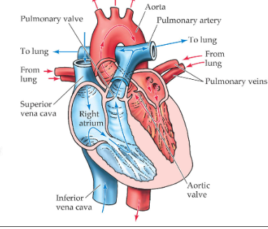
The heart
Muscular organ that pumps blood through the body
Blood vessels
Tubes that direct blood flow
Blood
Nutrient- rich connective tissue. It enables the transport of substances from the external enviroment to every cell in the body.
Consists of:
Plasma
Cells and plateles (fromed elements)
flow of blood
Path way for deoxygenated blood: Heart’s right atrium—> right ventricle—> pulmonary atery —> lungs.
And then, gas exchange
Pathway for oxygenated blood:
Left atrium —> left ventricle —> Aorta —Rest of the body + heart
Atrioventricular Valves
Tricuspid valve: Door between your right atrium and right venticle
Mitral valve: Door between your left atrium and left ventricle
Semilunar valves
Aortic valve: Opens when blood flows out of your left ventricle to your aorta
Pulmonary valve: Opens when blood flows from your right ventricle to your pulmonary artery
Ateries
Carry blood away from heart
Thick walls withstand high pressure
Capillaries
Smallest vessel
RBC’s travel in single file and slowly
Body cells are not bigger than 2 cell widths away from a capillary
Connect veins and ateries
Veins
Low pressure blood pressure returns to heart
Semi-lunar valves prevent backflow
Carries blood from the body cells to the heart. They carry waste.
Plasma
Fluid-suspension of proteins and dissolved nutrients
Erythrocytes (RBC’s)
Lack nuclei, ribosomes, & mitochondria
Carries O2 and CO2,
Leukocytes (WBC’S)
Cells of the immune system
Thrombocytes (Platelets)
Involved in clogging and not considered a true cell
Differences between Arteries Vs Veins
Flow direct: Arteries flow away from the heart, veins toward
Pulse: Present and the wall strech by pressure, veins have no pulse because they are too far from the heart
Walls: Arteries are thick, muscular, and elastic for blood pressure. Thinner, less muscular & elastic (can be permentantly streched)
Type of blood carried: Artieries, oxy and deoxy. Veins: Deoxy and oxy
Colour: Arteries: bright red and veins are dark red
Dialation & constriction: A: Easily archieved V:Less easily archieved
Valves: A: No V: Yes
Cause of blood flow: A: heart pumping, V: Contraction of skeletal muscles
Flood if served: A: Blood spurts, no clotting (high BP) V: Slow, even flow, and blood clots
The cardiac cycle
Contraction: systole, these are controlled by the heart itself
Relaxation: diastole
Blood pressure
Measures the force of blood on the walls of arteries
Measured with a sphygmomanometer
2 pressures measured:
Systolic: Highest, ventricles pushing blood
Diastolic: Lowers, chambers relax and fill with blood
Measured as systolic/ diastolic in mmHg
Health is 120/80
Hypertension
Sustained high blood pressure
Risks:
Organ damages
Blood vessel damage can cause an anerism (in heart= heart attack, in brain = stroke)
Two different types of strokes:
- Ischaemic: Where the blood is blocked
Haemorrhagic: A bleeding of an atery
Prevention:
Good diet
Medication (diuretics to reduce blood volume)
Aneurism
A weakening of an atery wall that creates a bulge or distention of the atery
Hypotension
Abnormall low bp (90/160)
Risks:
Light headedness, dizziness, weakness, fainting
Brain damage
Prevention: hydration
Orthostatic/ Postural hyoptension
Sudden blood pressure drop due to stand up from a lying or sitting position. Common
Circulatory system in animals
Not all animals have circulatory systems
Some organisms that are complex have two layers of cell in their body plan, which use diffusion through their skin.
Both internal & external tissue are in contact with an aqueous enviroment making diffusion an easy task
Close circulatory system
Where blood is entirely contained within vessels. (Arteries, veins and capillaries) and it circulates
Amphibians
They have three- chambered heart. 2 atria + one ventricle
Single ventricle: Pumps mixed blood to the lungs, skin , and the rest of the body.
Circulation: Double circulation system
Pulmonary circulation: Moves bloodbetween the heart and lungs/ lungs for oxygenation Gas exchange happens during circulation: pulmocutaneous circulation
Fish
Two chambered heart with 1 atrium and 1 ventricle
Atrium: Recieves deoxygenated blood from the body
Ventricle: Pumps doxygenated blood to the gills for oxygenation
Gill circulation
The atrium cellects blood that returned from the body and ventricle pumps the blood to the gills where gas exchange happens and the blood is reoxygenated
Open circulation
When blood is not enclosed in blood vessels. The fluid is pumped into a cavity called a hemocoel
Blood mixes with interstital fluid creating hemolymph which is pushed around the when the heart beats in the body
Ostia: the opening that allows blood to re-enter the heart after circulation
common in insects and mollusks
Insects
open circulation
Sinuses: Spaves within the hemocoel where hemolyph bathes the organs directly
Trachea: A network of tubes that provide direct air exchange and reduces the reliance on the circulatory system for oxygen delivery
Aorta: carries hemolymph from the heart to the body cavity in some anrthopods
Open system: circulatory system
Snails and clams
Hemolymph flows into hemocoel, the simple heart pumps through vessels
Closed system mollusks: cephalopods
Blood stays in veseels and they have a more complex heart system which supports active, high energy life style
Close vs open circulatory systems
Organs are not in direct contact of blood vs organs come in direct contact with blood
Blood flow can be regulated and pressure is high vs blood flow cannot be regulated and the pressure is low
Blood flows through closed spaces and have capillaries vs flows through open spaces and dont have capillaries
Materials transported by the blood arrives to the tissues through diffusion vs blood pumped by heart travels to the blood vessels and soak directly into cells returing then by different mechanisms
1 heart vs 1 or 2
Oxygenated blood can reach the extremeties of the body much faster vs can support a high activty body by burning less calories to move the same amount of oxygen
They can metabolize much faster because it lets them move, digest, and eliminate waste quicker vs takes more time to reach the extremeties of the body and is only feasible in smaller animals
Enviromental impact of using insects
Lower enviromental impact compared traditional livestock
Require less land, water, and food to grow and emit fewer greenhouse gases
Bugs in products
Shellac: Shiny coating on candies and apples, made from resin secreted by lac bugs
Carmine: red pigment from cochinal insects, used in foods like strawberry yogurt, candy, and cosmetics
Cricket flour: Sustainable and protein-rich flour in proteins bars, baked goods, and pasta.
Functions of the digestive system
Primary: Breaks down large molecule into smaller molecules for absorption
Secondary: Hold mutualistic bacteria that aid in digestion and waste excertion
Enzymes
Biological cataslysts that allows most chemical reaction carried out in a cells
Not change or used up
Bond molecules together to form new molecules or molecules apart
3 main enzymes
Amylase, Lipase, Protease
What are the enzymes that break carbohydrate down to glucose
Amylase
Sucrase-Isomaltase
Maltase,
lactase
Protein to Amino Acids
Enzymes used: Pepsin, Trypsin, Petidase breakdown into amino acids
Fatts to fatty acids/ Glycerol
Lipase breaking fats downing int fatty acids and Glycerol
Gastrointestial tract vs digestive system
Specific part of the digestive system. Its the continuous tube that runs mouth to BUTTHOLE
Digestive system: All organs involved in the process of digestion
Ingestion
Food ingested through the broke down through mastication
Main functions:
Ingests food
Bolus: Chews and mixes
Used amylase to breakdown of carbohydrates
Swallowing:
Tongue and plate press food into bolus
Tongue and throat muscles and guide bolus over to epiglottis into esophagus
Peristalsis pushes bolus to the stomach
Endoscopy
Internal observation of the upper digestive system
Colonoscopy
Internal observation of the large intestine
Sphincter and Ingestions
Sphincter: Circular muscles that controls movement through the body
Food passes through 2 sphincters on the way to the stomach
Upper esophageal sphincter (barrier to air)
Lower esophageal sphincter (barrier to acid)
Taste buds + Salivary Glands
Slivary glands:
Moistens and dissolves, allowing for swalling
contains carbs
Taste buds: Detecting nutrients in food we eat that trigger hormonal responses to regulate digestion, absorption, and appetite control
Mechanical digestion
The physical breakdown of food to high surface for chemical digestions. First in the mouth
Chemical digestion
Breaking down of nutrients into absorbable molecules (mostly via enzymes)
Happens in the mouth —> stomach —> small interstine
Digestion in the stomach
Main functions:
Store swallowed food and liquid
Chrun the food, liquid, and digestive juices produced by the stomach
Slowly empty its contents into the small intestine
Only a few substances such as water, aspirin, and alcohol can be abosrbed into blood directly from the wall of the stomach
Main enzymes: protease + lipase
Gastic Glands
Consists of:
Muscous cells that secrete alkain mucus to protect lining of the stomach
Chief cells
Parietial cells: Secretes HCL
Absorption in small intestine
Responsible for absorbing majority of nutrients into (bloodstream)
6m long, holds some bacteria that help in digestion
Big SA to boost absorption
SA is maximized by Villi and microvili
Villi
Finger-like projection on inner surface of small intestine
Microvilli
Hair-like projection of surface of villi. Absorption direct nutrients into bloodstream
Absoprtion in large intestine
From small intestine, chyme is a water, salty waste product
The ileocecal sphincter opens and allows the product to the large intestine
Waste is called stool or feces
Will absorb water and minerals, causes feces to solidify and clump
Microbiota of the large intestine
Harvesting energy from digested
Protecting aganist pathogens
Regulating immune function
Gut dybosis
When there is an imbalance of benefical bacteria in the gut
Infections: Chlostridium difficile, Crohn’s disease, and infalmmatory bowel disease
Elimination
Process of removing undigested food and waste products from the body
Rectume: Short length of the colon where feces for defaction
When the rectal wall streches, triggers the urge to eliminate feces from the body
Acieved by allowing/pushing feces through the anal sphincter (anus)
Birds digestive systems
Birds can eat a variety of foods (insects, worms, berries, seeds, etc.)
The crop stores food
Most chemical digestion occurs in the stomach
The gizzard is filled with gravel/pebbles the bird eats to mechanically digest food stuffs
Gastroesophageal Relux Disease
Occurs when the lower esophageal sphincter fails to close properly. This allows toach acid to flow backwards into the esophagus
Frequent exspoure to acid causes inflammation known as esophagitis and can to barrets esophagus (precancerous change in the tissues cell type)
Peptic Ulcers
Erosion of the lining
Caused by H. pylori bacteria and longer term pain medication use. Which depeletes the protective layer
Celiac Disease
An autoimmune disorder where the ingestion of gluten that propts the immune system to attack the small intestine
Nutient Malabsorption
Immune response that damages and flattens the villi.
The finger-like projections responsible for nutrient absorption which leads to malnutrition no matter how much food is consumed
Appendicitis
Inflammation of the appendix due to bacterial infection
Can ruprure if not removed
IBS
Causes cramping, abdominal pain, bloating, gas, diarhea/ constimation
No known cause
Treatment: Managed diet
Colon cancer
Cancer of the large intestin
Detected when there is blood in your stool
Treatment: Surgical removal of section and chemotheraphy
Colostomy bag
stoma is formed in abdominal wall that provides alternate exit for colon
FEcal matter is collected in a replaceable bad instead of passing through
Gallstones
Chloesterol or bilirubin in the bile hardens into stone-like deposits
The stones block the common bile duct, preventing bile from entering the duodenum. Needs to be removed with laproscopic surgery