PSB3340 Exam 2
1/93
There's no tags or description
Looks like no tags are added yet.
Name | Mastery | Learn | Test | Matching | Spaced | Call with Kai |
|---|
No analytics yet
Send a link to your students to track their progress
94 Terms
Sensory receptor cells
translate physical sensations to electrical signals
Sensory receptor organs
specialized to detect a certain stimulus (eyes, nose)
Receptor cells
within organ
convert stimulus to electrical signal
graded potential (not AP)
transducer
Sensory transduction
conversion of electrical stimulus to change in membrane potential (receptor cell, graded)
Receptor potentials
local membrane potentials specific in response to adequate stimuli from receptor cells
Receptor potentials
can result in the generation of APs OR release of a graded amount of NT
only impact voltage in a small local area of cell
summate
Restricted range of responsiveness of sensory systems
Responses range between individuals, lifespan, and species
like how we don’t have receptors that can pick up UV light
Range fractionation
Different cells have different thresholds for firing over a range of stimulus intensities
Stimulus intensities encoded by frequency of AP firing
Different cells cover different fractions of possible intensities (like how colors are split up into RGB)
diff. areas of skin responding to different amounts of pressure applied
Stimulus location
sensory touch and pain receptors in body map onto corresponding regions of the primary somatosensory cortex = somatotopic representation
differing intensities of mechanical pressure activate nociceptors (pain)vs. fine touch receptors
Coding
patterns of APs that reflect a stimulus
number and frequency of APs, rhythm of AP clusters
ex. spatial, temporal summation
Adaptation
Loss of response to sustained stimulus - reduction in AP frequency
Highlight changes
Tonic receptors
slow/no adaptation
no decr. in AP frequency
Phasic receptors
rapid adaptation
decr. in AP frequency
Suppression
Adaptation
Accessory structures (dog ears, eyelids) - before reaches sensory receptors
Top-down processing - suppression
Higher brain centers suppress some sensory inputs + amplify others
attention is most common!
memory, expectations etc.
Pathways
Each sensory system has a distinct sensory pathway through brain
distinct senses bc APs travel along separate nerve tracts = labeled lines
MOST pass through thalamus (NOT olfactory)
Terminate in cerebral cortex
General senses (somatosensory, visceral) along somatic or visceral afferent nerve fibers
Specialized via specific cranial nerves (optic, olfactory)
General sensory pathway
Receptors → thalamic relay nuclei → primary sensory cortex (varied) → secondary sensory cortex (varied) → association cortex
*smell does NOT pass through thalamus first
Receptive fields
space in which a stimulus will alter a neuron’s firing rate
excitatory center/inhibitory surround or inhibitory center/excitatory surround = edge detection
can change by experience
severed limb - cortical area devoted to that area shrinks, others expand
intentional stimulation = expand in cortical representation
Non-primary (association) cortex
receives main input from primary cortical area for specific sense
responds to a large range of stimuli over a larger portion of the environment
Attention
Mainly activated: posterior parietal lobe and cingulate cortex
Synesthesia
blurring of sensory modalities
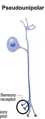
Pseudo unipolar neurons
Somatosensory receptors
All start as one appendage from soma
4 tactile receptors
Detect touch
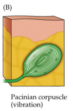
Pacinian corpuscles
Meissner’s corpuscles
Ruffini’s endings
Pacinian corpusles
“onion layered”
deeper than meissners/merkels
touch moves some layers relative to others, uncovering channels
vibration, transient stimulation
fast adapting
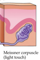
Meissner’s corpuscles
near surface of smooth skin
layers of overlapping disks moved by pressure - open underlying ion channels
light touch
fast-adapting
help perceive object forms
prevalent in fingertips, tongue, lips
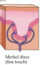
Merkel’s discs
fine touch (light, sustained)
slow-adapting
help perceive object forms
prevalent in fingertips, tongue, lips
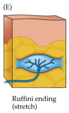
Ruffini’s endings/corpuscles
nerve fiber branches embedded in collagen
stretching collagen opens channels in embedded dendrites
sustained, deep stimulation
stretch
slow-adapting
somatosensory AND proprioceptive (body position)
Epidermis
outermost layer
dead cells
free nerve endings
Dermis
middle
Merkel disc
Meissner corpuscle
Hypodermis
Innermost layer
fat + connective tissue
anchors skin to muscles + help shape body
Pacinian corpuscle
Ruffini ending
Mechanoreceptors
Tactile touch sensors of skin
Respond to physical deformation of their membranes by opening mechanically gated ion channels (non selective)
Piezo ion channel protein
In tactile receptors
Opens when mechanically stretches and depolarizes the cell by letting cations enter
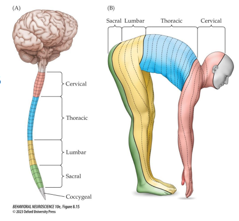
Dorsal column system
deliver touch info to brain
up spinal cord to medulla (brainstem) → crosses over midline → thalamus → primary somatosensory cortex
Dermatome
Region of skin innervated by a particular spinal nerve
Reflect quadruped
Primary somatosensory cortex (S1)
receive touch info from opposite side of body
homunculus - amount of tissue corresponds to how sensitive/densely enervated the area is not how large it is
Secondary somatosensory cortex (S2)
maps both sides of body in registered overlay
blends together info
Free nerve endings
Temperature, pain, itch
Different nerve endings produce different receptor proteins to respond to temp changes, chemicals, pain, itch
Thermoreceptors
Free nerve endings
Cold = more numerous, superficial
TRPM8 - alsp methol
Warm
TRPV1 receptor - also capsaicin (chile) - polymodal receptor
TRPM3 - even higher temps (no capsaicin)
Itch free nerve endings
Respond to histamine (NT) or chloroquine (released by mast cells in skin)
Activates TRP receptors + terminate in the pons = itch sensation
Nociceptors
Pain
Intense pressure
Hear
Chemicals released by destroyed cells
Extreme pain will be nociceptors AND free nerve endings
Mutation in gene that codes for one of its voltage gated Na channels causes insensitivity another chronic pain
High temps/pain = large, myelinated axons to conduct signals rapidly
Tissue damage
= “inflammatory soup” that sensitize or excite terminals of nociceptors
Peptides
Lipids
Neurotransmitters
Neurotrophins
Substance P
Promotes inflammation
Vasodilation → more blood flow → attract immune cells to site
Anterolateral (spinothalamic) system
Delivers pain + temp to brain
Info crosses midline in the spinal cord (FIRST) → thalamus
More poorly localized than if it ascended through the dorsal column before crossing midline (like fine touch, vibration)
Neuropathic pain
Phantom limb pain
Microglia overexciting pain pathways in limb
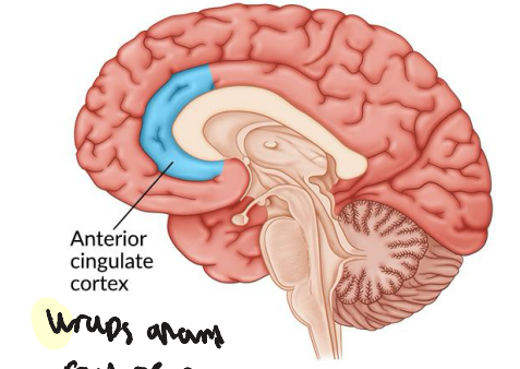
Anterior cingulate cortex (ACC)
Immediate emotional consequences of pain
Social rejection also activates this area
Periaqueductal gray (PAG)
Area in midbrain involved in pain perception
Main source of endogenous opioids
PAG neurons send endorphin-containing axons to medulla and spinal cord
Placebo releases endogenous opiates
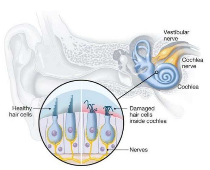
Hair cells
Sensory receptors cells for hearing
Mechanoreceptors - increases/decreases in air pressure
In organ of corti in cochlea
Amplitude
Perceived as loudness
Frequency
Perceived as pitch
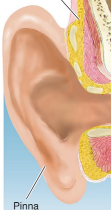
Pinna
External ear
funnels sound
outer ear
ear wax
antifungal
water resistant
antibacterial
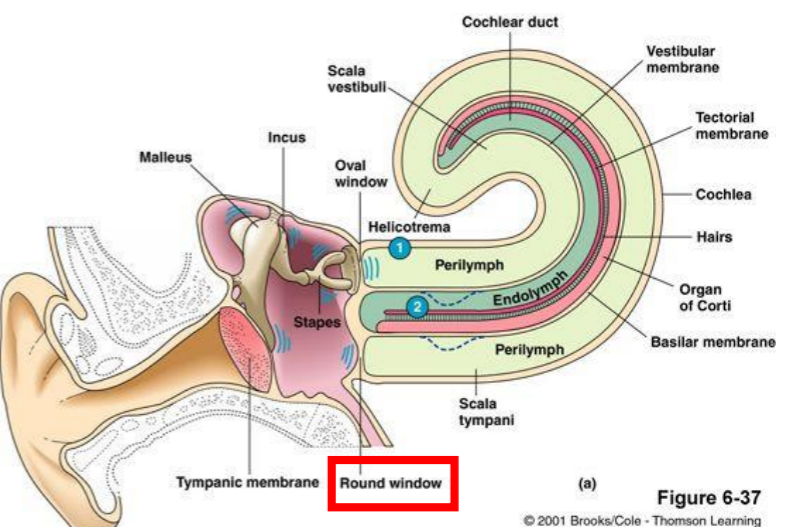
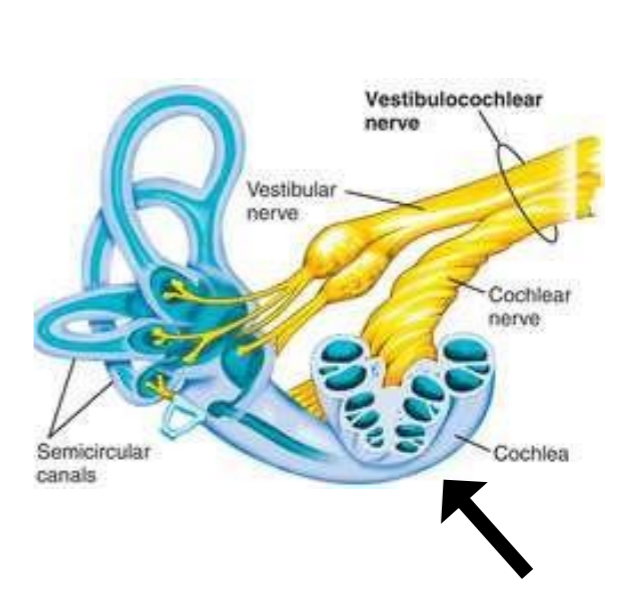
Cochlea
Fully developed at birth
Tympanic membrane
ear drum
beginning of middle ear
concentrates sound energies
connected to the oval window by 3 ossicles
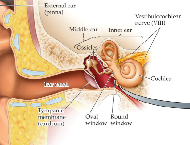
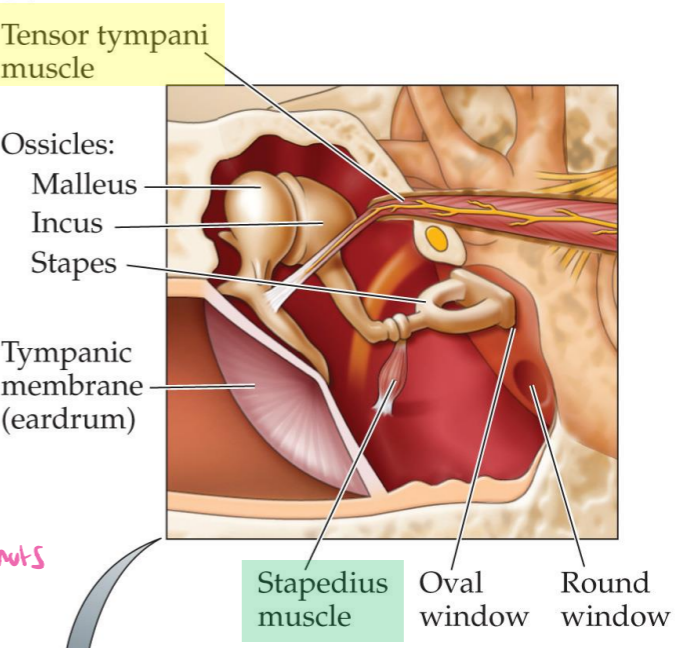
Ossicles
Mechanically multiply force through lever action
Larger tympanic membrane to smaller oval window
Malleus - hammer
Incus - anvil
Stapes - stirrup
Acoustic reflex
2 muscles in middle ear can contract to inhibit vibration in the ossicles - help prevent damage
Have delay
Tensor tympani - attached to malleus
Stapedius - attaches to stapes
Cochlea
3 parallel canals
Scala vestibuli
Scala media - middle; contains organ of conti
Scala tympani
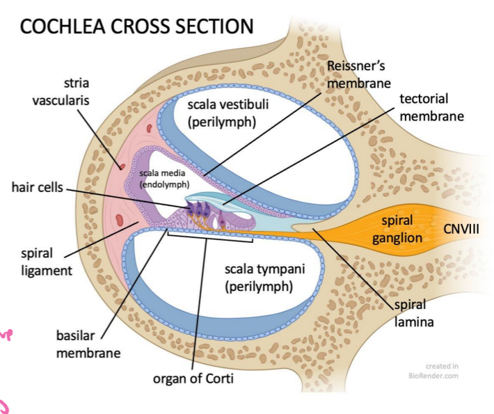
Scala vestibuli
Cochlea channel
At the oval window, vibrations from stapes transferred to fluid here
Scala media
Isolated from other layers
Endolymph with different ionic makeup than perilymph
Contains receptor system → organ of Corti → hair cells convert sound to neural
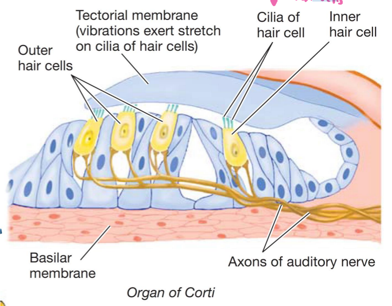
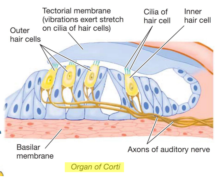
Organ of Corti
Contains hair cells - embedded in basilar membrane
Sound vibrations cause basilar membrane to oscillate like waves
Scala tympani
Cochlea channel
Round window - pressure release valve so sound waves can travel through perilymph
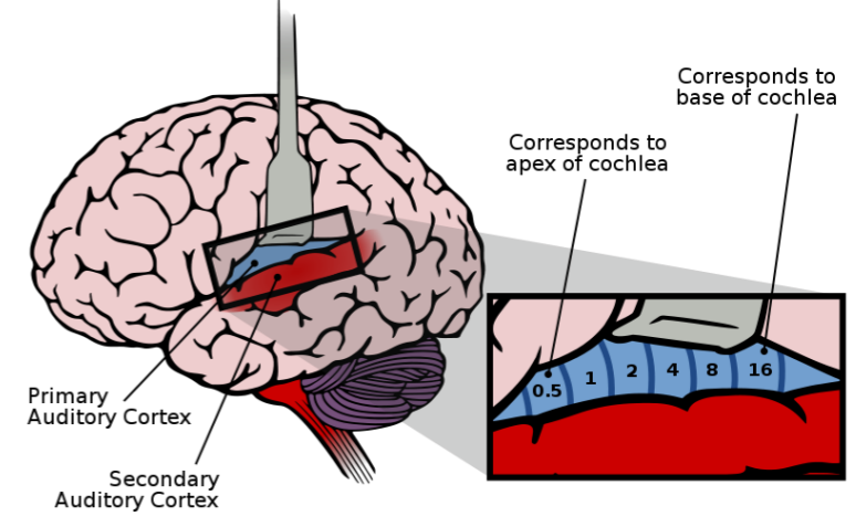
Tonotopic organization
Diff. sound frequencies map to diff. areas of cochlea
Low frequency - displaces near end, round window
High frequency - displaces base of basilar membrane, near oval window
Place coding
Near base = treble
Near apex = bass
Temporal coding
Firing pattern of auditory neurons help encode frequency
Volley principle
groups of neurons fire in turn (during refractory period) to create high frequency sounds
mastoid process
Bone in back of jaw
add low frequency sound to our own voice when we hear it directly
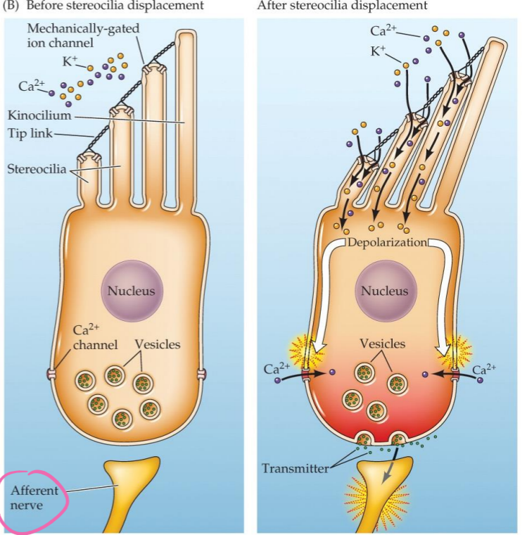
Stereocilia - hair cells
Extend into tectorial membrane
Tip links connect stereocilia - tension pulls stereocilia together - opening ion channels
don’t produce APs themselves - release graded amounts of NTs (glutamate) - afferent nerve produces AP
K+ depolarizes (endolymph has high K+ concentration; perilymph has low) → Ca+ channels open → NT released → cochlear nerve (afferent) AP
Inner hair cells
auditory signal transduction
lack causes deafness
most afferent connections to brain
each innervated with approx. 10 nerve fibers for diff. loudness levels
efferent (top down) to inner ear cells help suppress loud noises by reducing excitability of afferent nerve (slow down rate of APs)

Outer hair cells
sound amplification
lack causes hearing impairment
efferent (top down) change lengths of OHCs to fine-tune cochlea to help discriminate frequencies
hyperpolarize - lengthen - more sensitive
depolarize - shorten - less sensitive
Vestibulocochlear nerve (CN VIII)
Afferent (to brain) axons of both hair cells become cochlear nerve which combines with vestibular nerve = Vestibulocochlear nerve
Auditary + vestibular info
Binaural
Two ear information
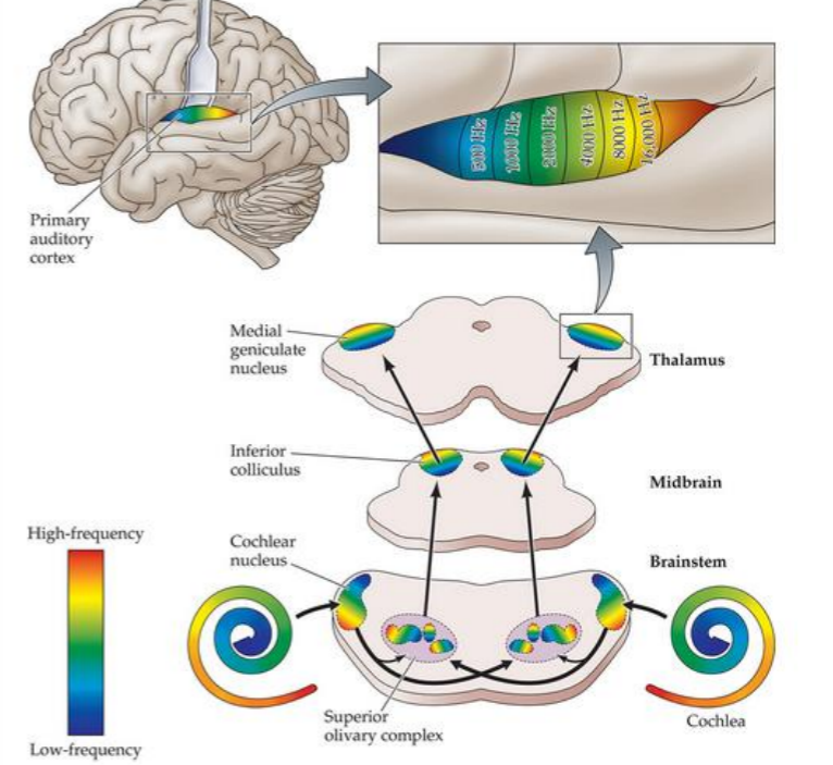
Most info from each ear projects to cortex on opp. side of brain
Begins in the brainstem superior olivary nucleus
*Auditory pathway
S uperior olivary nucleus
L ateral lemniscus
I nterior colliculus
M edical geniculate nucleus
To superior temporal gyrus
All levels have tonotopic organization (arranged in a map according to frequencies to which they respond)
Tuning curves
Show a cell’s response to various frequencies
Sound frequency/pitch discrimination increases along hierarchy of system
Primary auditory cortex (A1)
Part of temporal lobes
Located within the lateral sulcus
Involved in lip reading - integrating sight/sound info
Dorsal auditory stream
Where?
Ventral auditory stream
What?