Exam 5 (questions I dk
1/142
There's no tags or description
Looks like no tags are added yet.
Name | Mastery | Learn | Test | Matching | Spaced | Call with Kai |
|---|
No analytics yet
Send a link to your students to track their progress
143 Terms
On axial CT images, which muscle of the neck is seen most posteriorly?
sternocleidomastoid
erector spinae
posterior scalene
longus capitis/longus colli
erector spinae

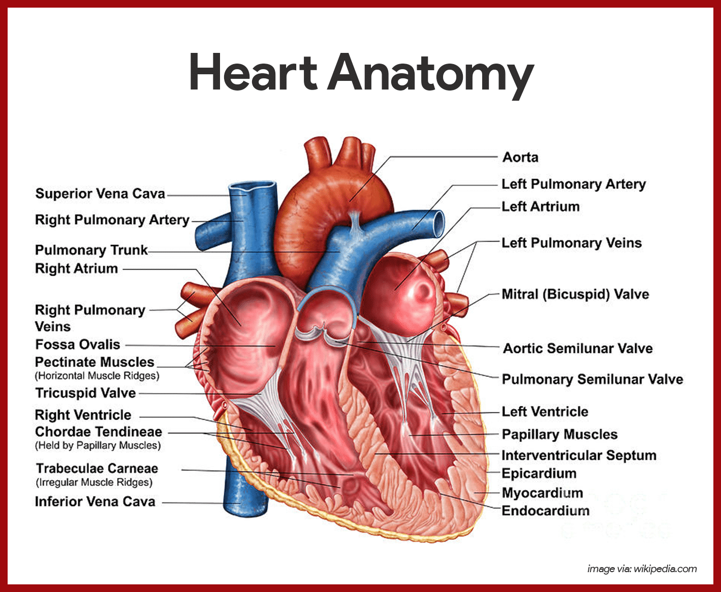
The lungs are supplied with blood by the:
aorta
pulmonary artery
bronchial artery
carotid artery
pulmonary artery
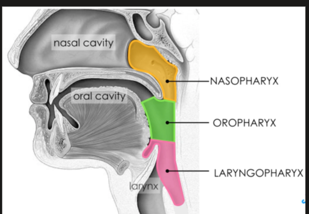
What divides the nasopharynx and the oropharynx:
soft palate
epiglottis
auditory tube
tonsils
soft palate
FYI
Epiglottis separates: oropharynx and hypopharynx
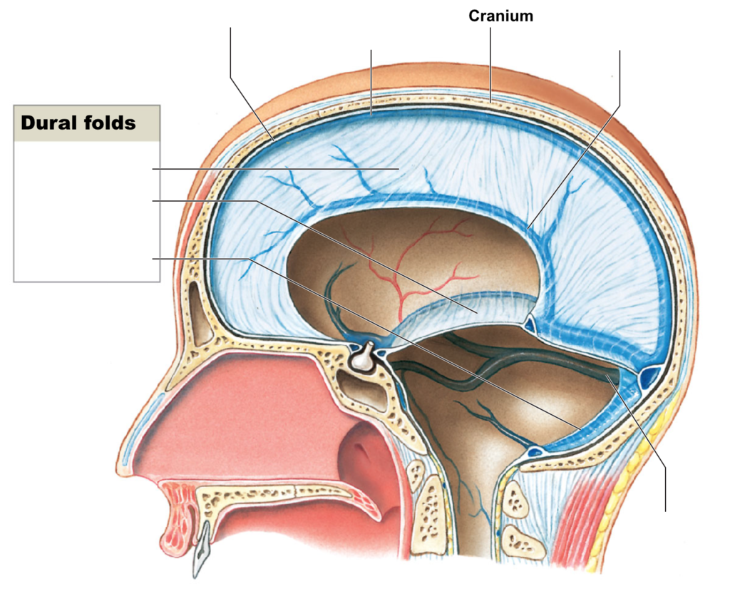
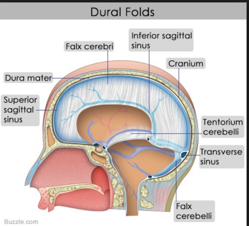
The fold of dura that divides the right and left hemispheres of the cerebrum is :
corpus callosum
falx cerebri
tentorium
pia mater
falx cerebri
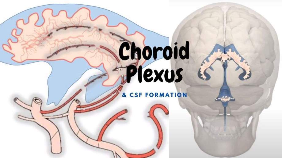
Cerebro-spinal fluid is formed in the:
corpus callosum
subarachnoid space
choroid plexus
arachnoid
choroid plexus (by ependymal cells)
The part of the body which forms an angle between the upper and lower eyelids is called:
lacrimal sac
orbit
canthus
nasion
canthus
The final branches of the bronchi are the:
pulmones
bronchus
lobules
alveoli
alveoli
Primary bronchi→ secondary (lobar) bronchi→ tertiary (Segmental) bronchi→ bronchioles (microscopic)→ alveoli
The smallest unit of protoplasm that is capable of independent existence is the:
atom
nucleus
cell
amoeba
cell
Reasoning
protoplasm: the colorless material comprising the living part of a cell, including the cytoplasm, nucleus, and other organelles.
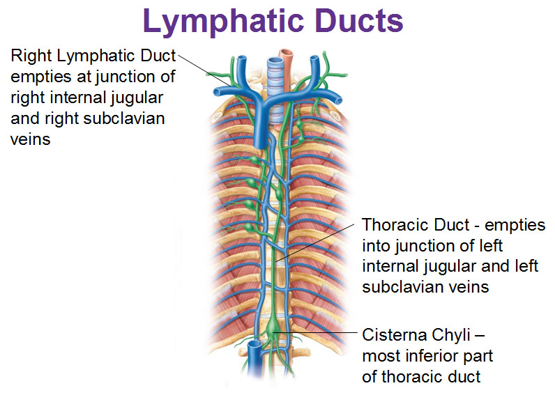
The thoracic duct begins in the abdominal cavity at the level of :
T11
L2
L4
L5
S1
L2
Reasoning:
Cuz it begins at cisterna chyli
Other sructures at L2
pancreas head (body at L1)
Spinal cord ends (L1-L2)
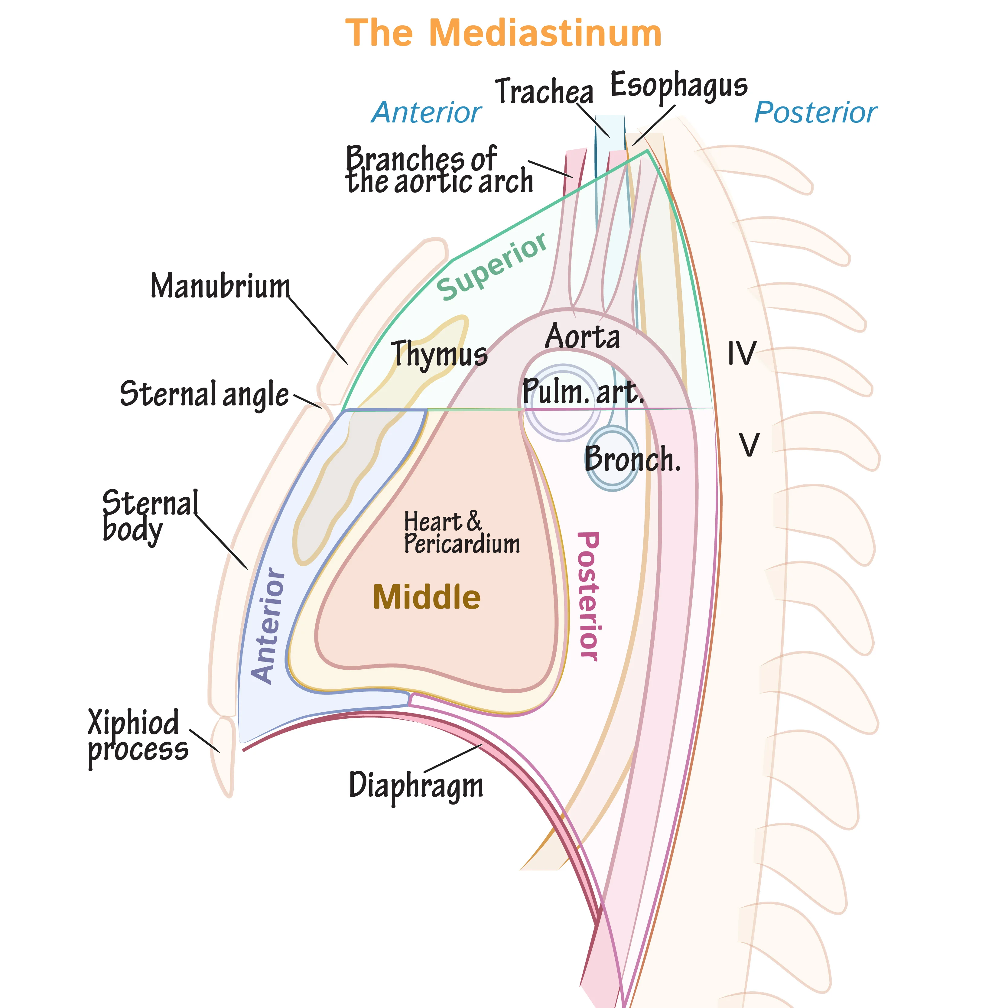
Which of the following structures is not in the mediastinum:
paratracheal nodes
cisterna chyli
esophagus
vena cava
thymus
cisterna chyli
Reasoning:
Cuz it is at the level of L2
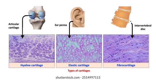
List the 3 types of cartilage
1. Hyaline cartilage
Most common
Found in: nose, trachea, ribs (costal), articular surfaces, fetal skeleton
2. Elastic cartilage
Flexible, contains elastic fibers
Found in: ear (pinna), epiglottis, auditory tube
3. Fibrocartilage
Tough, dense, resists compression
Found in: intervertebral discs, pubic symphysis, menisci
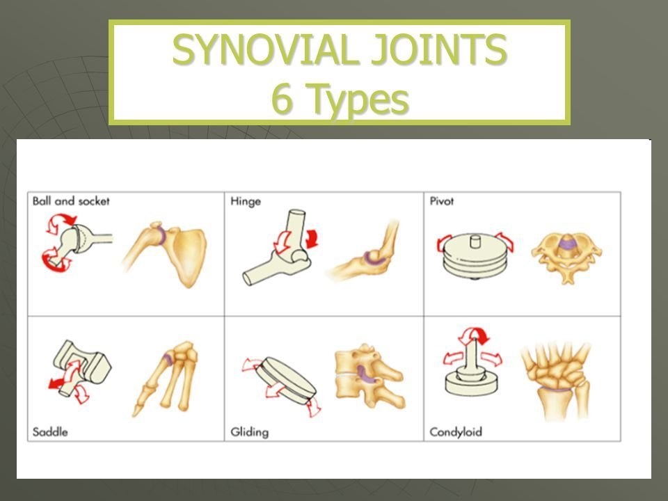
The 3 Functional Classifications (movement‑based) of joints
Synarthroses — immovable joints
(e.g., skull sutures)Amphiarthroses — slightly movable
(e.g., pubic symphysis, intervertebral discs)Diarthroses — freely movable
(these are your synovial joints)
The 3 Structural Classifications (what they’re made of)
Fibrous joints → usually synarthroses
Cartilaginous joints → usually amphiarthroses
Synovial joints → always diarthroses
Fu CkS
6 Types of Synovial (Diarthrodial) Joints
Hinge (elbow, knee)
Pivot (atlas/axis)
Ball‑and‑socket (hip, shoulder)
Saddle (thumb CMC)
Condyloid (wrist)
Gliding/Plane (intercarpals)
Prince Harry Pulled Charles’ Saddle Bag
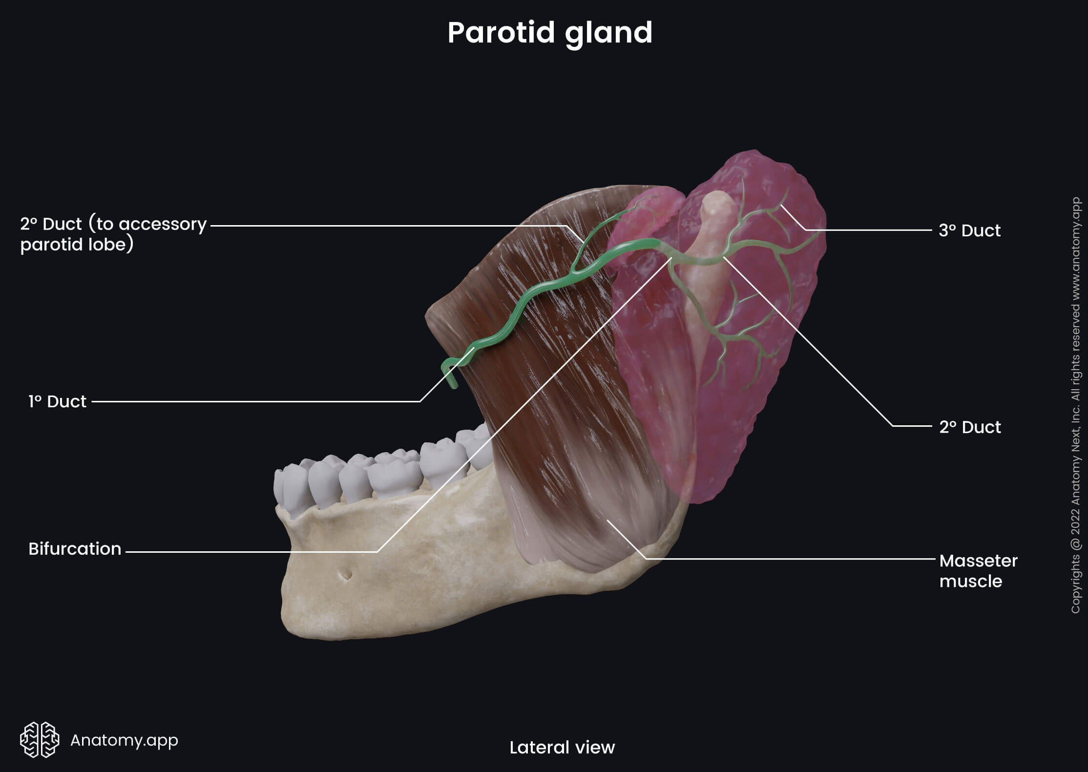
Stensens duct drains:
submandibular salivary glands
parotid salivary gland
sublingual salivary glands
cisterna chyli
cisterna magna
parotid salivary gland
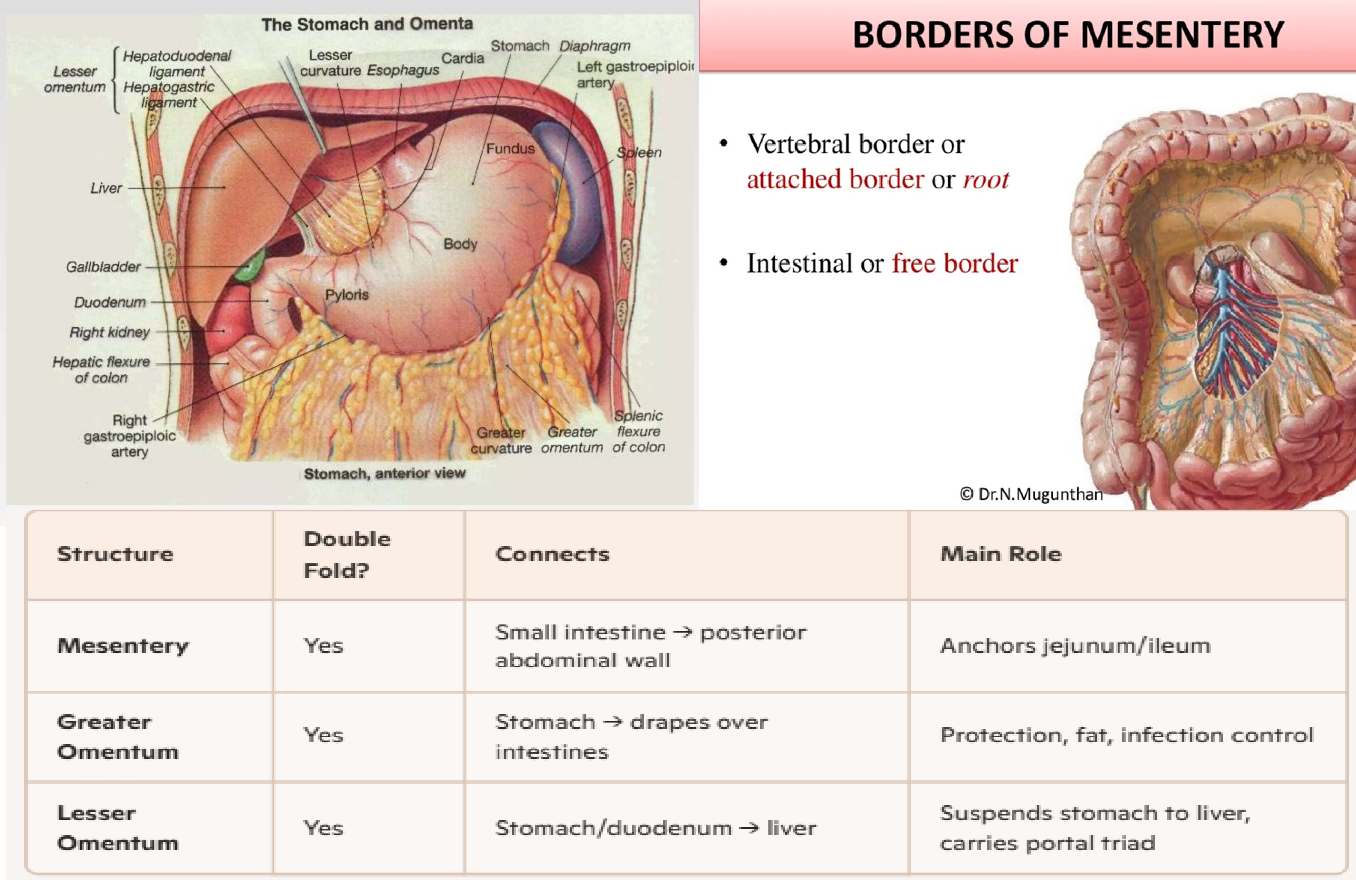
The double-fold of peritoneum which anchors the small intestine to the posterior abdominal wall is:
lesser omentum
greater omentum
mesentery
mucosa
none of these
mesentery
Reasoning:
The main difference between omentum and mesentery is that omentum is a fatty blanket hanging down in front of all the intestines, whereas mesentery is the supporting tissue to both small and large intestines.
Furthermore, omentum originates from the visceral peritoneum while mesentery originates from the parietal peritoneum.
Moreover, the two main types of omentum are the greater and the lesser omentum while the two types of the mesentery are the dorsal and ventral mesentery.
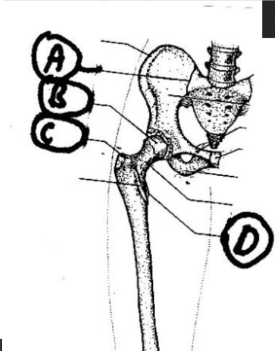
Identify from the drawing below, the lesser trochanter:
A
B
C
D
D
FYI:
A: SI joint
B: Femoral head-Acetabulum
C: Greater Trochanter

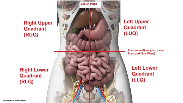
The spleen lies in the ___________ quadrant of the abdominal cavity:
right upper
left upper
right lower
left lower
left upper
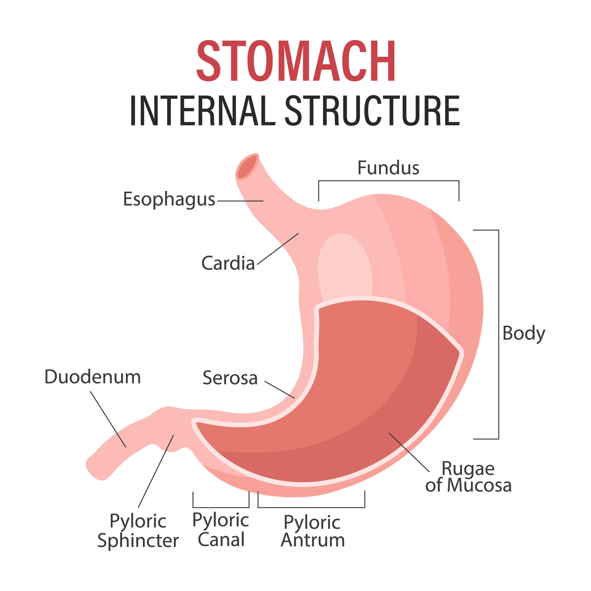
The lesser curvature of the stomach is on the ________ border:
medial
lateral
anterior
posterior
none of these
medial
A group of cancer cells that have invaded a blood vessel and have become detached is known as a:
thrombus
metastasis
tumor embolus
neoplasm
tumor embolus
Reasoning:
Thrombus- stationary
embolus- moving
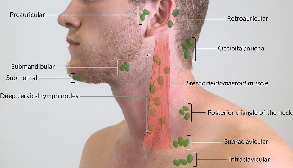
The scalene node lies:
below the clavicle
above the clavicle
at the pelvic brim
superior to the diaphragm
posterior to the manubrium
above the clavicle
Reasoning:
Scalene Node
Left supraclavicular node
Virchow’s node
The most distal of the following list of structures of the large bowel is:
cecum
ascending colon
descending colon
sigmoid colon
appendix
sigmoid colon
The true vocal cord is called the:
larynx
supraglottis
glottis
subglottis
glottis
FYI:
Larynx is the cartilage that protects it
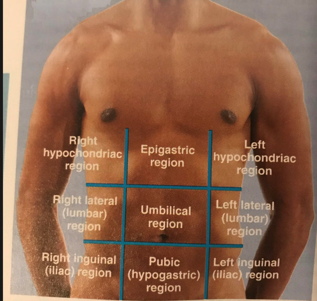
list the 9 regions of the body

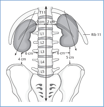
Between what vertebra do kidneys sit
T11-L3
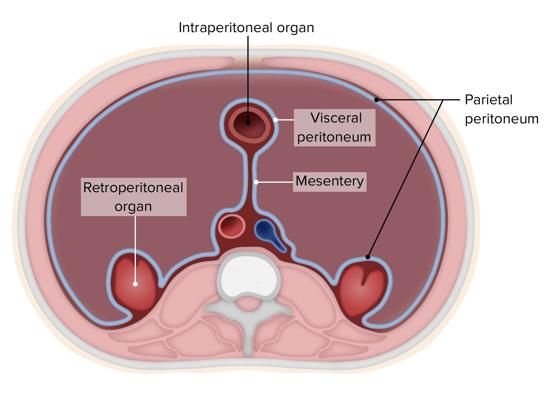
Organs in retroperitoneal vs intraperitoneal cavity
intraperitoneal- Digestive organs, spleen
Retroperitoneal- blood vessels, kidneys & their structures, esophagus, rectum
In which region is it
Cecum
Stomach (fundus)
Pancreas (body)
Right kidney
Sigmoid colon
Appendix
Spleen
Transverse colon
Bladder
Liver (right lobe)
Small intestine loops
Gallbladder
Left kidney
Pancreas (head)
Stomach (general)
Uterus
Ascending colon
Descending colon
Duodenum (1st–3rd parts)
Adrenal glands
Splenic flexure
Hepatic flexure
Cecum → Right Iliac
Stomach (fundus) → Left Hypochondriac
Pancreas (body) → Epigastric
Right kidney → Right Lumbar
Sigmoid colon → Left Iliac / Hypogastric
Appendix → Right Iliac
Spleen → Left Hypochondriac
Transverse colon → Umbilical
Bladder → Hypogastric
Liver (right lobe) → Right Hypochondriac
Small intestine loops → Multiple (mostly Umbilical, Lumbar, Iliac)
Gallbladder → Right Hypochondriac
Left kidney → Left Lumbar
Pancreas (head) → Umbilical
Stomach (general) → Epigastric
Uterus → Hypogastric
Ascending colon → Right Lumbar
Descending colon → Left Lumbar
Duodenum (1st–3rd parts) → Epigastric
Adrenal glands → Epigastric
Splenic flexure → Left Hypochondriac
Hepatic flexure → Right Hypochondriac
The organ which lies in the left hypochondrium posteriorly, close to the 9-11th ribs and whose medial end is 5cm from the midline is the:
kidney
stomach
colon
spleen
D. spleen
Double check
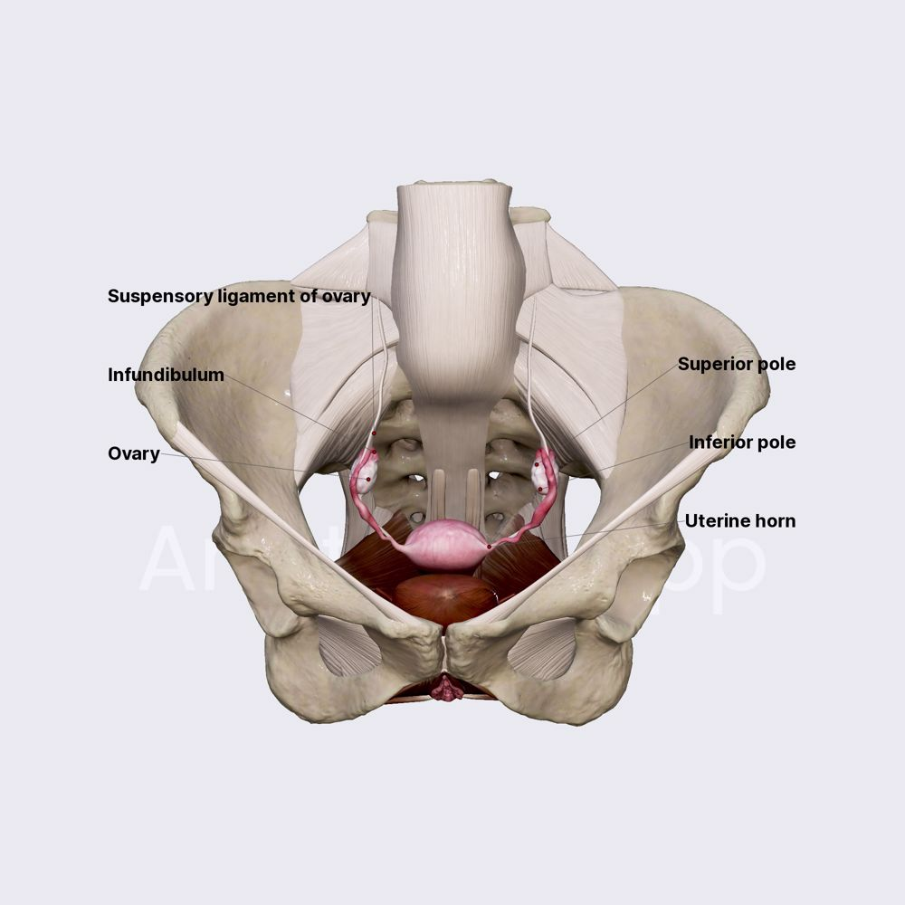
Which of the following organs is situated immediately medial to the lateral vertical planes of the anterior superior iliac spine:
kidneys
uterus
ovaries
bladder
ovaries
Erythropoiesis takes place in the:
spleen
red bone marrow
thymus gland
yellow bone marrow
red bone marrow
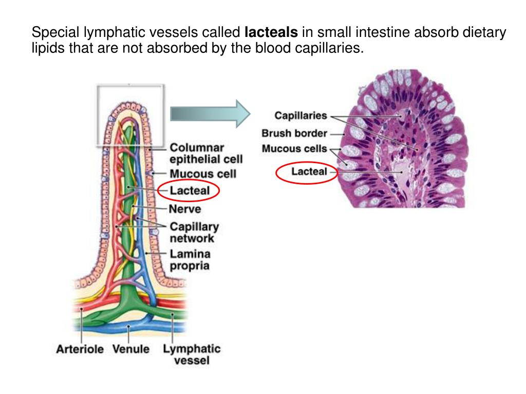
The lymphatic vessels in the villi of the small intestine are called:
lacteals
crypts
sinusoids
cisterns
lacteals
One of the chief variables influencing normal blood volume is:
the amount of body fat
heart size
the number of anastomoses in the circulatory system
liver size
the amount of body fat
Reasoning:
Blood volume is tied to lean body mass, not total body weight.
Muscle is highly vascular → needs more blood
Fat tissue is poorly vascular → needs much less blood
So:
👉 People with more body fat have lower blood volume per kilogram
👉 People with more lean mass have higher blood volume per kilogram
This makes body fat percentage one of the chief variables affecting total blood volume.
The protein released from body cells following viral invasion, and which acts to defend other body cells, is:
Prothrombin
Keratin
Interferon
Glucagon
Interferon
Reasoning:
Prothrombin- clots
keratin: hair and nails
Glucagon: when glucose is gone, stores sugar
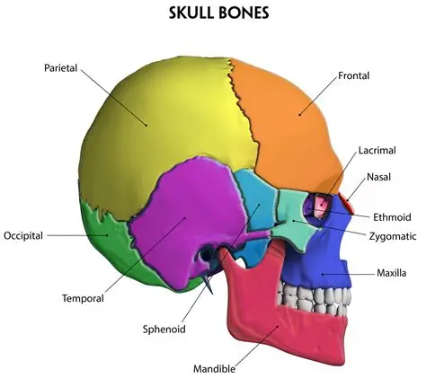
The bone which serves as the keystone in the architecture of the cranium is the ___________ bone:
Occipital
Temporal
Frontal
None of these
none of these
Reasoning:
The keystone bone is the “sphenoid bone”
It articulates with all other cranial bones
It sits centrally and helps hold the skull together
As a person grows older, their skeletal muscles undergo a process called:
Atony
Atrophy
Fibrosis
Hypertrophy
Atrophy
Reasoning:
-trophy: nourishment, growth, or development of cells, tissues, or organs
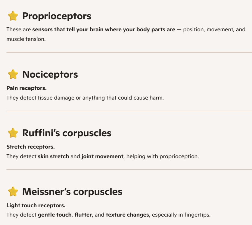
Pain receptors are also called:
Proprioceptors
Nociceptors
Ruffini's corpuscles
Meissner's corpuscles
Nociceptors
Hint:
noxious plant- bad for you nociceptors detect that
Ruffini- stretch (Mr. Fantastic stretched to Ruiffini a drink)
messi- light touch (messi’s got that light touch)
Which type of tissue serves as a protective barrier and for moving substances into and out of the blood?
Epithelial
Connective
Muscle
Nervous
Epithelial
Reasoning:
Forms protective barriers (skin, lining of organs, GI tract)
Lines blood vessels (endothelium)
Controls movement of substances into and out of the blood
(diffusion, filtration, secretion, absorption)
This includes things like capillary walls, which are made of simple squamous epithelium — extremely thin to allow exchange.
The liquid environment around cells is called:
blood plasma
cellular plasma
interstitial fluid
environment fluid
interstitial fluid
Because the esophageal wall is thin, ______________ metastases from a lesion of the esophagus occurs early---I. Local; II. Distant; III. Lung
I
I, II
I, II, III
II, III
I
reasoning:
Esophageal skip metastases can extend up to ~5 cm from the primary tumor.
Key “occurs early”
The patient's most common complaint prior to diagnosis of a soft tissue sarcoma is:
A bruise
A painless lump
A painful lump
An ulcerated area on the skin
Painless lump
What is the single most important factor in the outcome of soft tissue sarcoma patients?
histologic grade of tumor
The less differentiated they are the worse it gets
Soft tissue sarcoma etiologies
Previous RTT- breast & HD
Von Recklinghausen (aka Neurofibromatosis) (many tumors)
Li-Fraumeni syndrome (P53)
RV Life -isn’t soft!
Kruckenberg's tumor is a malignant tumor of the :
Gastrointestinal tract
Uterus and vagina
Ovary which is frequently secondary to a malignancy of the gastrointestinal tract
Apex of the lung which is frequently secondary to a malignancy
of the breast.
3.Ovary which is frequently secondary to a malignancy of the gastrointestinal tract
reasoning:
Wikipedia
A Krukenberg tumor refers to a malignancy in the ovary that metastasized from a primary site, classically the gastrointestinal tract, although it can arise in other tissues such as the breast.
GI→ ovary
The principle mode of metastatic spread of sarcomas is via:
lymphatics
bloodstream
direct extension
peritoneal implantation
bloodstream
The principle mode of metastatic spread of carcinomas is via:
lymphatics
bloodstream
direct extension
peritoneal implantation
lymphatics
Carcinoma is a malignant tumor of :
connective tissue
epithelial tissue
neural tissue
endothelial tissue
epithelial tissue
Sarcoma is a malignant tumor of---I. connective tissue; II. neural (crest) tissue; III. endothelial tissue
I, II
II, III
I, III
I, II, III
I, II, III
DOUBLE CHECK
Reasoning:
Connective tissue (bone, cartilage, fat, muscle) → YES
Endothelial tissue (blood vessels, lymphatics) → YES
Examples: angiosarcoma, Kaposi sarcoma
The period of time between cells becoming cancerous and uncontrolled cellular growth leading to a tumor mass is the:
period of independence
latent period
period of induction
period of carcinogenesis
latent period
Which of the following tumors is most likely to metastasize via the transportation route?
Cancer of the ovary
Cancer of the bladder
Cancer of the rectum
Cancer of the kidney
Cancer of the ovary
Reasoning:
transportation route: blood route , i,e, a sarcoma
bladder- transitional cell carcinoma (most common)
rectum-
kidney- RCC (renal cell carcinoma
Ovary - adenocarcioma
Most common pathologies of the digestive tract:
Esophagus: SCC
Stomach: 1.adeno, 2.lymphoma
Duodenum & Jejunum: adeno
Ileum: carcinoid
Colon: adeno
Rectum: SCC, carcinoid
Anal: SCC
FYI:
rectum and stomach have 2
Options are SAC of SHlT: SCC, Adeno, Carcinoid
Which of the following CNS tumors is NOT associated with total spinal irradiation (TSI) as a treatment option? (just list the ones that are instead)
Medulloblastoma -best answer
Ependymoma
Pineal Blastoma (pinealoma)
Oligodendroglioma
Which of the following is not a CNS tumor that usually requires a lumbar puncture as part of the diagnostic work-up? (list the ones that require it instead)
Medulloblastoma
High-Grade Ependymomas
Leukemia
To exclude Meningeal involvement
The examination of cells recovered from secretions, tissue washings, sputum, vaginal secretions, or exudates is known as:
electron microscopy
exfoliative cytology
needle aspiration
tissue culture
Exfoliative cytology is a minimally invasive diagnostic technique that examines cells shed from body surfaces or collected by brushing or scraping to detect abnormalities, infections, or malignancies.
Mets chart
Carcinogenesis may be a multistage process occurring over many years. Rank the following stages in chronologic order---I. Dysplasia; II. Promotion; III. Carcinoma in situ; IV. Initiation; V. Invasive carcinoma
II, I, IV, III, V
IV, I, II, III, V
IV, II, I, III, V
IV, II, I, V, III
IV, II, I, III, V
Even if you don't remember all of this, you know this has to end with invasive carcinoma, and you may/should remember that this starts with initiation. Just knowing those two narrows it down to only two choices left
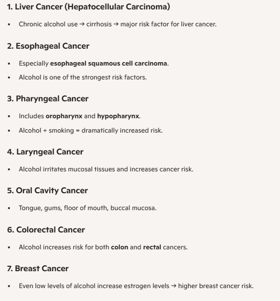
Excessive alcohol consumption has NOT been associated with which type of cancer?
buccal mucosa
liver
breast
lung
Hodgkin's Lymphoma
HD
Also kids get HD and they don’t drink
Mnemonic: BELL COP
Breast
Esophageal
Liver
Larynx
Colon & Rectal (colorectal)
Oral cavity
Pharynx: Oropharynx & Hypopharynx
FYI for smoking related: LOL PB Sandwiches Are Cool
Lung
Oral cavity
Larynx, pharynx, esophagus (LPE)
Pancreatic
Bladder & Kidney
Stomach
AML (adult + child)
Cervical
Any benign epithelial tumor, especially of the skin or mucous membranes is known as a/an:
adenoma
papilloma
lipoma
nevus
papilloma
types of benign tumors
The objective of a staging system are to do all of the following EXCEPT:
assist physicians in treatment planning
assist pathologists in establishing a tissue diagnosis
give an indication of prognosis
assist in evaluation of treatment results
assist pathologists in establishing a tissue diagnosis
Of the following, which tumor is not benign?
chromophobe adenoma
basophilic adenoma
eosinophilic adenoma
pineoblastoma
pineoblastoma
Osteogenic sarcoma usually effects the long bone:
metaphysis
epiphysis
diaphysis
periosteum
metaphysis
the growth plate
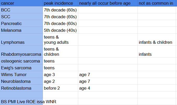
The age at which Wilm's tumor is most likely to occur is _________ years.
1-5
5-10
10-17
18-25
1-5
Extra avg. age for all tumors (onc)
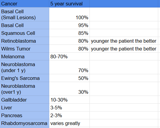
Extra: 5 year survivals for all cancers (onc 1)
BS RW Men
ENGish LP Racist
Hodgkin's lymphomas constitute approximately _% of all malignant lymphomas.
20
40
50
80
20
Reasoning:
The rest are NHL: 80% of all lymphomas
80% NHL
20% HD
Tumors of the upper third of the esophagus constitute about ___ % of esophageal tumors.
20
30
40
50
20
FYI:
The vast majority of esophageal tumors are lower esophagus
L‑M‑U = 60‑30‑10”
How is the esophagus divided
Upper 1/3 (cervical): C6-T2: Cricoid Cartilage - Manubrium
Middle 1/3: T2-T8: Manubrium - Hilum
Lower 1/3 (thoracic): T8-T10(/11): Hilum- GE (gastroesophageal) Junction
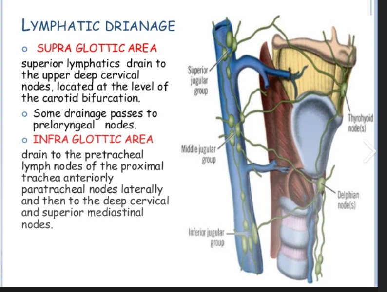
Ca's of the supraglottis mets chiefly to the:
lungs
brain
lymph nodes
bones
lymph nodes
Female most common pathologies:
Ovarian
Endometrium
Vaginal
Cervical
Ovarian- epithelial
Endometrium-adeno
Vaginal- SCC
Cervical- SCC
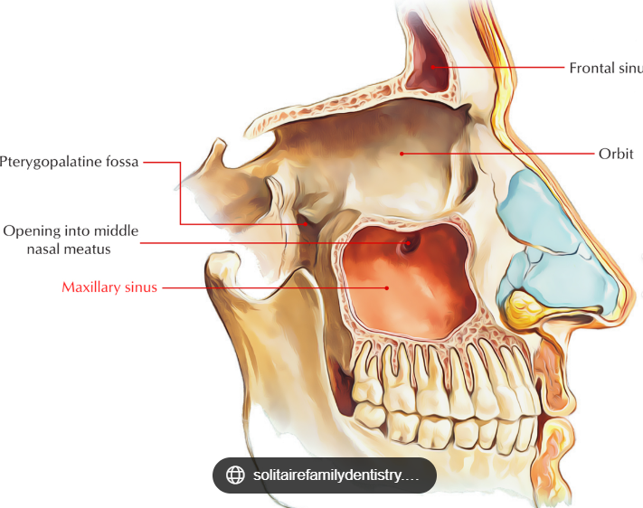
Ca of the maxillary antrum spreads by means of:
local invasion
lymphatic spread
hematologic spread
distant mets
local invasion
Reasoning:
The maxillary sinus has:
Thin bony walls
Very rich local connections to the orbit, nasal cavity, palate, pterygopalatine fossa, and infratemporal fossa
Relatively sparse lymphatic drainage compared to other head & neck sites
Because of this anatomy, the tumor tends to erode bone early and extend directly into nearby structures.
Which type of lung ca is most clearly related to cigarette smoking?
small cell ca
squamous cell ca
adeno ca
large cell ca
squamous cell ca
Hint:
Smoking Squamous
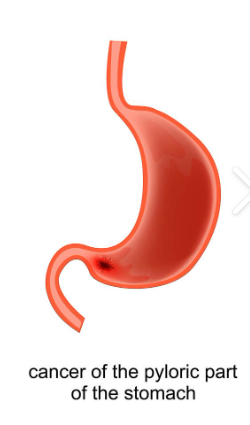
Approximately 3/4 of stomach cancer occurs near the:
pylorus, or on the lesser curvature side of the antrum
pylorus, or on the greater curvature side of the antrum
body of the stomach
fundus of the stomach
pylorus, or on the lesser curvature side of the antrum
Reasoning:
Distal part of lesser curvature (maybe cuz H pylori)
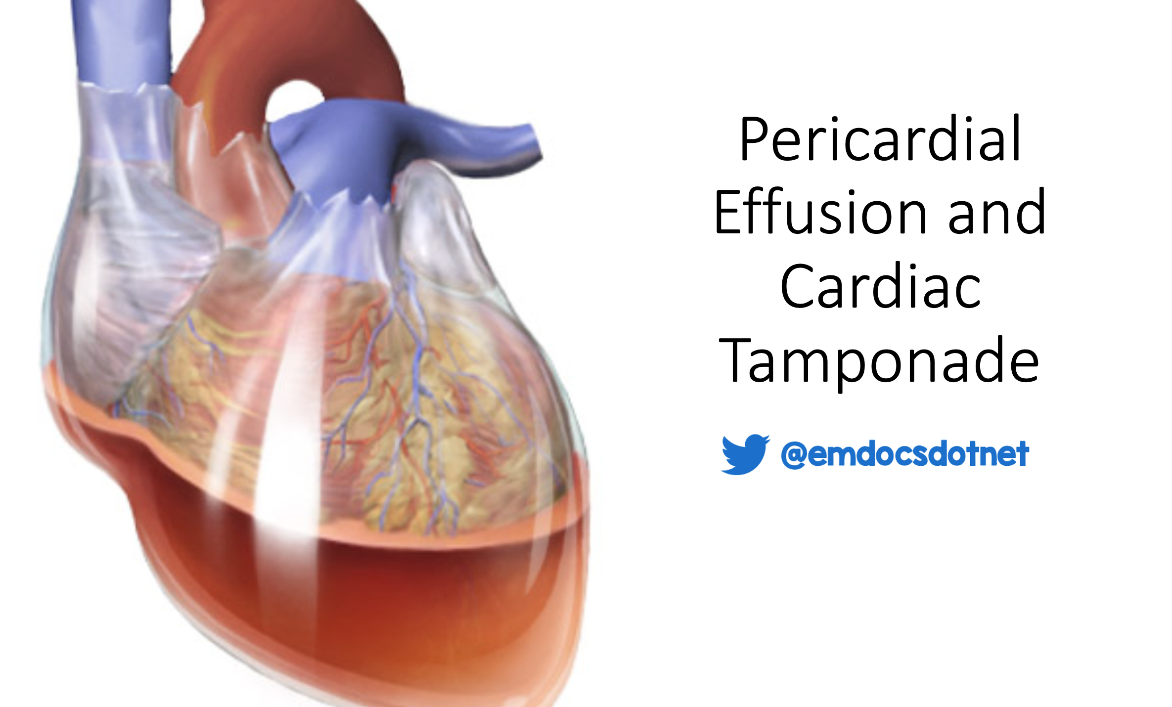
A patient with what type of ca is at risk for developing a cardiac tamponade?
lung
Hodgkin’s Disease
colon
brain metastases
Lung
Reasoning:
lung cancer is the one most classically associated with developing a cardiac tamponade.
The pericardium sits directly next to the left lung and mediastinum.
Certain lung cancers — especially left‑sided, adenocarcinoma, or small‑cell — can:
1. Directly invade the pericardium
Tumor grows into the pericardial sac → fluid accumulates → tamponade.
2. Spread to pericardial lymphatics
Lung cancer commonly metastasizes to mediastinal nodes → blocks lymphatic drainage → pericardial effusion → tamponade.
3. Cause malignant pericardial effusion
Cancer cells seed the pericardium → fluid builds up rapidly.
Malignant testicular tumors originate in the:
germ cells
spermatic cord
glandular tissue
Leydig cells
germ cells
Reasoning: That is why they are called Seminomas
The most common lung ca is:
adeno carcinoma
large cell carcinoma
mesothelioma
oat cell carcinoma
adeno carcinoma
Seminomas are more common in the _______ decade of life.
second
fourth
seventh
eighth
fourth
Men in their 30s-40s
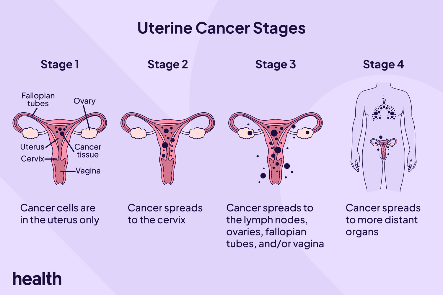
Ca of the uterus usually spreads by:
local invasion
hematologic
lymphatic
coelomic
local invasion
Reasoning:
Probably why it has a really good prognosis
Lesions of the piriform fossa show early spread to the __________ lymph nodes.
mediastinal
infradiaphragmatic
pelvic
cervical
cervical
Piriform fossa is #1 cancer of the hypopharynx
The peak incidence of breast ca occurs at about age:
20
30
65
90
65
reasoning: 65-70
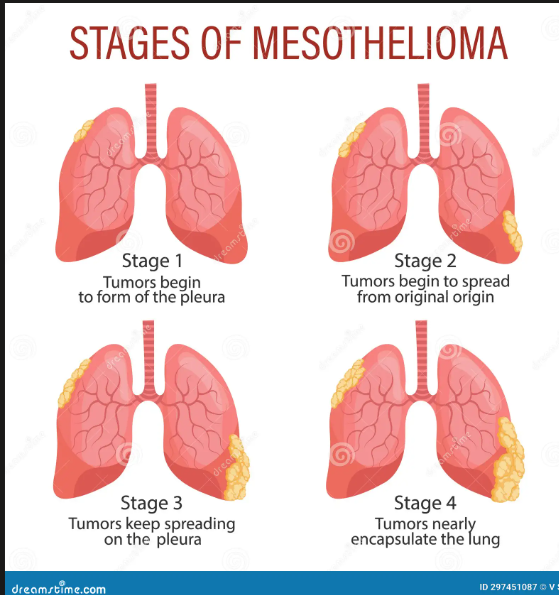
Mesotheliomas are tumors of the:
pleura
brain
bronchi
lymph nodes
pleura
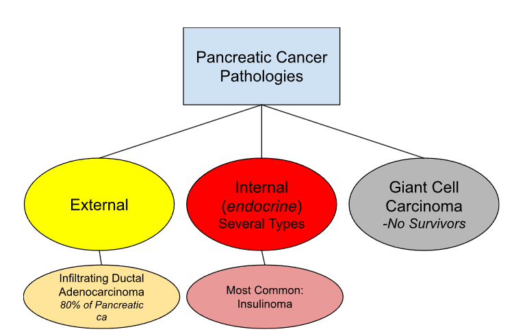
The point of origin of ca of the pancreas is usually the:
pancreatic duct
head of the pancreas
tail of the pancreas
Islets of Langerhans
head of the pancreas
reasoning:
point of origin: most common cancer location in pancreas
Pancreas spinal level
L1-L2
pancreas pathologies
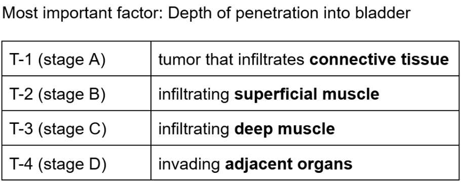
Which of the following statements about bladder ca is incorrect?
it is among the top 5 most common ca in men
it shows an increased incidence in analine dye workers
there is a strong relationship with smoking
cure rates are generally 85% or greater
cure rates are generally 85% or greater
reasoning:
Bladder cancer overall 5‑year survival ≈ 75–80%
Muscle‑invasive disease has much worse survival than superficial disease
Bladder cancer staging
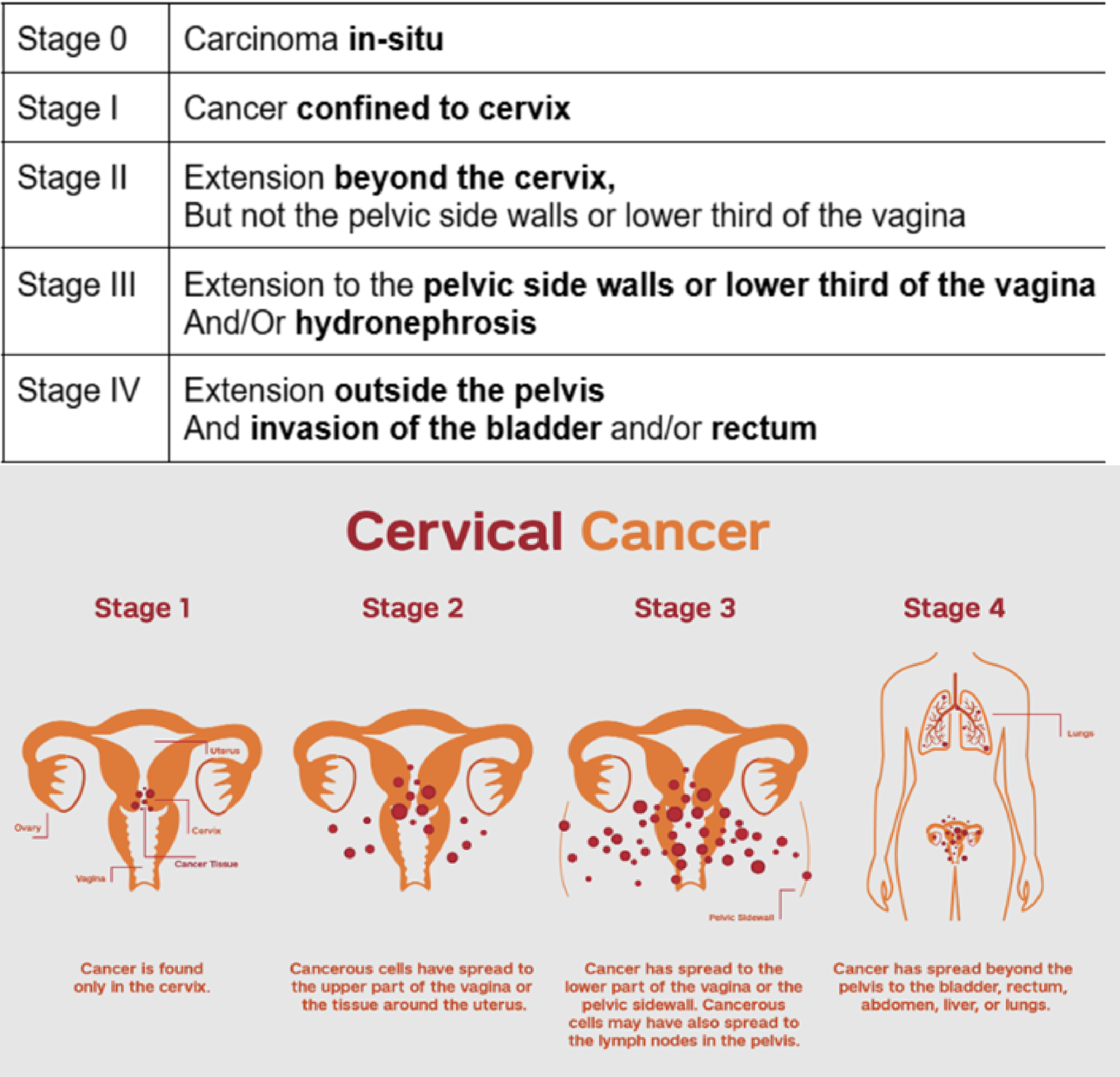
According to the FIGO system, a patient with a lesion that extends into the lower vagina is classified as stage:
I
II
III
IV
III
Cervical cancer staging
List all the pap smear classifications and what disorder or other bit of information they represent:

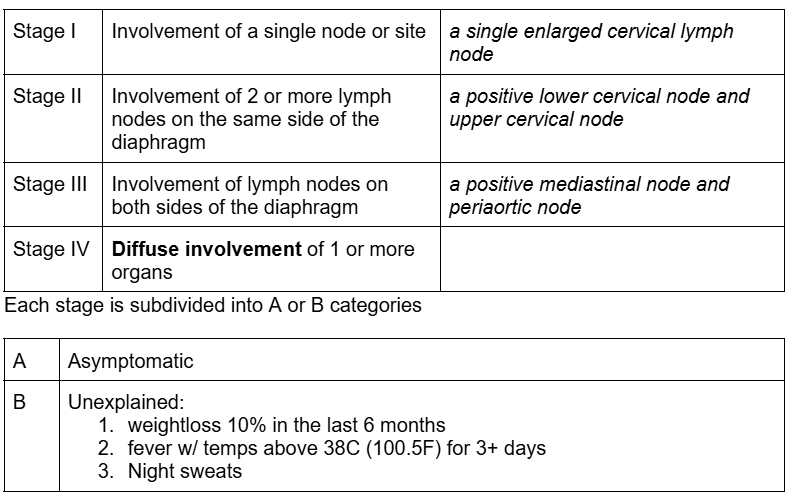
Ann Arbor Staging System
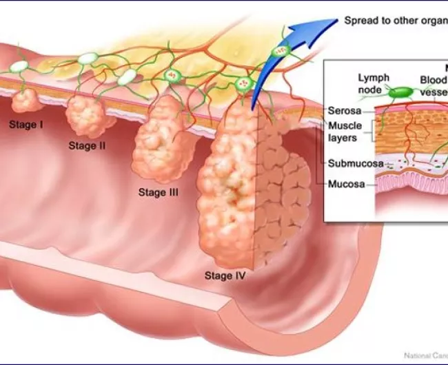
4 stages of colorectal cancer TNM staging system
Stage I (or A): Invasion into the submucosa
Stage II (or B): Invasion into the serosa
Stage III (or C): Invasion into the serosa w/ + nodes
Stage IV (or D): Any invasion w/ distant mets
Laryngeal cancer staging for Glottis “true vocal cord” tx
T1 Confined to cord + normal cord mobility
T2 Supra- or sub-glottic extension + normal or impaired cord mobility
T3 Confined to larynx + fixed vocal cords
T4 Extension beyond larynx and/or Cartilage destruction
Breast cancer staging
Tis- carcinoma in-situ
T1- tumor 2cm or less
T1a - no fixation to underlying fascia
T1b - fixation to underlying fascia
T2 - tumor greater than 2 cm, but less than 5 cm (2-5cm)
T3 - tumor greater than 5 cm
T4 - tumor of any size with direct extension to the chest wall or skin
Rai staging system for CLL
Stage 0: lymphocytosis only (excessive # of lymphocytes)
Stage 1: Stage 0 + Lymph Node Enlargement
Stage 2: Stage 1 + Splenic Involvement
Stage 3: Stage 2 + Anemia
Stage 4: Stage 3 + Thrombocytopenia
FYI: lymphocytosis is high # of lymphocytes in blood
With vaginal and cervical cancers, there is an increased risk of clear cell adenocarcinoma and abnormalities o the stratified epithelium in women whose mothers (during pregnancy) use which of the following drugs:
Diethlstilbestrol (DES)
Dihydrotestosterone (DEET)
Dysmenorrheal (DMH)
Deoxyribonucleic acid (DNA)
Diethlstilbestrol (DES)
The most common histologic type for small bowel cancer is:
Squamous cell
Hepatic sarcoma
Adenocarcinoma
Transitional cell carcinoma
Adenocarcinoma
The most common histologic type for cancer of the stomach is:
Squamous cell
Adenocarcinoma
Lymphoma
Transitional cell carcinoma
Adenocarcinoma
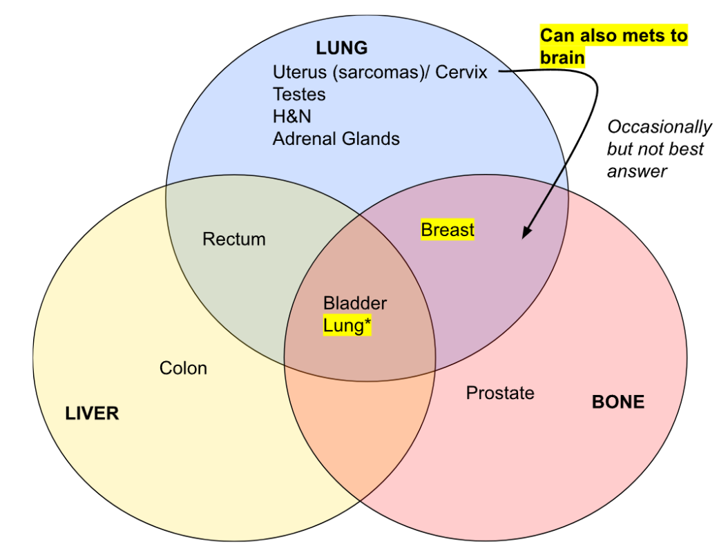
Common sites of metastases for colon cancer are: 1. Liver; 2. Lung; 3. Peritoneum; 4. Bone
1,3
2,3
2,3,4
1,2,3
2,3,4
Reasoning:
BUT REALLY IT SHOULD BE 4
In Hodgkin disease, involvement of several nodal regions of both sides of the diaphragm accompanied by localized involvement of an extralymphatic site is stage:
II
II E
III
III E
III E
In the treatment of Waldeyer ring for nonHodgkin lymphoma, the fields’ delineation closely resembles that of carcinoma of the:
Supraclavicular fossa nodes
Orbit
Pelvis
Nasopharynx
Nasopharynx
A young woman has swelling in her lower neck for suspected HD. A chest x-ray also revealed mediastinal adenopathy. She had not experienced any fever, night sweats, or weight loss. Staging would be:
I A
II A
II B
III A
II A