Lab 8: Special Senses
1/63
There's no tags or description
Looks like no tags are added yet.
Name | Mastery | Learn | Test | Matching | Spaced | Call with Kai |
|---|
No analytics yet
Send a link to your students to track their progress
64 Terms
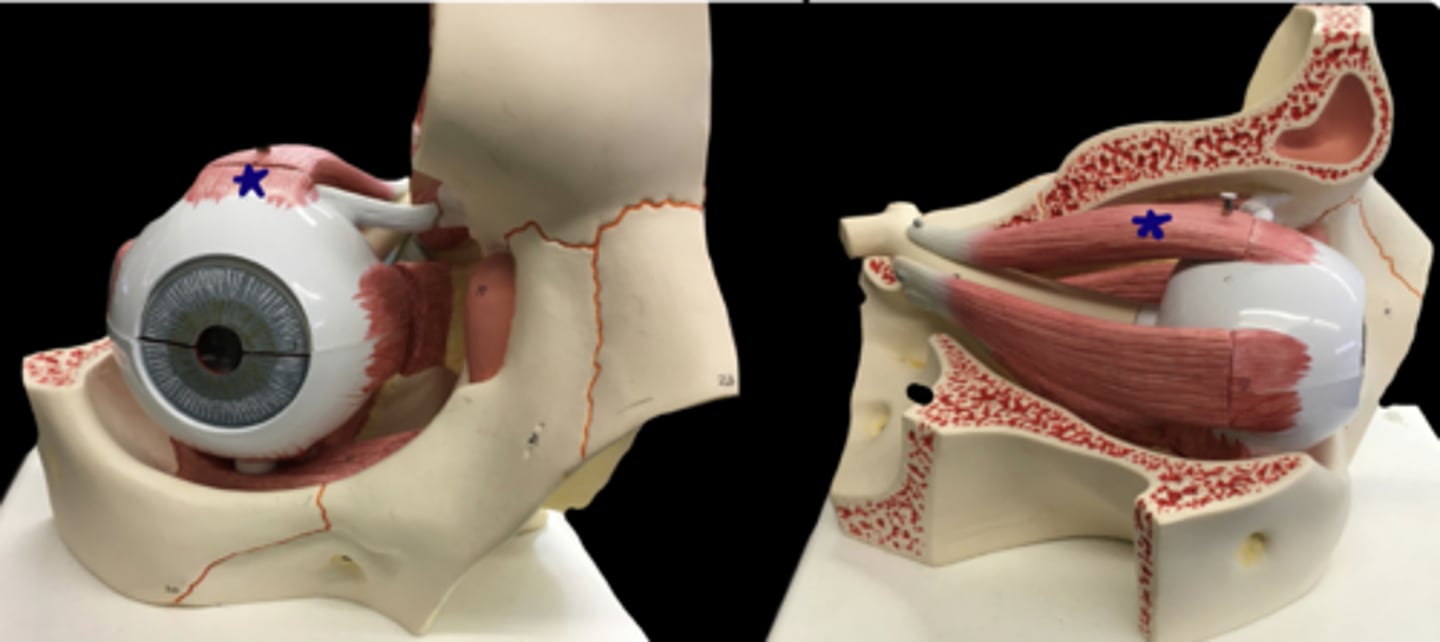
superior rectus muscle
top of the eye muscle
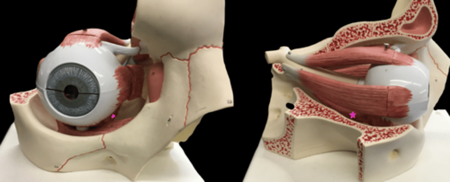
inferior rectus muscle
bottom of eye muscle
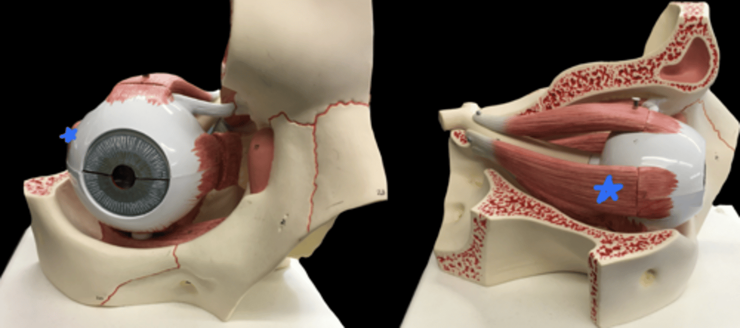
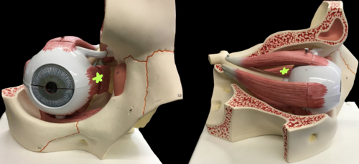
lateral rectus muscle
lateral side eye muscle
medial rectus muscle
medial side eye muscle
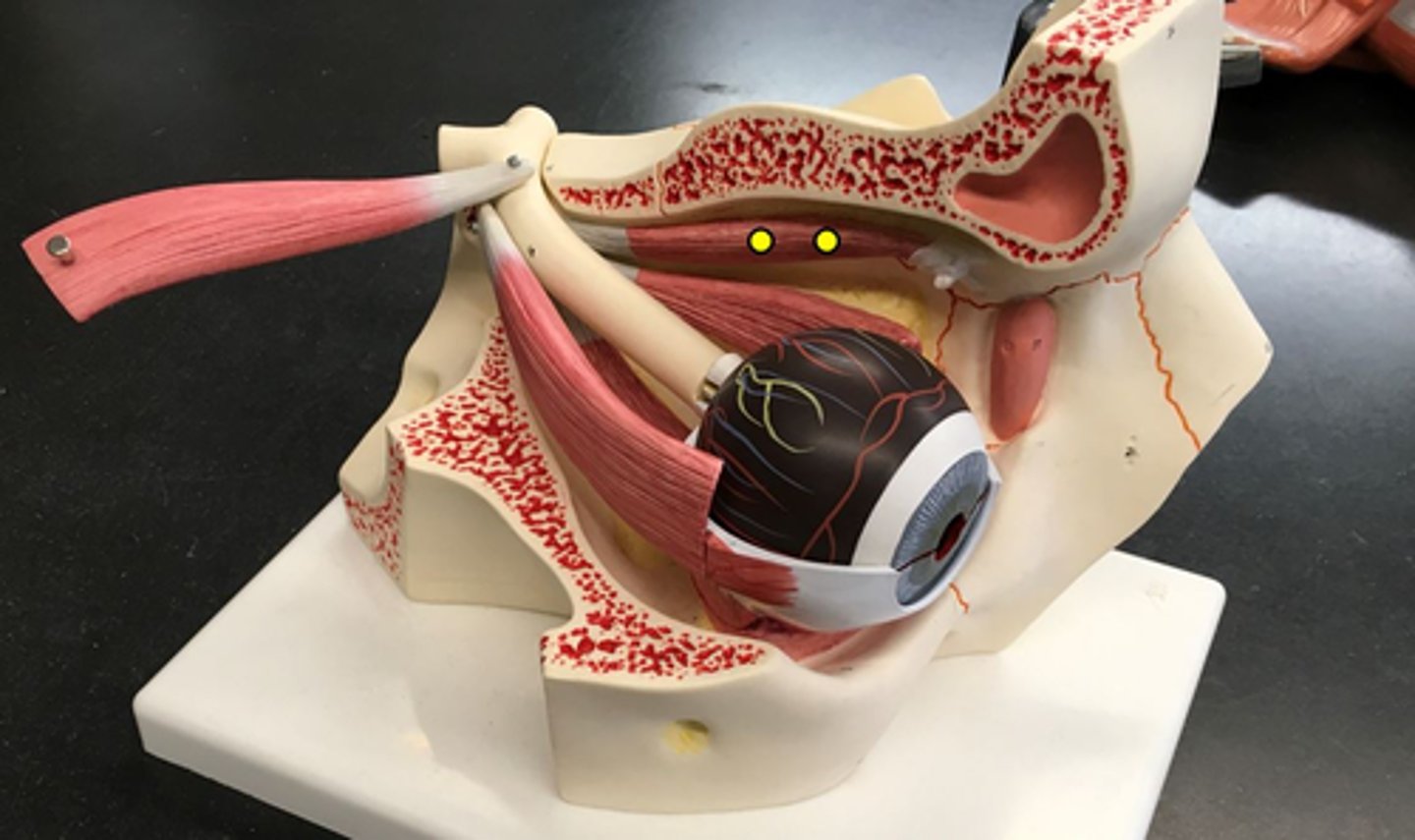
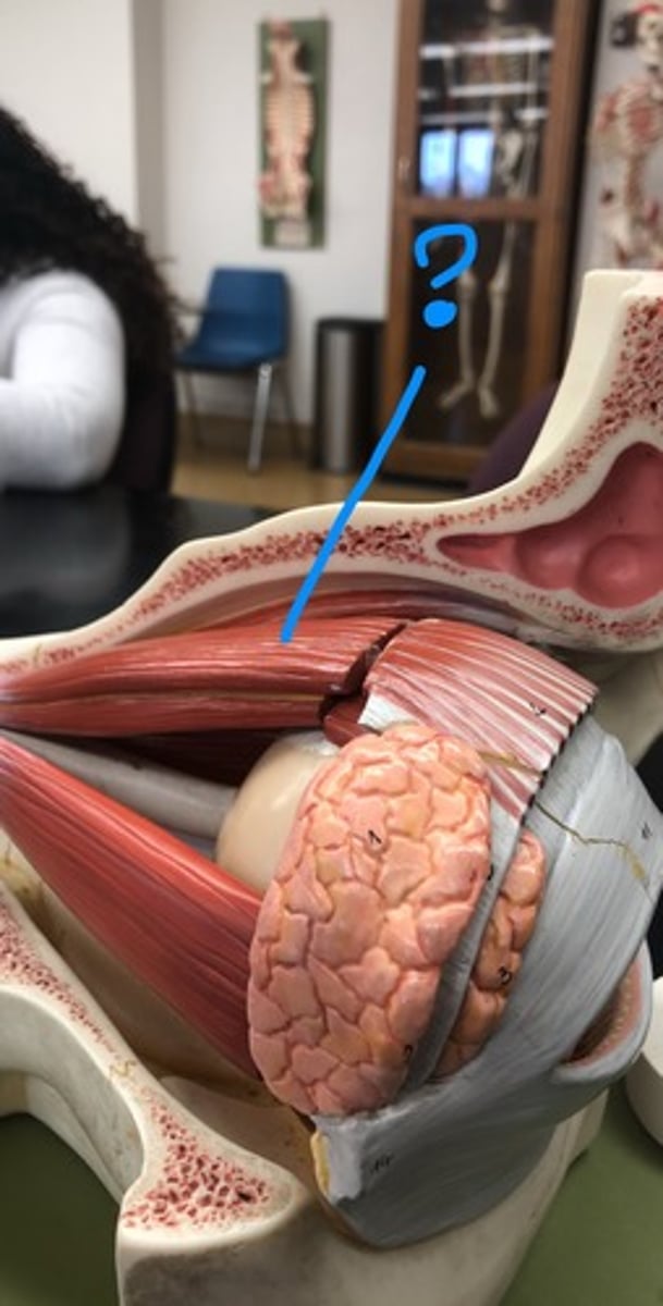
superior oblique muscle
Muscle that is more medial and connects to the trochlea of the eye
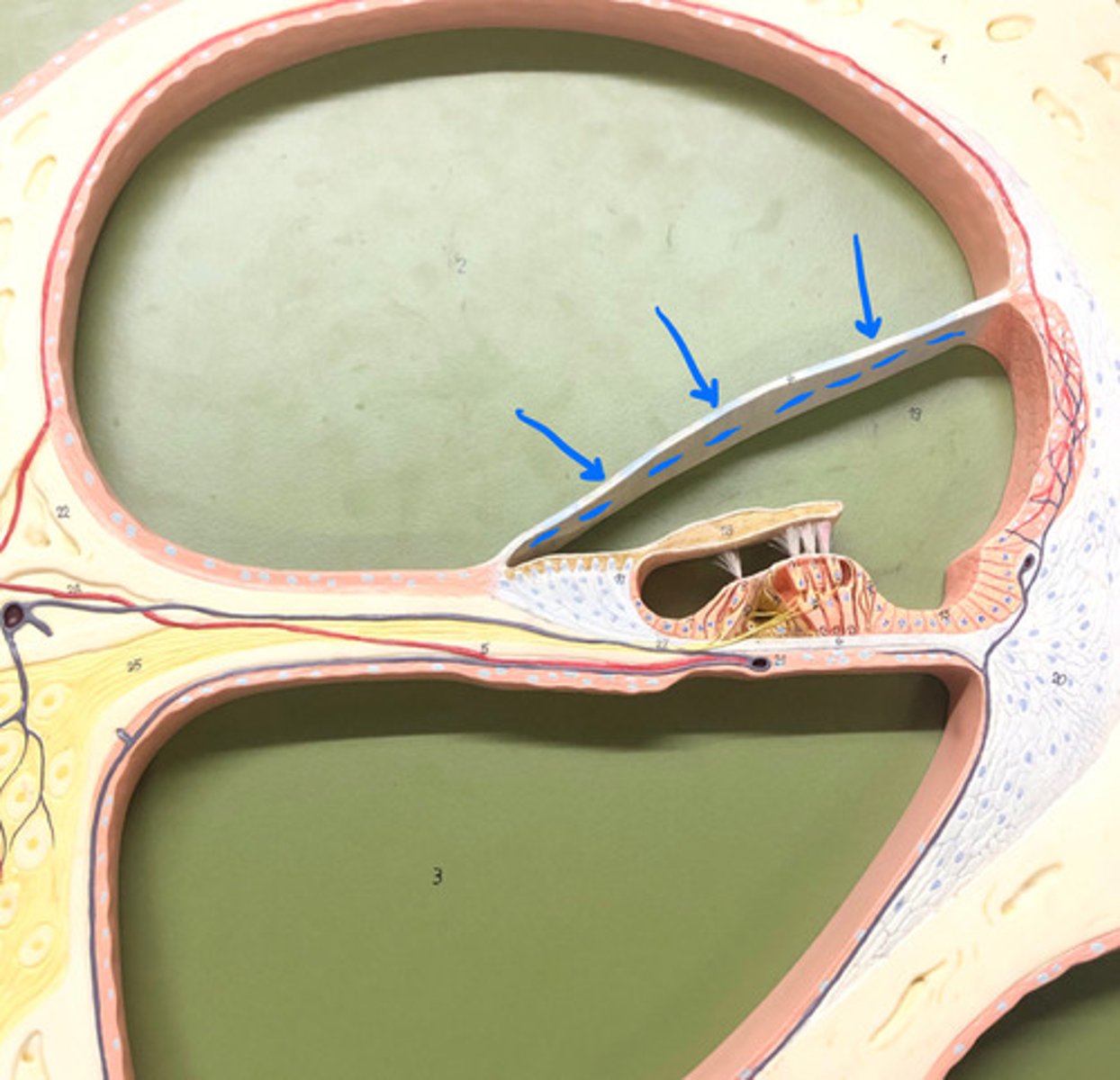
trochlea of eye
ligamentous strap
- blue
levator palpebrae superioris
connects to eyelid
inferior oblique muscle
goes around the bottom of the eye
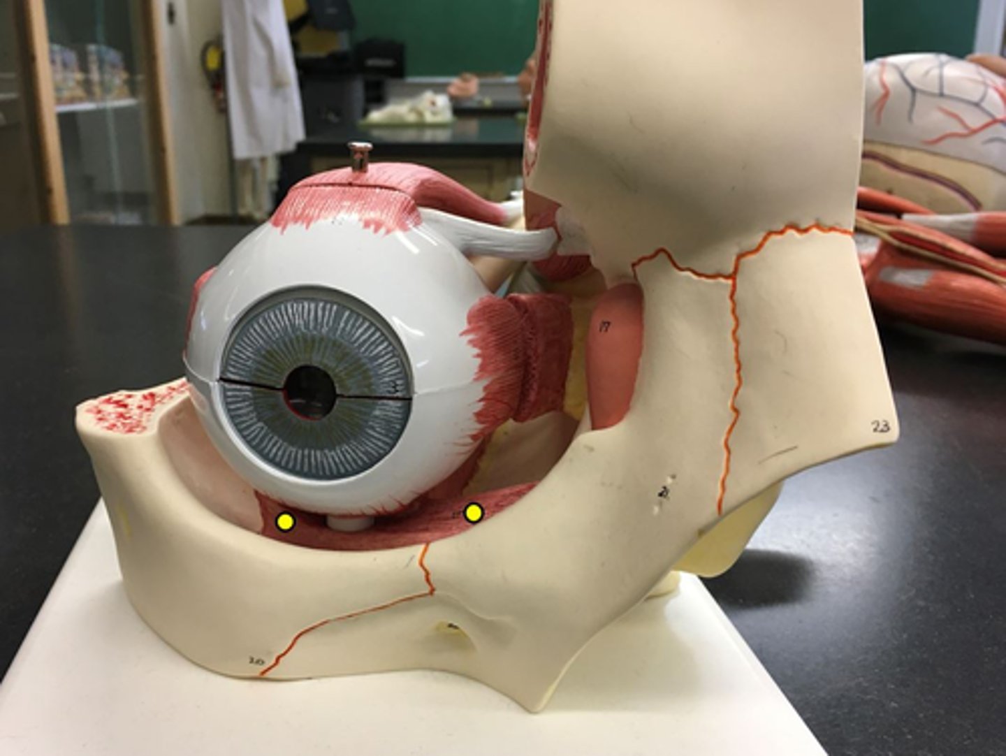
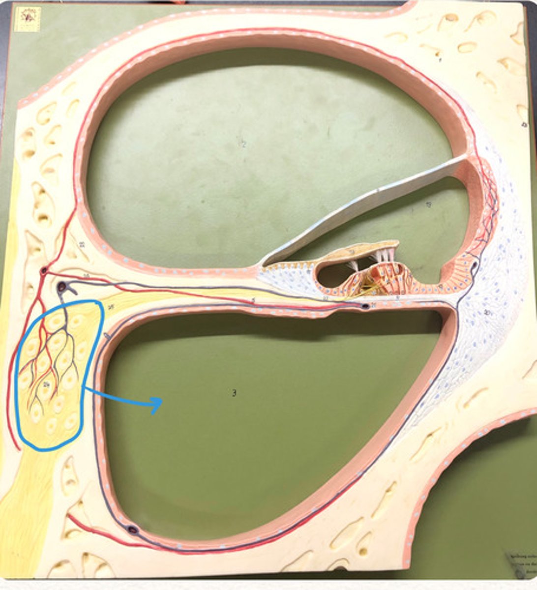
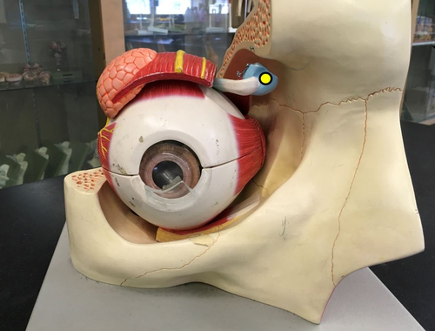
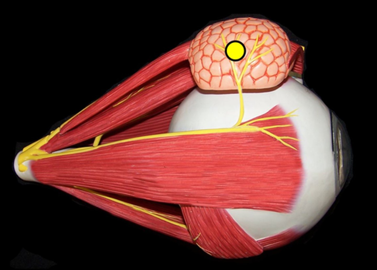
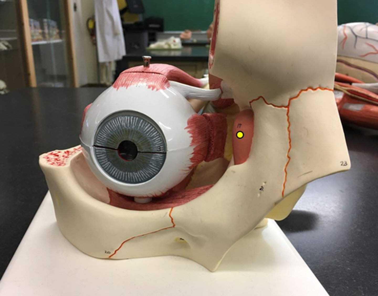
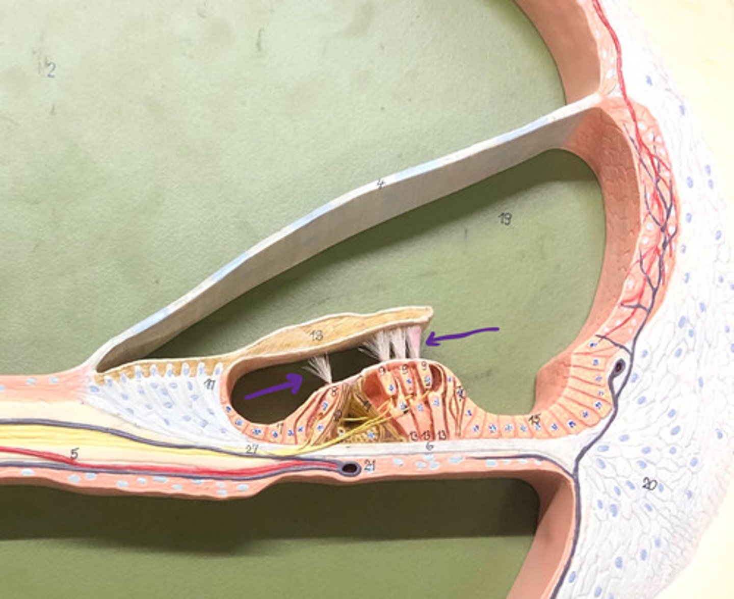
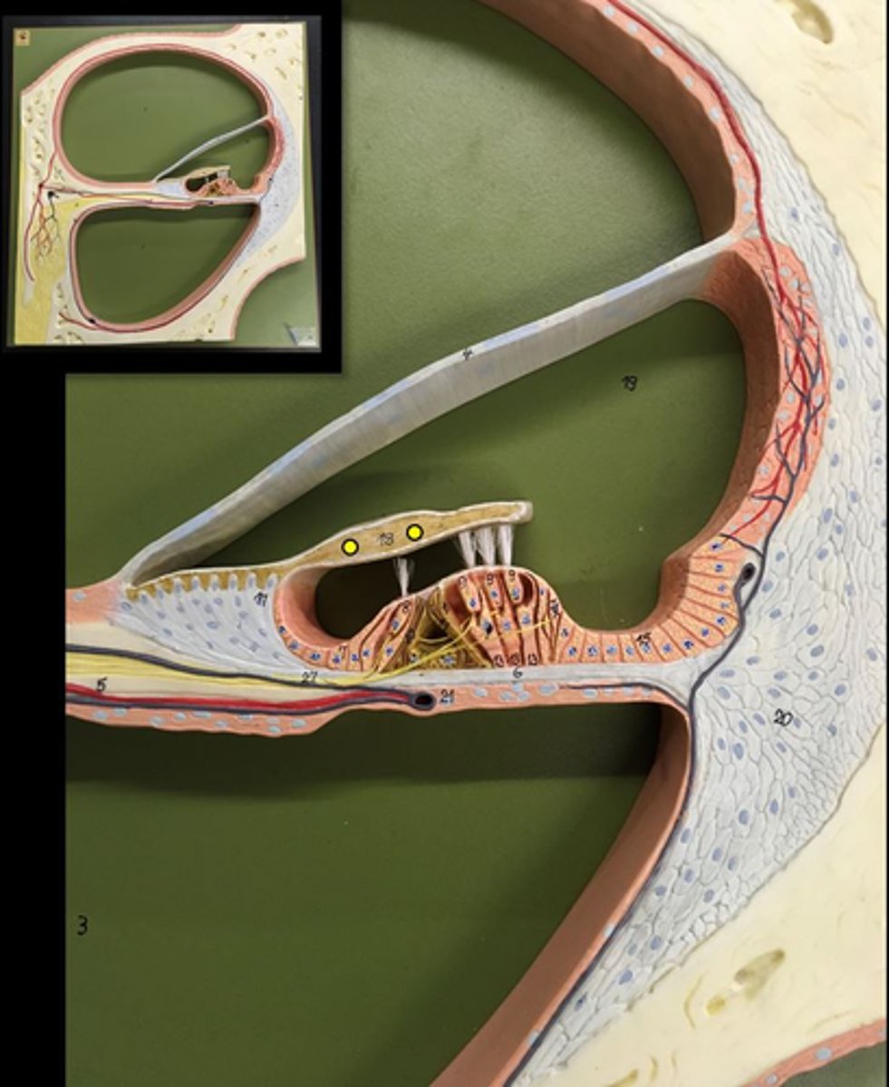
lacrimal gland
Gland that sits on the top off the eye laterally
lacrimal sac
an enlargement of the upper portion of the lacrimal duct
nasolacrimal duct
Name this passageway
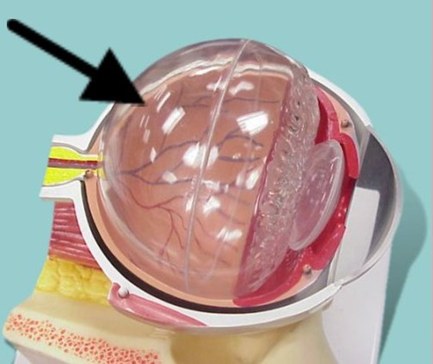
vitreous chamber
The big eye "hole"
- the "space"
vitreous body
The clear ball inside the eye model
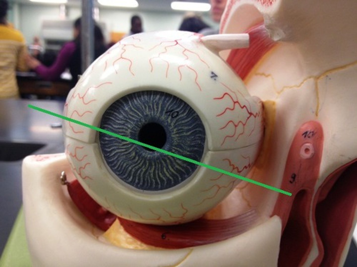
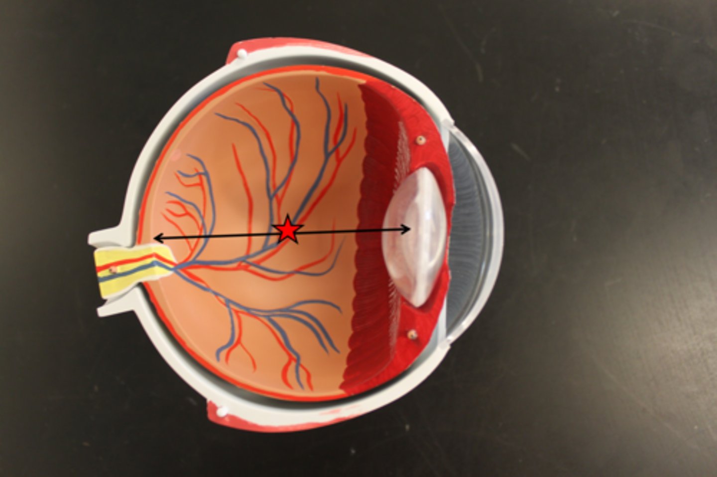
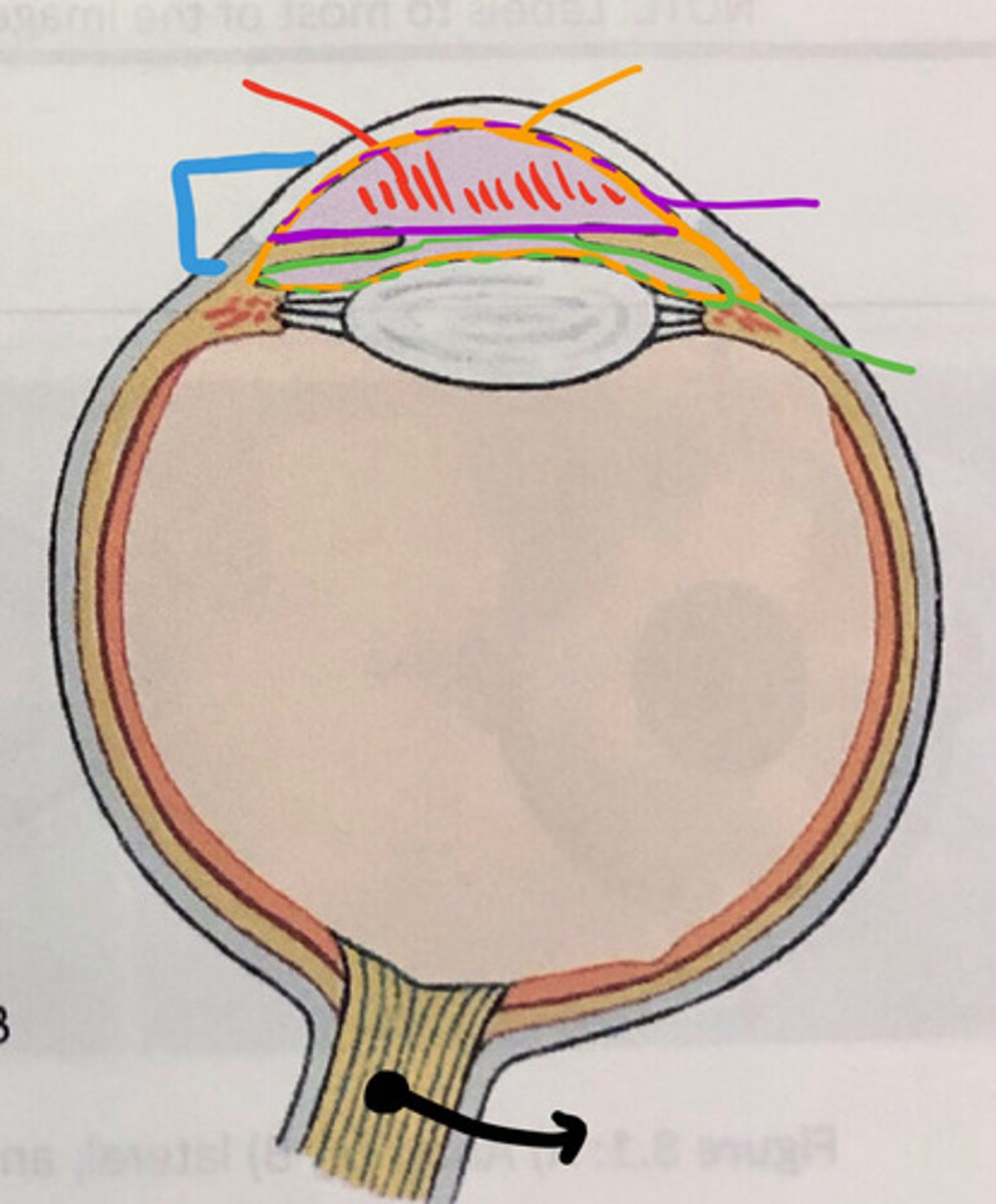
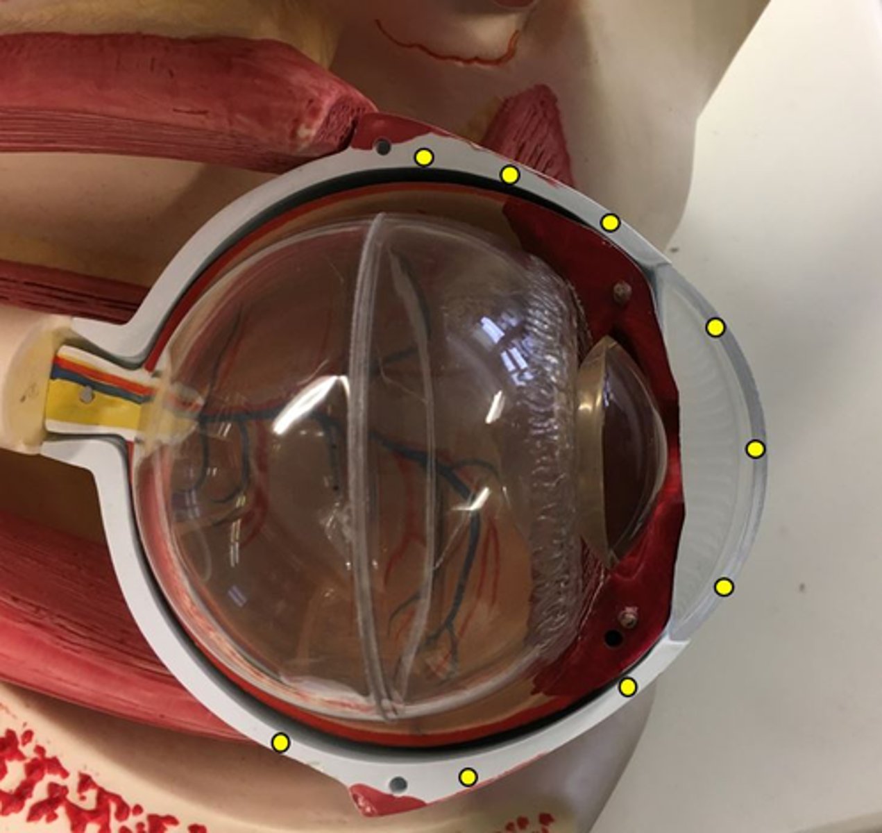
anterior cavity
From the cornea to the lens
- Orange lines
* Name this REGION*
aqueous humor
FLUID in the eye, found between the cornea and the lens
anterior chamber
from the cornea to the iris
-Purple line
Name this space

posterior chamber
from the iris to the lens
-green line
Name this space

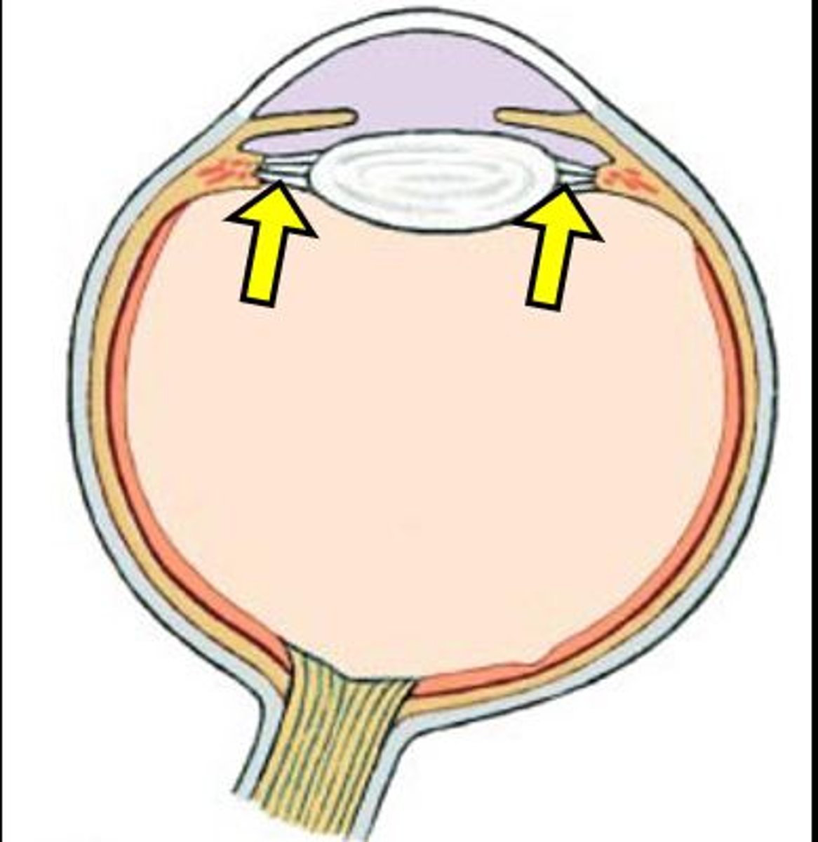
lens
clear "football" in the eye
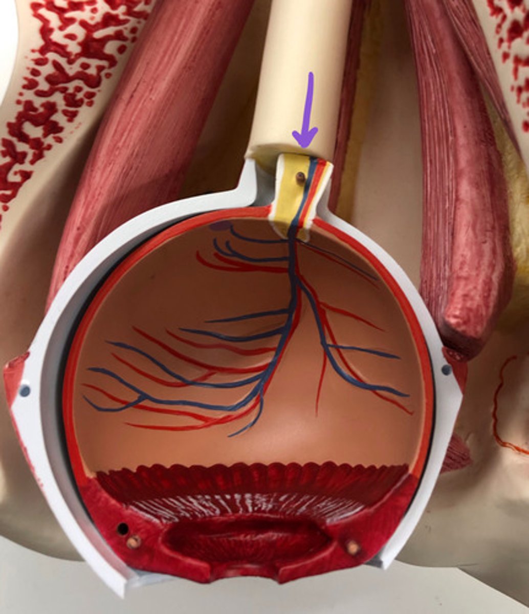
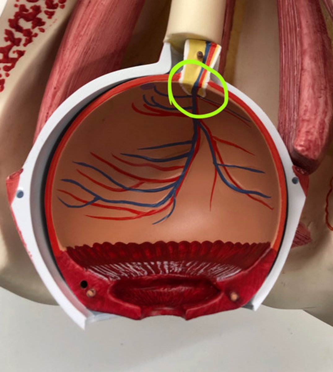
optic nerve II
nerve that goes to the eye
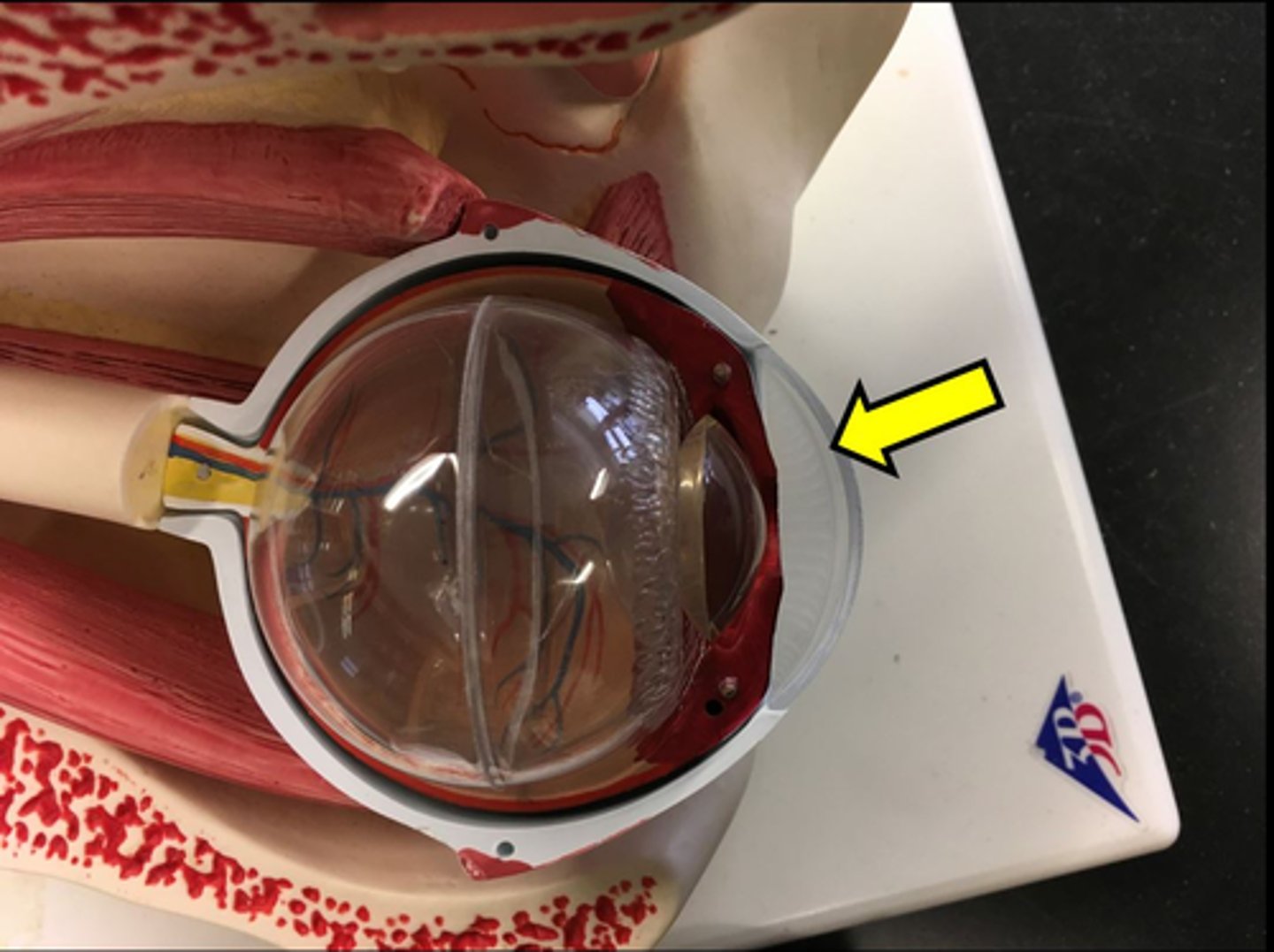
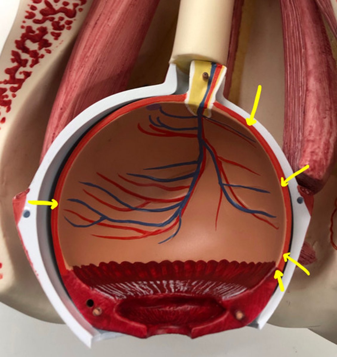
fibrous tunic
the cornea and sclera together
Name this layer
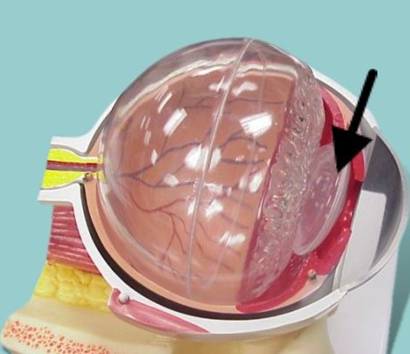
uvea (vascular tunic)
iris, ciliary body, choroid
- lies beneath the sclera
Name this layer
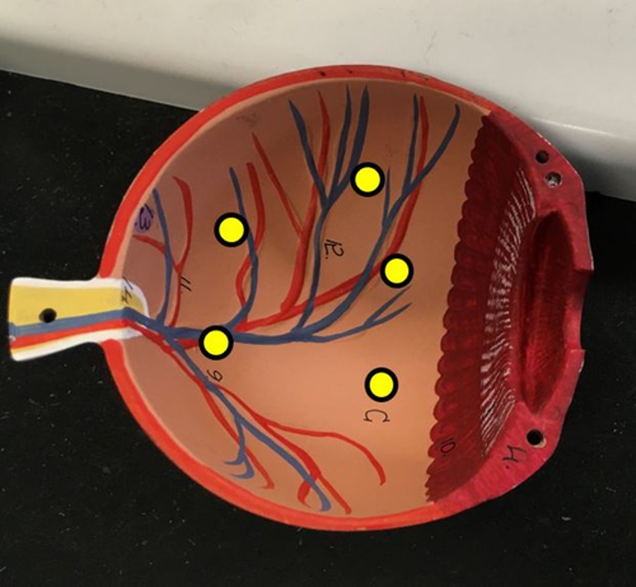
retina
inner most layer of the eye
- orange with blood vessels
Name this layer
sclera
the white of the eye
cornea
clear part in the front of the eye
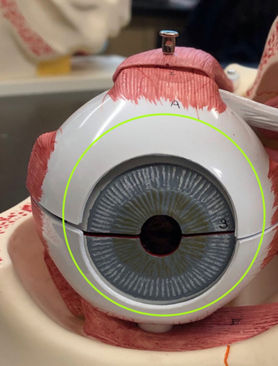
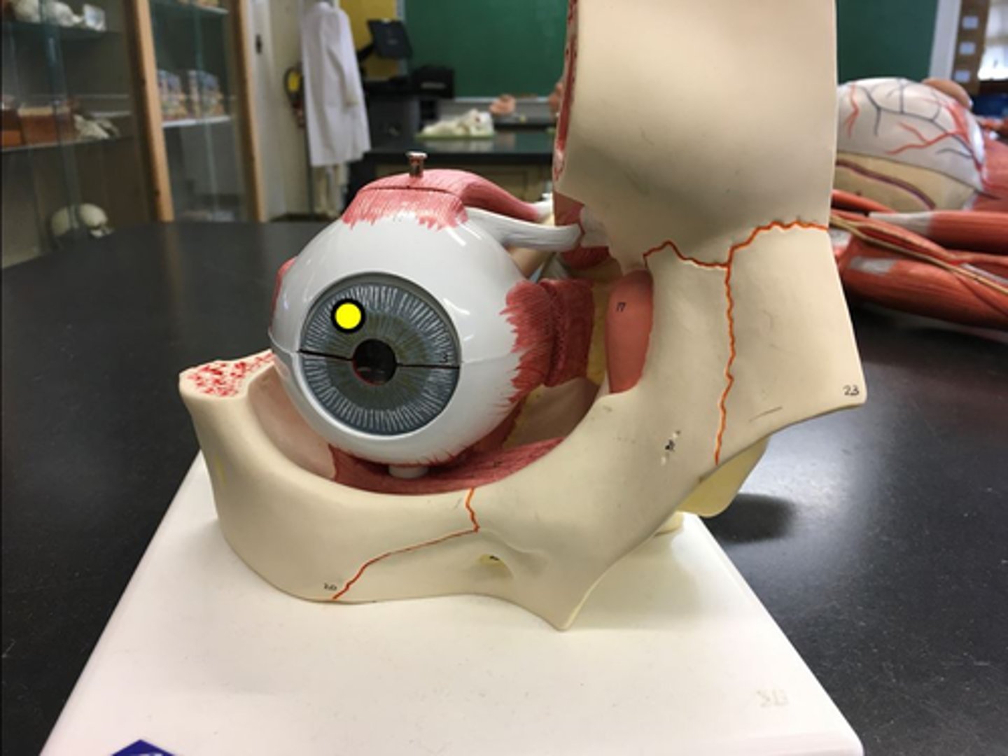
iris
colored part of the eye
pupillary constrictor muscles
More on the interior of the iris
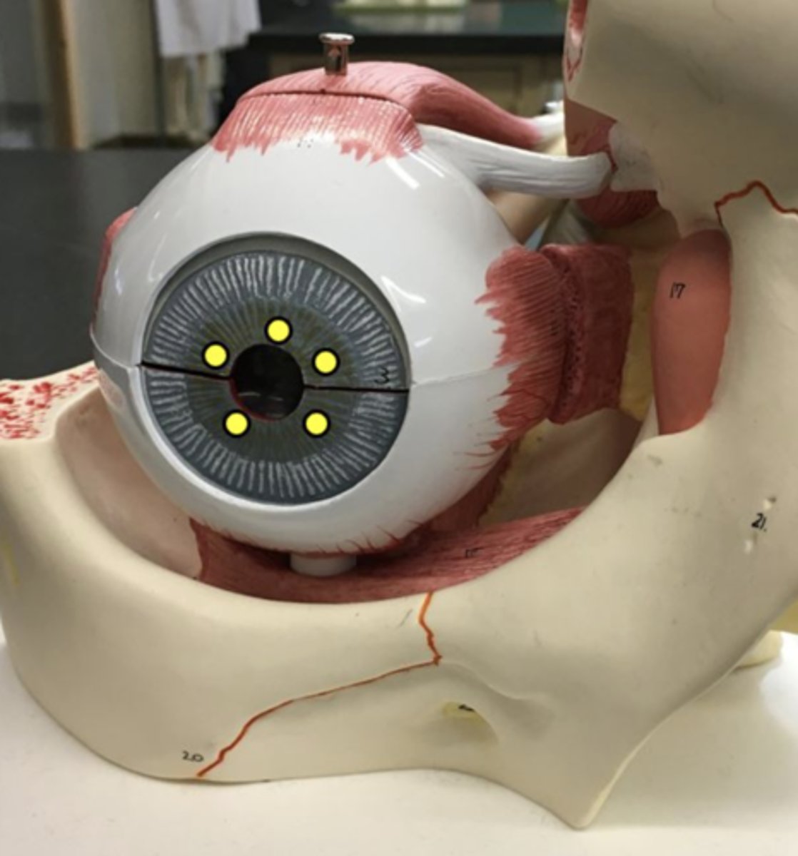
pupillary dilator muscles
Muscles on the outside of the iris
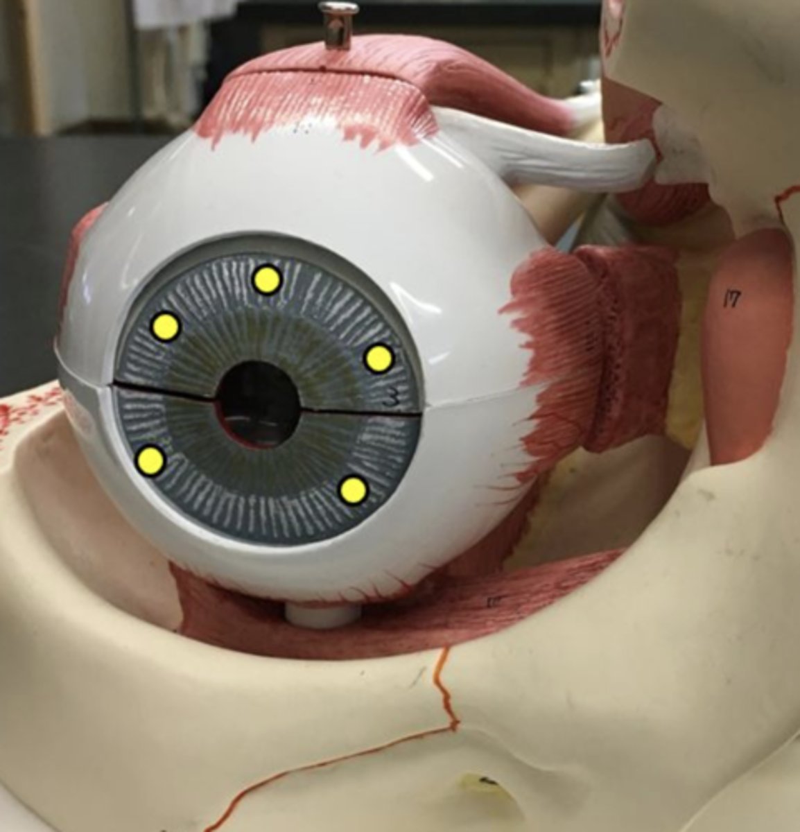
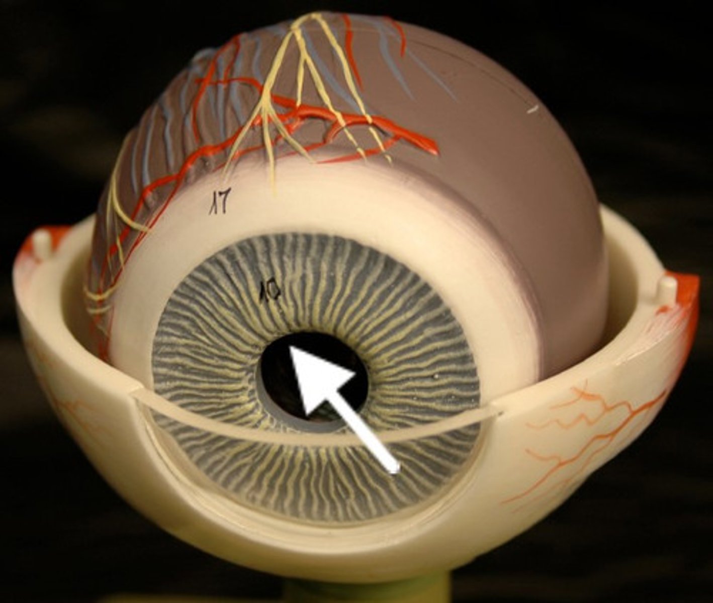
pupil
opening in the eye
Name this space
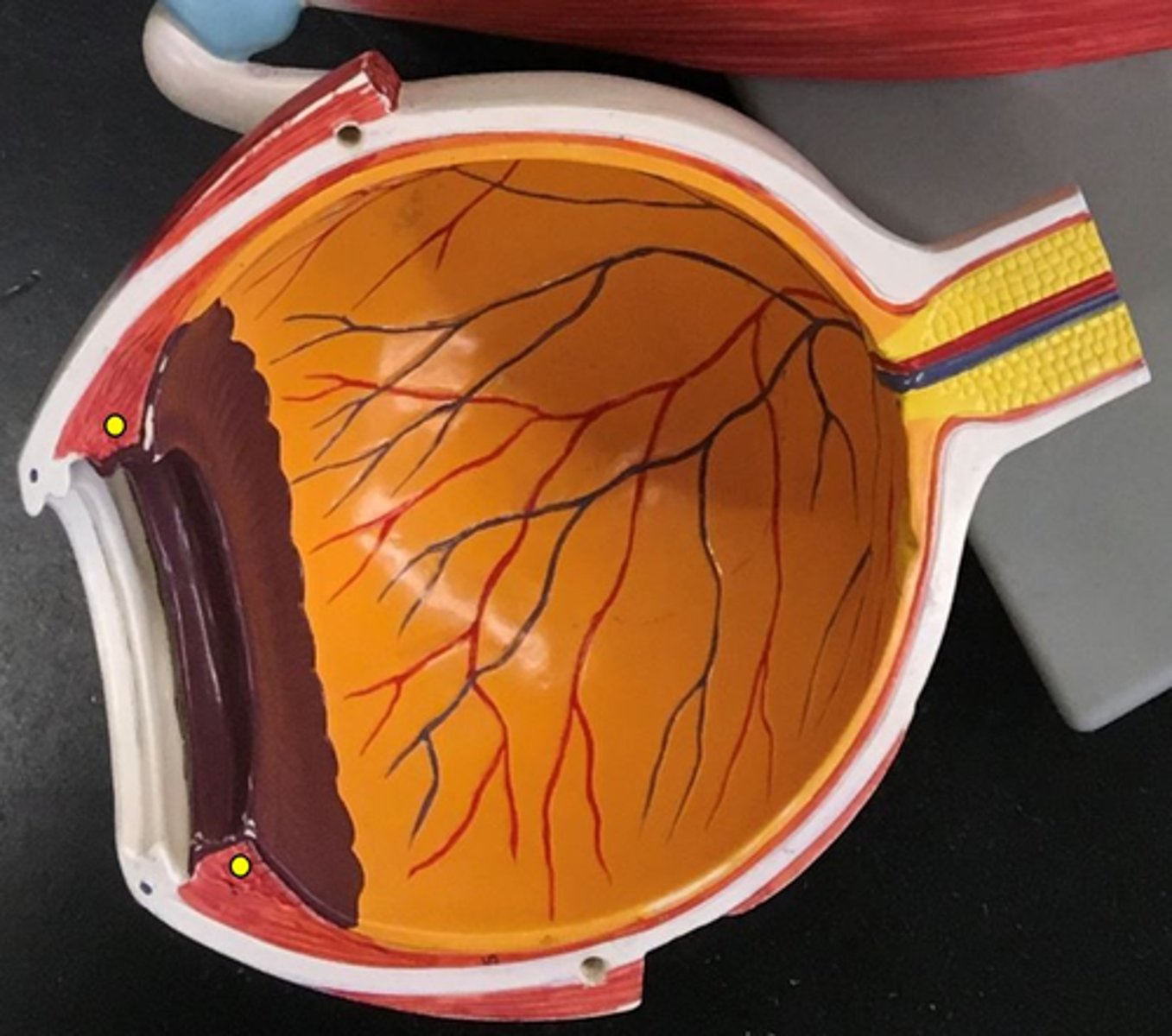
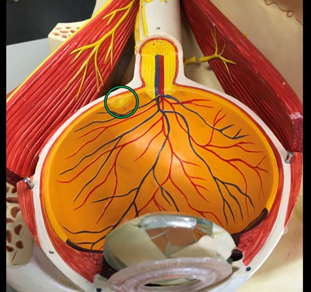
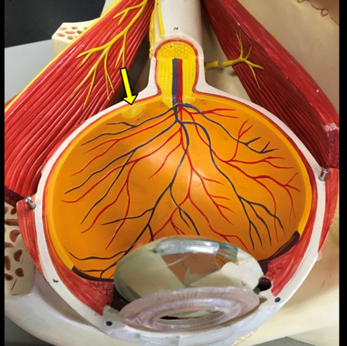
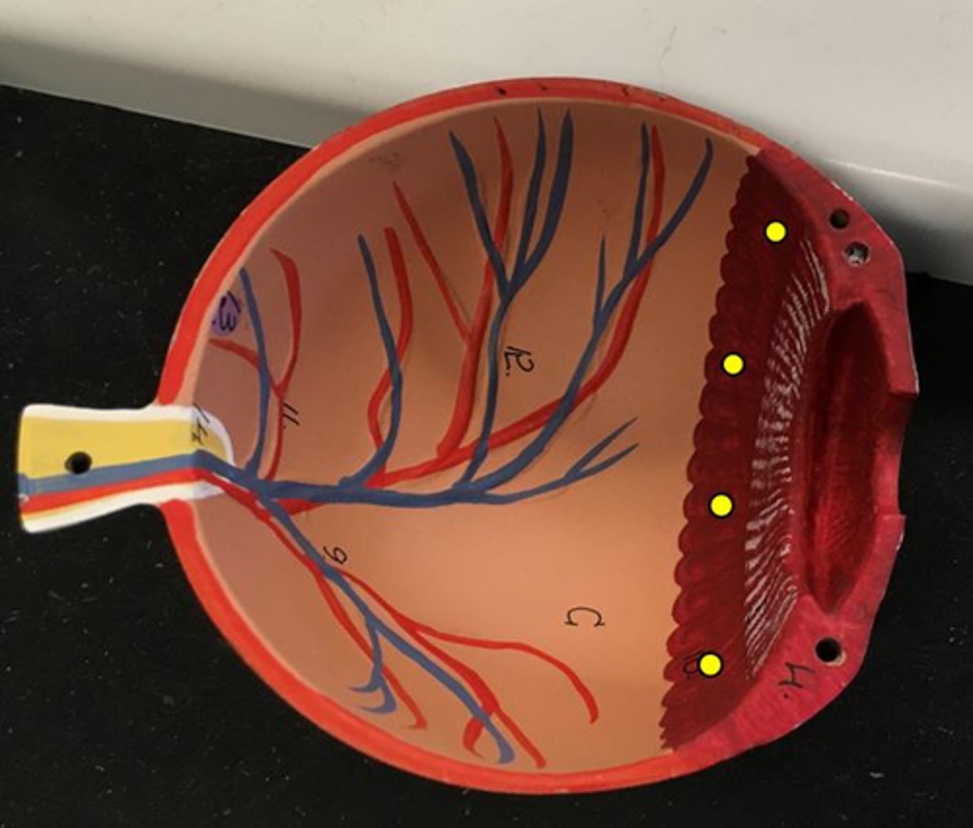
ciliary body
- Red dots that are attached to the iris
ciliary zonule
Ligaments that connect the lens and the iris
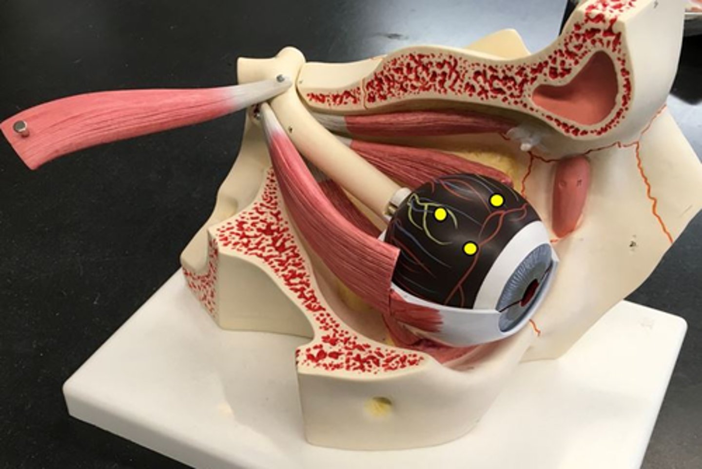
choroid
second layer around the eye -Red on model
connects to the iris
yellow arrows
macula lutea
blind spot in the eye
- purple on some models
fovea
indentation within the macula lutea
yellow arrow
Name this depression
optic disc
where the optic nerve comes into the eye
ora serrata
"flower in the eye"
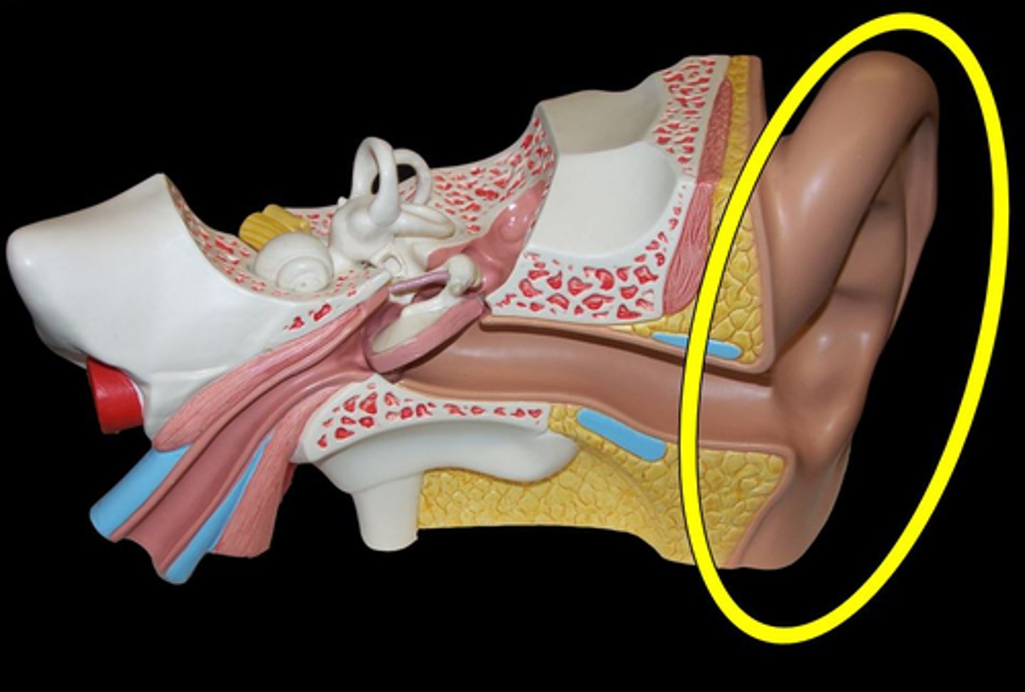
pinna
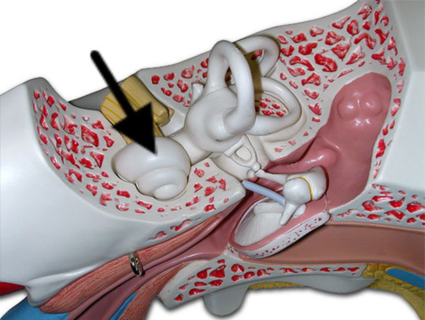
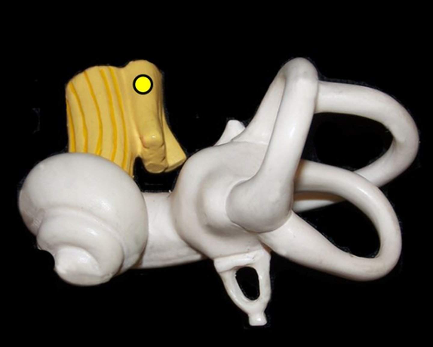
Outermost portion of the ear.
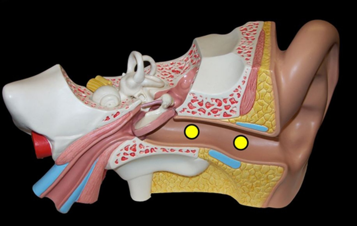
external acoustic meatus
Name this passageway
tympanic membrane
blue arrow
-removable
"ear drum"
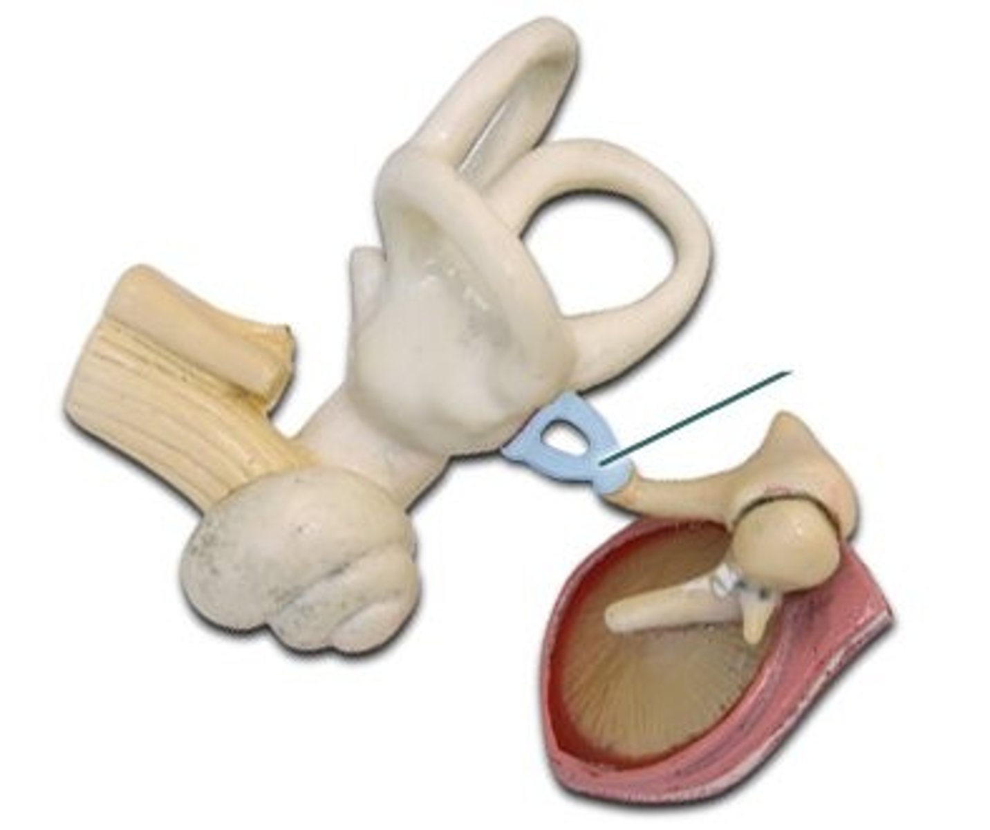
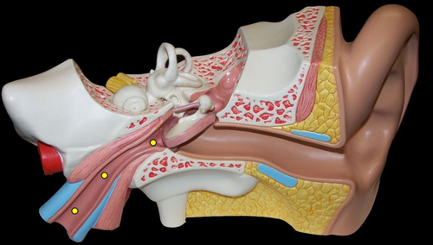
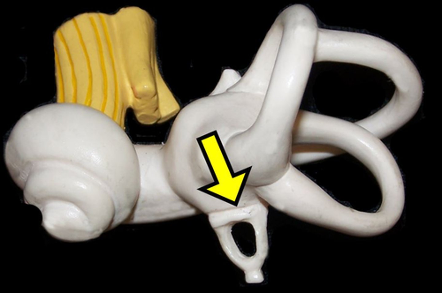
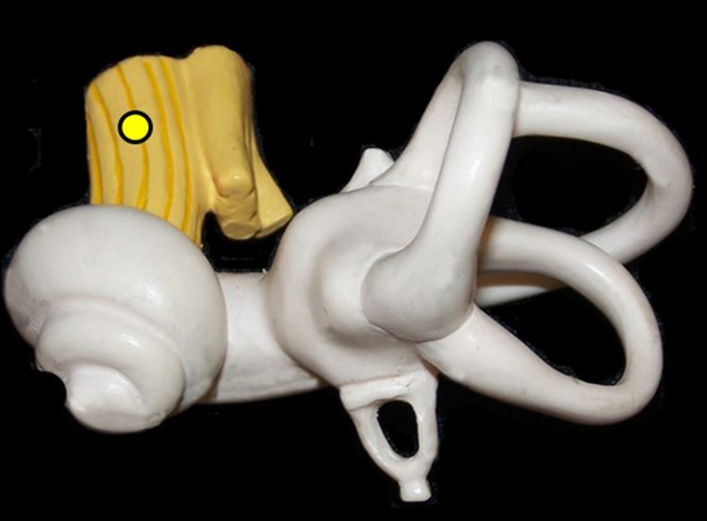
auditory ossicles
1, 2 & 3
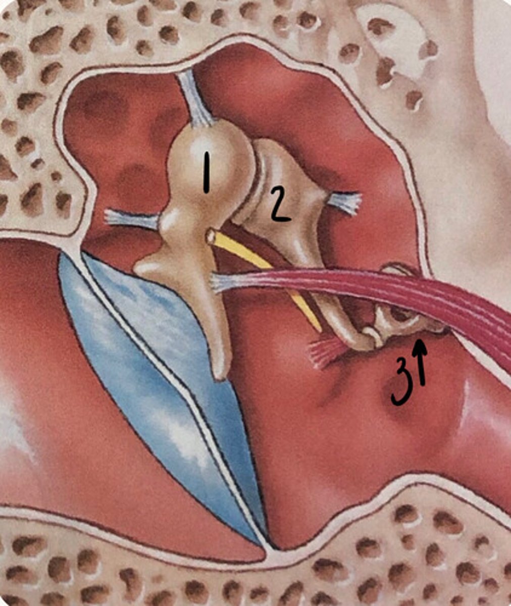
malleus
1
attached to the tympanic membrane
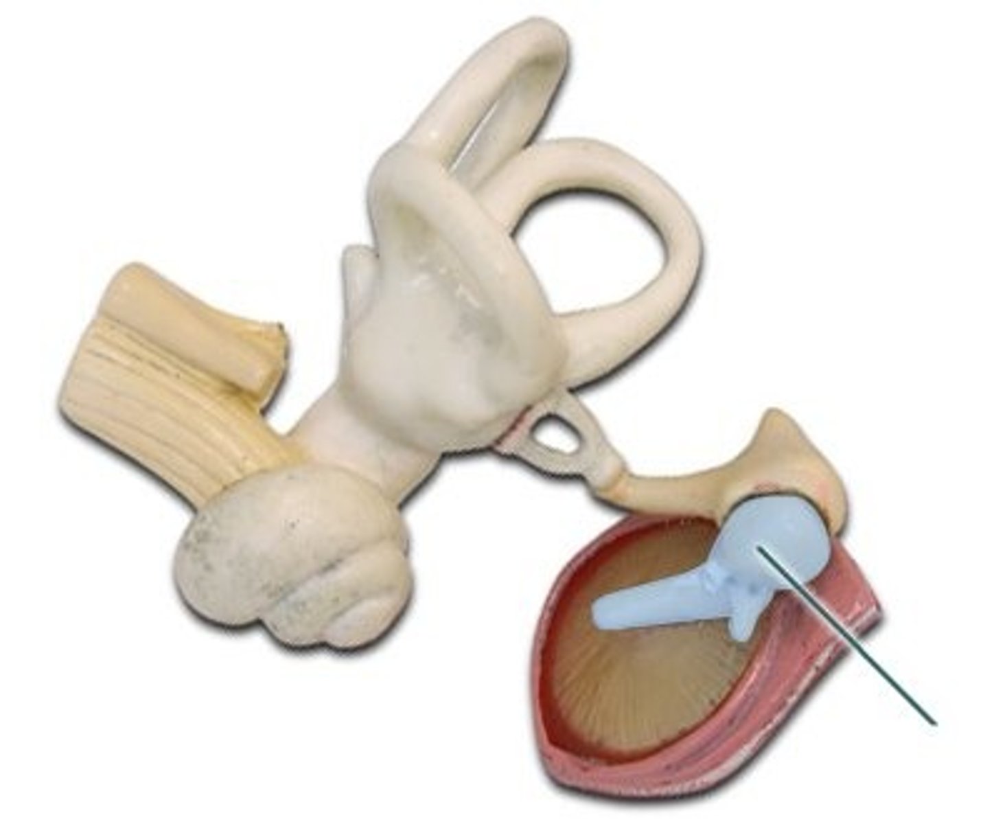
incus
second ossicle of the middle ear
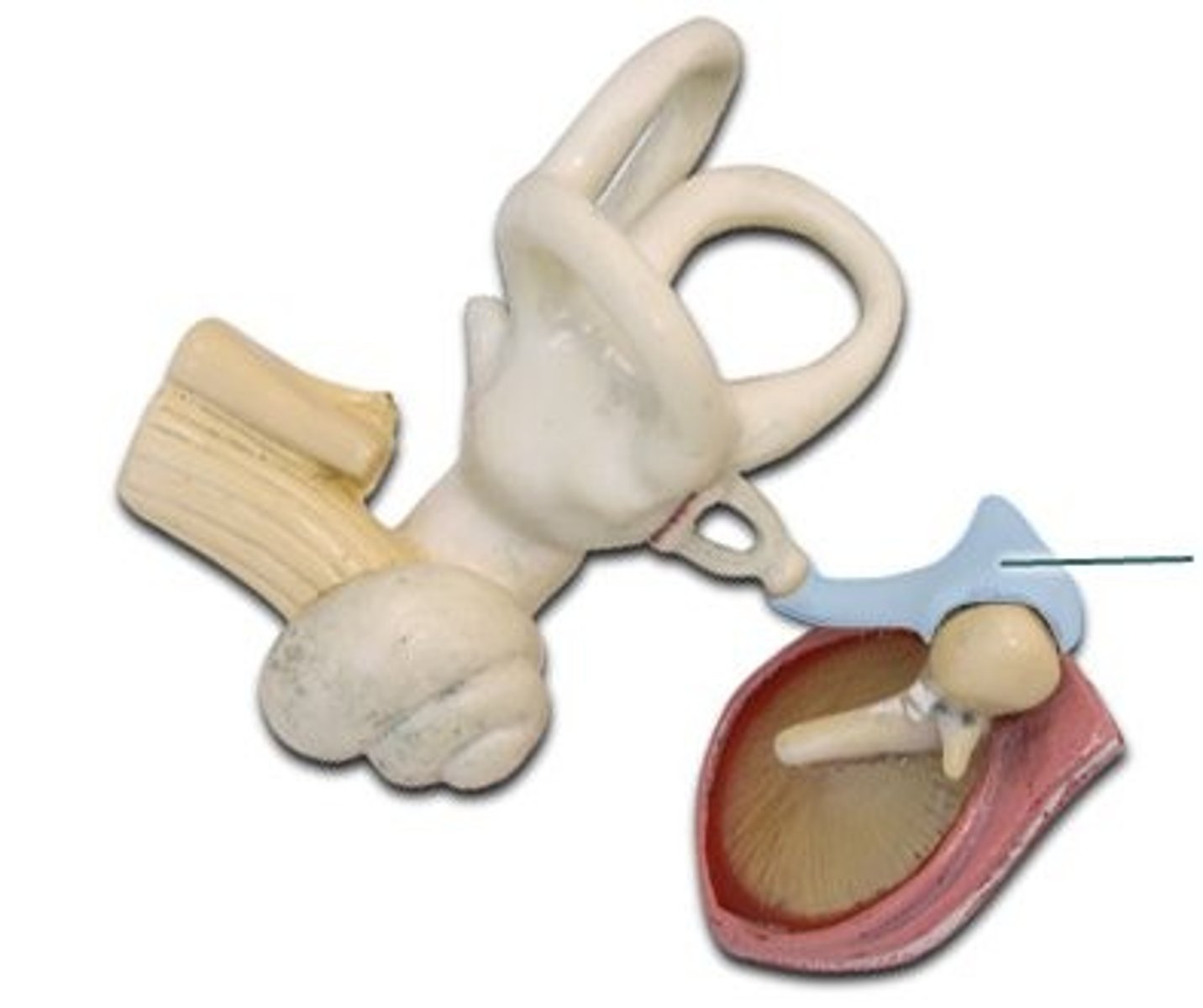
stapes
"stirrup"; inner of the 3 ossicles of the middle ear
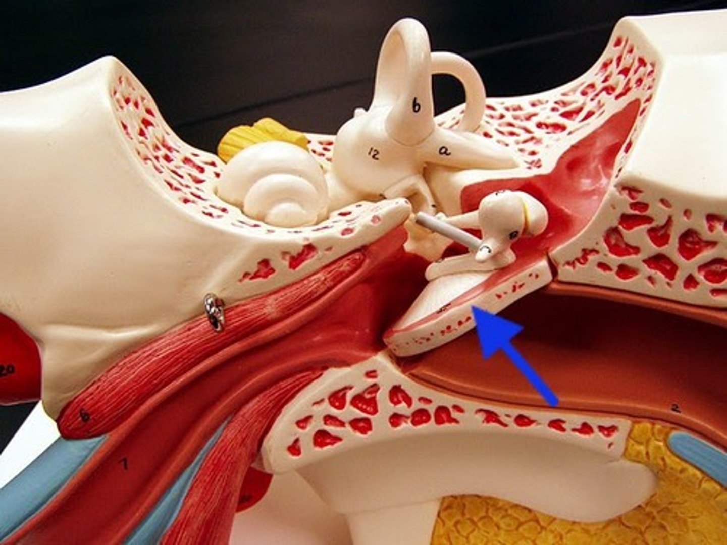
auditory tube
Name this passageway
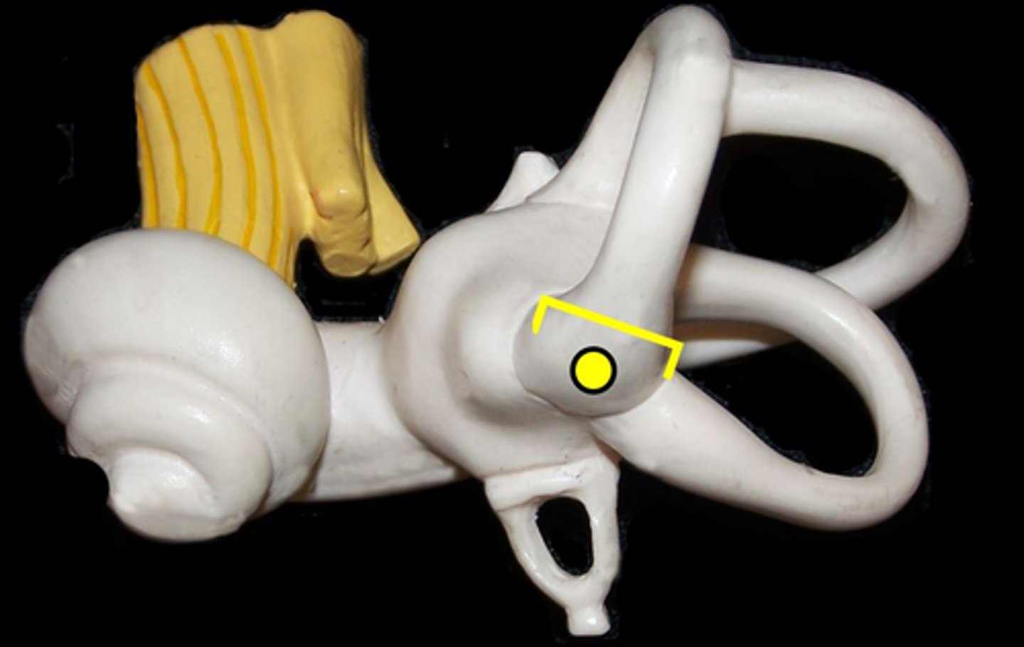
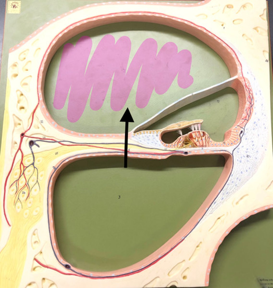
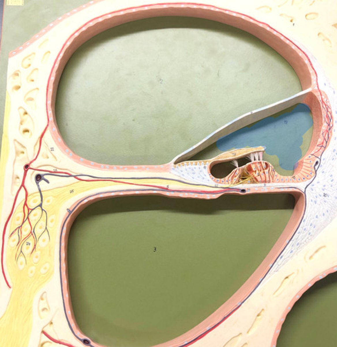
bony labyrinth
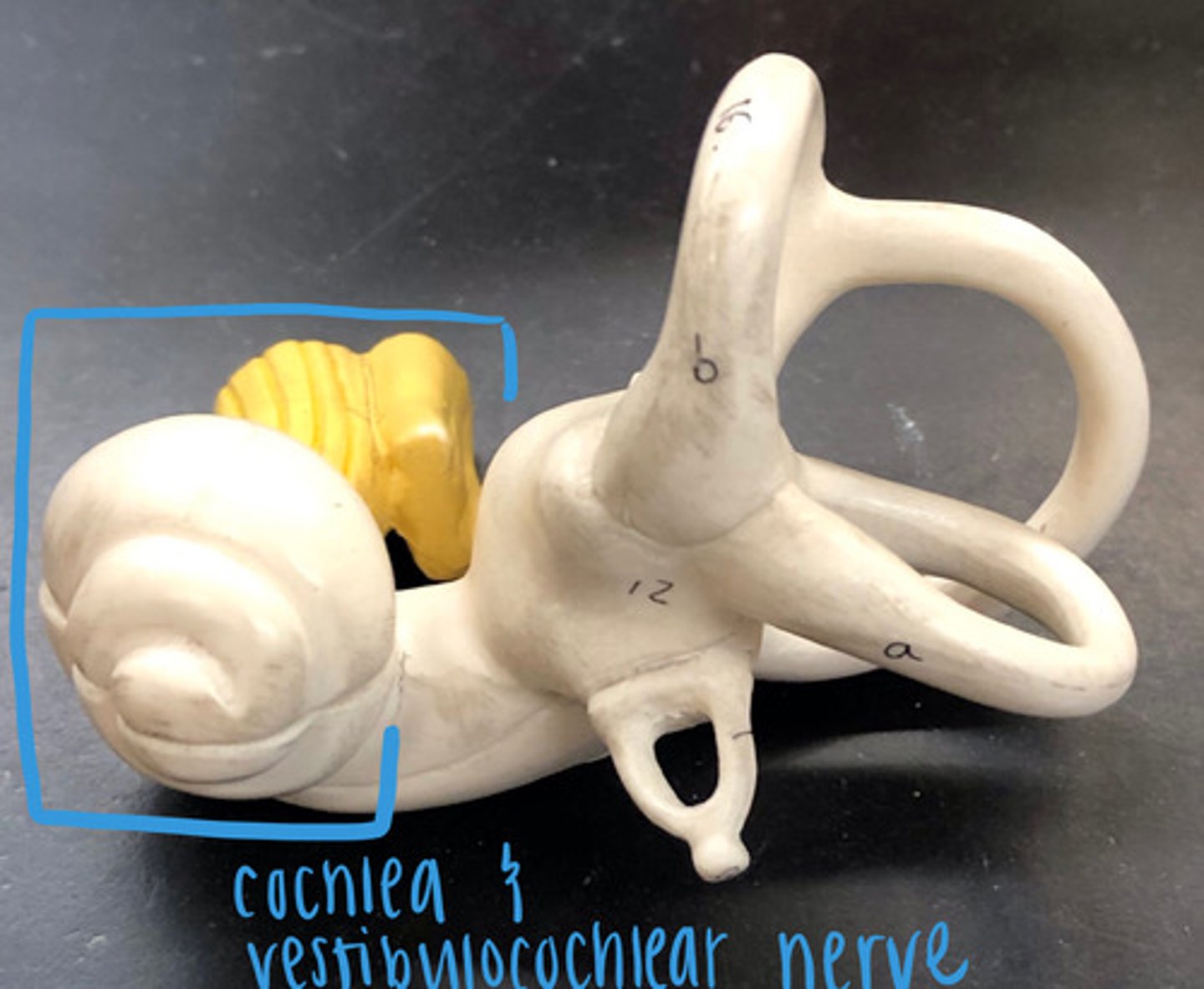
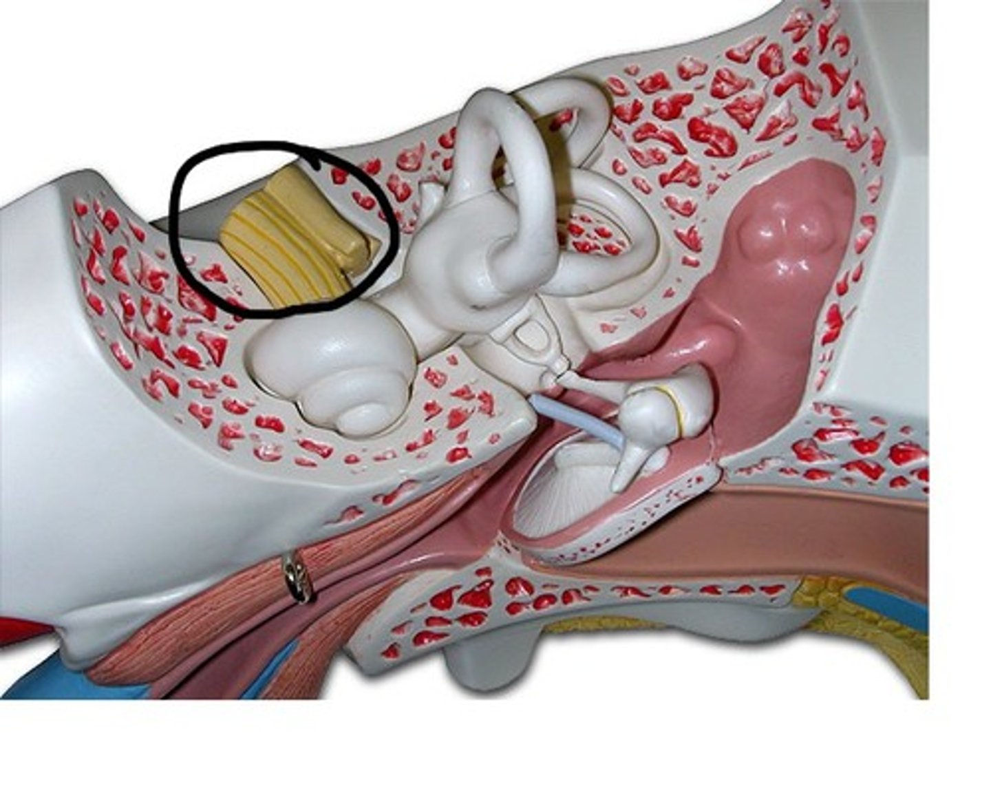
vestibulocochlear nerve VIII
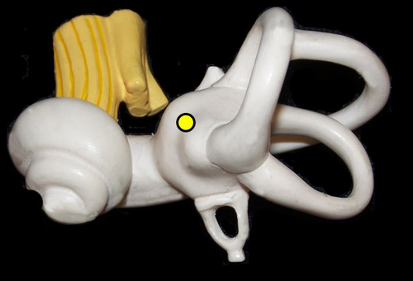
vestibule
the area between cochlea & semicircular canals.
anterior semicircular canal
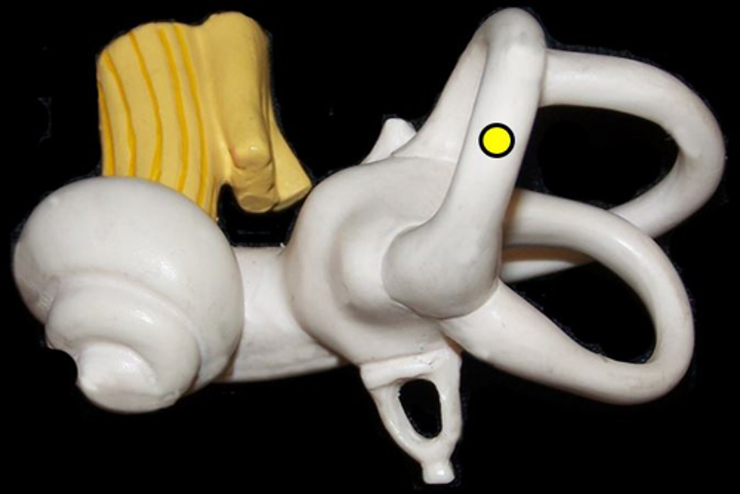
posterior semicircular canal
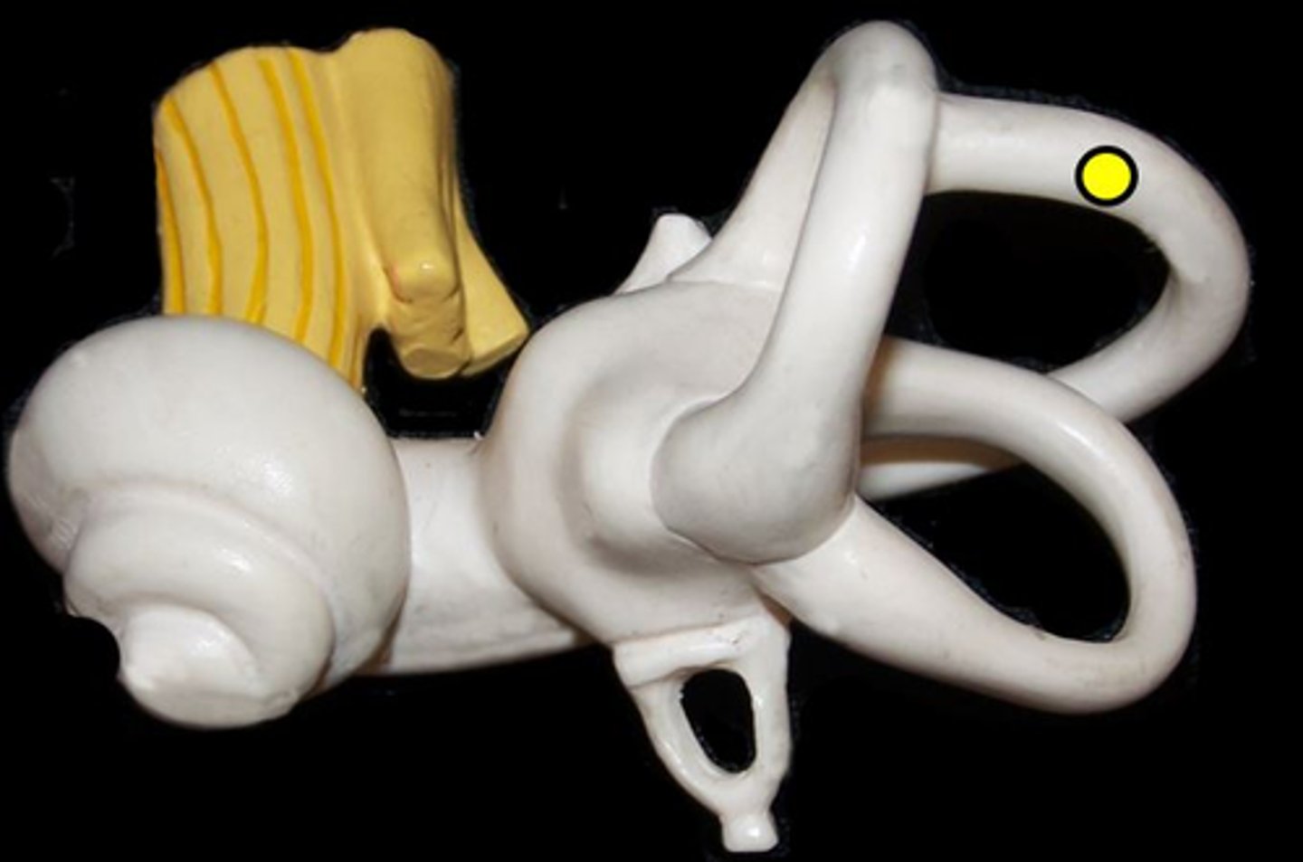
lateral semicircular canal
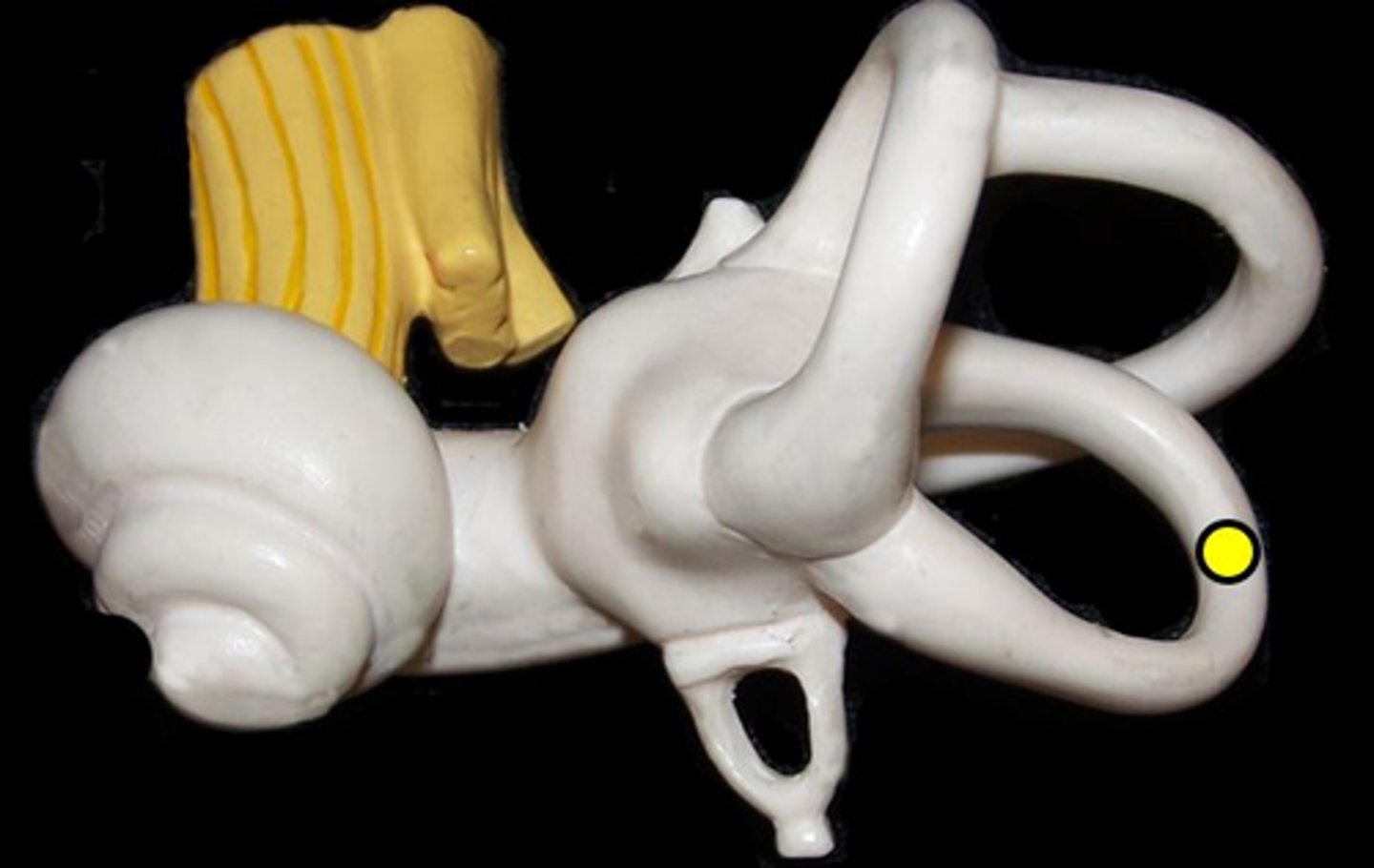
ampulla
enlarged regions at the bottom of the semicircular canals
cochlea
"snail shell"
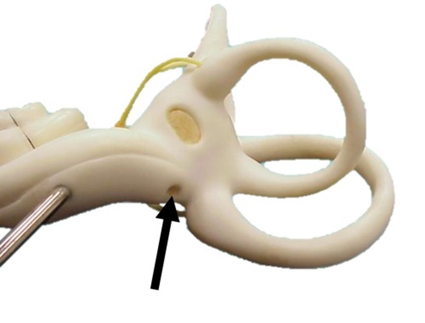
oval window
Opening under the stapes
Name this opening
round window
Cochlear branch of vestibulocochlear nerve VIII
vestibular branch of vestibulocochlear nerve VIII
vestibular duct
Name this space
cochlear duct
Name this space
blue
organ of Corti
Name this structure
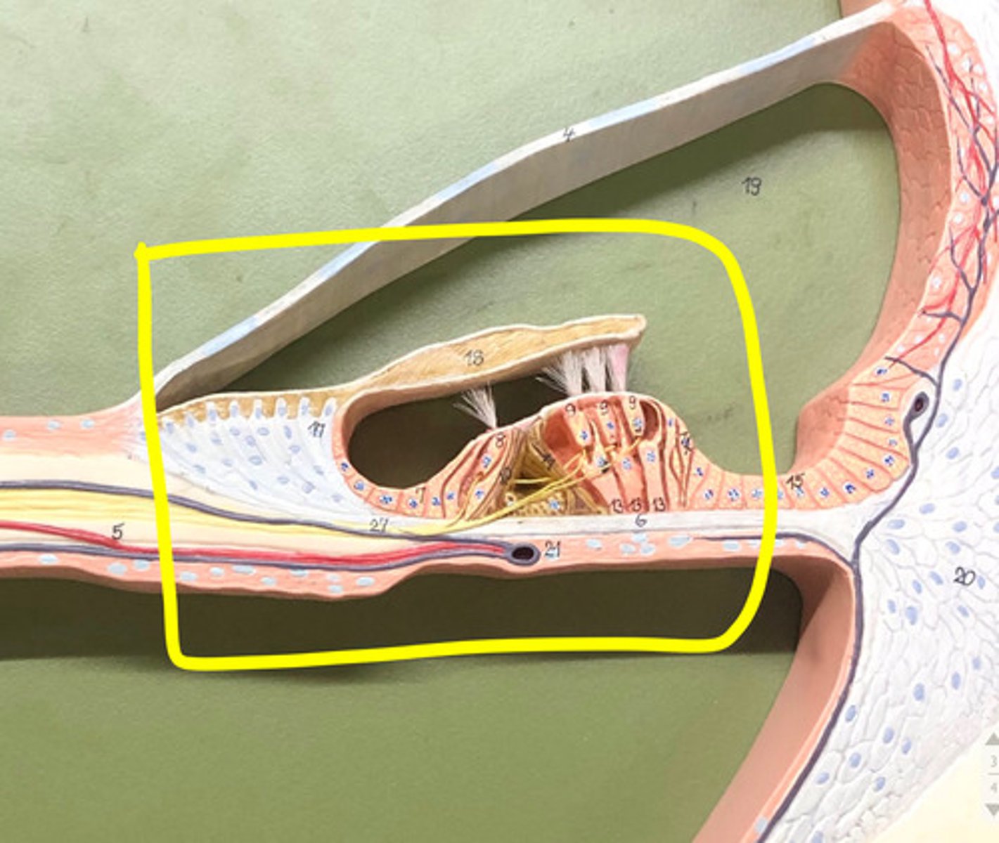
hair cells
tympanic duct
Name this space (blue)
vestibular membrane
basilar membrane

tectorial membrane
spiral ganglion