a and p final exam vocab
1/296
Earn XP
Description and Tags
do or die motherfucker
Name | Mastery | Learn | Test | Matching | Spaced | Call with Kai |
|---|
No analytics yet
Send a link to your students to track their progress
297 Terms
anatomy
study of the human body
physiology
study of function of different parts of the body
pathophysiology
study of functional, molecular, and cellular changes occurring within the body due to disease and injury,
sign
visible indication of illness
symptom
unseen effects of illness
homeostasis
self reg process
receptor
homeostatic control mechanism general component
control center
triggers effectors via nerves or hormones to bring variable back to set point
effector
acts in resposne to a stimulus
positive feedback loop and example
output of a system amplifies an initial change, driven away from the equilibrium
contractions for birth
negative feedback loop and example
self reg control syste, that acts to reduce, reverse, and counteract any deviation from a set point
blood glucose level, insulin release
anterior/ventral
front
posterior/dorsal
back
medial
toward midline
lateral
away from midline
intermediate
between
ipsilateral
same side, right leg and arm
contralateral
opp sides of body, right arm and left leg
proximal
closer to point of origin
distal
farther from point of origin
superficial
surface
deep
deeper than surface
cross section
study of internal body structures thru 2d slices
frontal plane
front and back portions
longitudinal plane
horizontal plane thru middle
midsagittal plane
equal left and right halves
oblique plane
angled plane
parasagittal plane
unequal left and right halves
transverse plane
superior and inferior halves
Major chemical elements of the body
carbon, hydrogen, oxygen, nitrogen. CHON
Ionic bond
attraction bn ions, electrons transferred from one atom to another
stronger than covalent
covalent bond
shared electrons bn atoms,
hydrogen bond
attraction of opp charged polar molecules or parts of molecules
weak bonds
formed when hydrogen atom is attracted to neg atom
formed w/in molecules → shape of protein, DNA
acids vs bases
acids: electrolytes that dissociate to release hydrogen ions in water
bases: release ions that can combine with hydrogen ions, the thing thatll pair with hydrogen released by acids
pH
concentration of H+ solution
role of buffer systems in homeostasis and example of buffer reaction
converts strong acids/bases into weaker ones to reg pH
dehydration synthesis
water removed to make bonds
hydrolysis
water added to break bonds
carbohydrates and function
quick source of energy
stored in liver and muscles as glycogen
manufacture cell structures
3 classes: monosacch, disacch, polysacch
monosaccharide and example
simple sugar, glucose
disaccharide and example
two monosach, sucrose
polysaccharide and example
10-100s of monosacch, glycogen
lipids and function
energy storage, insulation, protection
fatty acids
sat: single covalent bond
unsat: one of more carbons have a double bond
trans: mod unsat
steroids (cholesterol)
four interlocking rings
starting material for synthesis of vit d
phospholipids
makes up membrane
Process of amino acid formation from proteins, use terms carboxyl and amino ends, peptide bonds, polypeptide.
Amino acids are joined together by covalent bonds called
peptide bonds that connect the amine group of one amino
acid to the carboxyl group of another amino acid
enzyme and example and trends in naming
Globular proteins that act as biological catalysts
• Highly specific
• Very efficient
• Regulated by cellular controls
• Names usually end in –ase and are often named for the
reaction they catalyze
• Example: hydrolase, oxidase, amylase, lipase
formation of nucleic acids from nucleotides, especially nucleotide structure.
Composed of nucleotides: a nitrogen base (A, C, T, G, U), a
pentose sugar (ribose, deoxyribose), and a phosphate group
• Shape held together by hydrogen bonds – can be denatured
just like proteins
structure and function of ATP, why it’s a high energy molecule.
cytoplasm
consists of all the cellular contents bn the plasma membrane and the nucleus. Has two compartments cytosol and organelles.
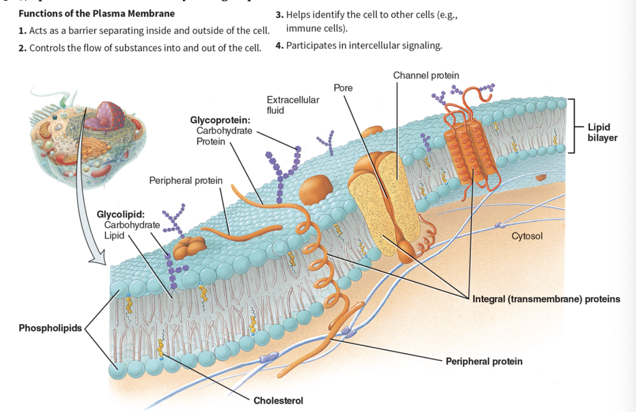
plasma membrane function
forms the cell’s flexible outer surface, separates the cells internal environment from the external. Selective barrier that regulates the flow of materials. Lipid bilayer
cytosol
the fluid portion of the cytoplasm, also called intracellular fluid
nucleus and function
houses most of the cell’s DNA. Within the nucleus, each chromosome, a single molecule of DNA associated with several proteins contains genes that control most aspects of cellular structure and function.
cytoskeleton
elaborate network of rods that run throughout cytosol. provides cell shape and organization. Assists in movement and anchoring of cell components. Incudes microfilaments, microtubules, and intermediate filaments.
centrosomes
cell center, located near the nucleus, contains centrioles = a pair of barrel shaped micro tubular organelles at right angles to each other.
cilia
whiplike, motile extensions on some cell surfaces (eg respiratory tract cells) - sweeping motion to move substances (like mucus)
flagella
longer extensions that move the whole cell (tail of sperm)
microvilli
minute, fingerlike extensions of plasma membrane that project from surfaces of select cells. (ex.: intestinal and kidney tubule cells). Used to increase surface area for absorption.
ribosomes
site of protein synthesis, consists of two subunits: made of proteins and ribosomal RNA (rRNA).
Two forms:
free: free floating in cytosol
membrane-bound; attached to membrane of ER
Rough Endoplasmic reticulum
surface appears rough - covered with membrane bound ribosomes.
Used to synthesize proteins that are secreted from cell.
proteins enter cisterns and are modified as they move through fluid-filled tubes. Final protein enclosed in vesicle and sent to golgi complex for further processing
Smooth endoplasmic reticulum
network of looped tubules continuous with rough ER. Contains enzymes in its membranes that function in:
lipid metabolism, detox of certain chemicals, conversion of glycogen to glucose. Storage and release of Ca, sarcoplasmic reticulum is specialized smooth ER found in skeletal and cardiac muscle cells,
golgi complex
stacked and flattened membranous sacs. Modifies, concentrates, packages proteins and lipids received from ER to: transport materials out of cell, inserts directly into plasma membrane, or hold contents in vesicle until needed.
lysosomes
spherical membranous bags containing digestive enzymes (acid hydrolases); made by golgi complex.
release inside injured cells causes cells to self digest (autolysis)
digests ingested bacteria, viruses, and toxins
degrades nonfunctional organelles
metabolic functions: break down and release glycogen; break down and release ca 2+
membrane fluidity
many membrane lipids and proteins move easily in the bilayer.
cholesterol stabilizes membrane and reduces fluidity.
membrane permeability
selectively permeable,
liquid bilayer ALWAYS permeable to small, nonpolar, uncharged molecules
transmembrane proteins (channels or transporters) increase permeability
macromoleculkes only able to pass by vesicular transport
electrochemical gradient
the combined influence of the concentration gradient and the electrical gradient on movement of a particular ion.
concentration gradient
difference in chemcial concentration on eahc side of the membrane
electrical gradient
difference in ion concentration bn one side of the plasma membrane and the other
-creates electrical charges on the membrane
central dogma of biology
DNA → RNA → Protein
passive membrane transport
requires no energy input
simple diffusion
facilitated diffusion
osmosis
active membrane transport
requires ATP
primary and secondary transport
vesicular transport- endocytosis (receptor-mediated, phagocytosis, pinocytosis/bulk-phase), exocytosis, transcytosis
diffusion
2 types: simple and facilitated
type of passive transport, natural movement of molecules from areas of HIGH concentration to areas of low concentration = moves down the concentration gradient.
Equilibrium reached when no net movement of molecules
simple diffusion
form of passive transport, influenced by: steepness of concentration gradient, temp., mass of diffusion substance, surface area, diffusion distance.
nonpolar lipid-soluble (hydrophobic) substances diffuse directly through phospholipid bilayer. eg; oxygen, carbon dioxide, steroid hormones, fatty acids.
facilitated diffusion
form or passive membrane transport
transmembrane proteins help solutes that are too polar or too highly charged move through the lipid bilayer.
2 kinds;
channel-mediated, eg, K+
carrier-mediated, eg glucose
osmosis
movement of a solvent like water (not particles) across a selectively permeable membrane. Moves from areas of LOW solute (high water) concentration to areas of HIGH solute (low water) concentration.
Water diffuses through:
lipid bilayer
aquaporins
Osmolarity
measures the concentration of the total number of solute particles in solvent. More solutes = less water.
tonicity
ability of a solution to change the shape or tone of cells by alerting the cells’ internal cell function.
primary active transport
energy directly from ATP hydrolysis.
substances transported: polar or charged solutes.
Energy changes shape of a transporter protein which pumps a substance across a plasma membrane AGAINST its concentration gradient, LOW TO HIGH concentration.
secondary active transport
energy obtained indirectly from ion gradients created by primary active transport.
Substances transported: Na+, K+, Ca2+, H+, I-, Cl-, and other ions
Energy stored (in a hydrogen or sodium concentration gradient) is used to drive their substances AGAINST their own concentration gradients.
Vesicular transport
transport of LARGE particles, macromolecules, and fluids across membrane in vesicles (membranous sacs)
Example: muscle contraction, release of acetylcholine.
endocytosis
transport into cell.
3 different types: receptor-mediated, phagocytosis, pinocytosis/bulk-phase.
Substances transported: solutes in extracellular fluid
exocytosis
transport out of cell
substances transported: neurotransmitters, hormones, and digestive enzymes
transcytosis
transport into, across, and then out of cell.
Substances transported: antibodies, across endothelial cells. A common route for substances to pass bn blood plasma and interstitial fluid.
phagocytosis
cell eating, cell engulfs large solid particles.
pinocytosis
cell takes in small amount of extracellular fluids
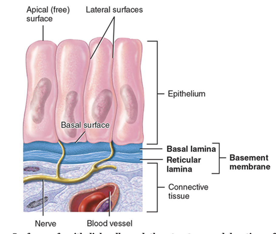
general features of epithelial tissue
consists of cells arranged in continuous densely packed sheets, in either singe or multiple layers. AKA epithelium.
Many cell junctions are present.
Cells attach to a basement membrane.
Avascular (no blood supply) but does have a nerve supply
Mitosis occurs frequently.
Main functions of ET
protection, absorption, filtration, excretion, secretion, and sensory reception.
classification of ET, first name
indicates number of layers:
single = 1 layer,
diffusion, osmosis filtration secretion, absorption
Pseudo-stratified = 1 layer that looks like many, deceitful
similar functions as simple ET
often have cilia or goblet cells (mucus production)
Stratified = 2+ layers
protection
Classification of ET, second name
indicates cell shape
squamous
flattened and scale like, rapid passage
cuboidal
box like cube, secretion and absorption
columnar
tall columns; protection, secretion, absorption
Transitional
changes shape from cuboidal to squamous and back: allows for stretch/distension
Stratified epithelia naming
shape can vary in each layer, so layer is named after the shape in apical layer.
Simple squamous ET
location: alveoli (lungs)
function: filtration, diffusion, secretion, controls vessels permeability.
simple cuboidal ET
location: thyroid glands
Functions: secretion and absoprtion
simple columnar ET
location: gallbladder
functions: secretion, absorption
non-ciliated columnar ET
location: lines GI tract
function: secretion and absorption
pseudostratified ET
location: lining of nasal cavity
function: secretion, movement with cilia