Toth Neuroanatomy I
1/55
There's no tags or description
Looks like no tags are added yet.
Name | Mastery | Learn | Test | Matching | Spaced | Call with Kai |
|---|
No analytics yet
Send a link to your students to track their progress
56 Terms
Central Nervous System
brain and spinal cord
integration and control system
interprets sensory input and dictates motor output
Peripheral Nervous System
portion of nervous system outside CNS
consits mainly of nerves that extend from brain and spinal cord
Spinal
Cranial
Spinal Nerves
to and from spinal cord
Cranial Nerves
to and from brain
enteric nervous system
walls of gastrointestinal tract also contain neurons called the enteric nervous system
PNS two divisions:
Sensory (afferent) division
Motor (efferent) division
Sensory (afferent) division’s divisions:
somatic sensory fibers
visceral sensory fibers
somatic sensory fibers:
convey impulses from skin, skeletal muscles and joints to CNS
visceral sensory fibers:
convey impulses from visceral organs to CNS
Motor (efferent) division:
transmits impulses from CNS to effectors
muscles and glands
Motor (efferent) division two divisions:
somatic nervous system
autonomic nervous system
nervous tissue two principle cell types:
neuroglia
neurons
neuroglia:
glial cells
small cells that surround and wrap delicate neurons
neurons:
nerve cells
excitable cells that transmit electrical signals
FOUR MAIN NEUROGLIA SUPPORTS CNS neurons
astrocytes
ependymal cells
microglial cells
oligodendrocytes
Astrocytes
most abundant
versatile
higly branched of glial
Astrocytes Functions:
support and brace neurons
play role in exchanges between capillaries and neurons
guide migration of young neurons
control chemical enviorment around neurons
respond to nerve impulses and neurotransmitters
participate in information processing in brain
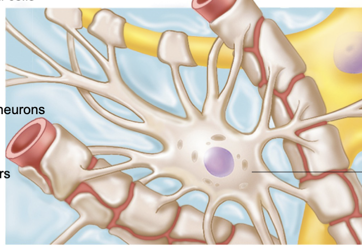
astrocyte
microglial cells
clean up
small, ovoid cells with thorny processes that touch and monitor neurons
migrate toward injured neurons
can transform to phagocytize microorganisms and neuronal debris
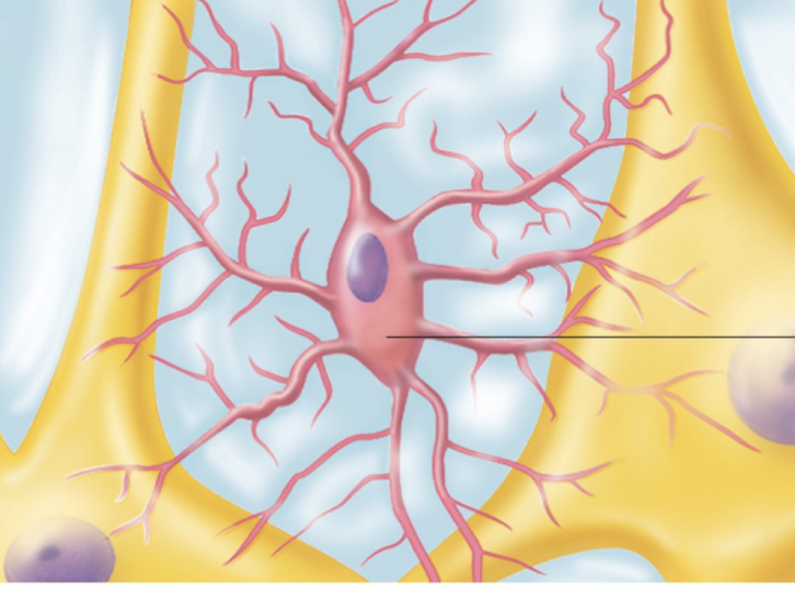
microglial
ependymal cells
range in shape from squamous to columnar
may be ciliated
cillia beat to circulate CSF
lines the central cavities of the brain and spinal cord
form permeable barrier between CSF in cavities and tissue fluid bathing CNS cells
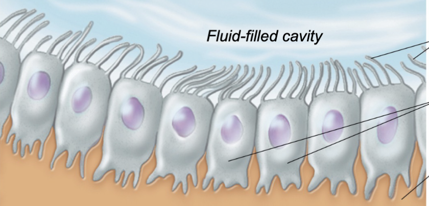
ependymal cells
oligodendricytes
produce myelin sheath
branched cells
processes wrap CNS nerve fibers, forming insulating myelin sheaths in thicker nerve fibers
Neuroglia of the PNS two types:
satelite cells
schwann cells
satelite cells
surround neuron cell bodies in PNS
function similar to astrocytes of CNS
schwann cells (neurolecmmocytes)
surround all peripheral nerve fibers and form myelin sheaths in thicker nerve fibers
similar function as oligodendricytes
vital to regeneration of damaged peripheral nerve fibers
Neurons:
nerve cells
are structural units of nervous system
large, highly specialized cells that conduct impulses
all have a cell body and one or more processes
special characteristics of neurons
extreme longevity (lasts a persons lifetime)
amitotic, with a fe exceptions
high metabolic rate: requires continuous supply of oxygen and glucose
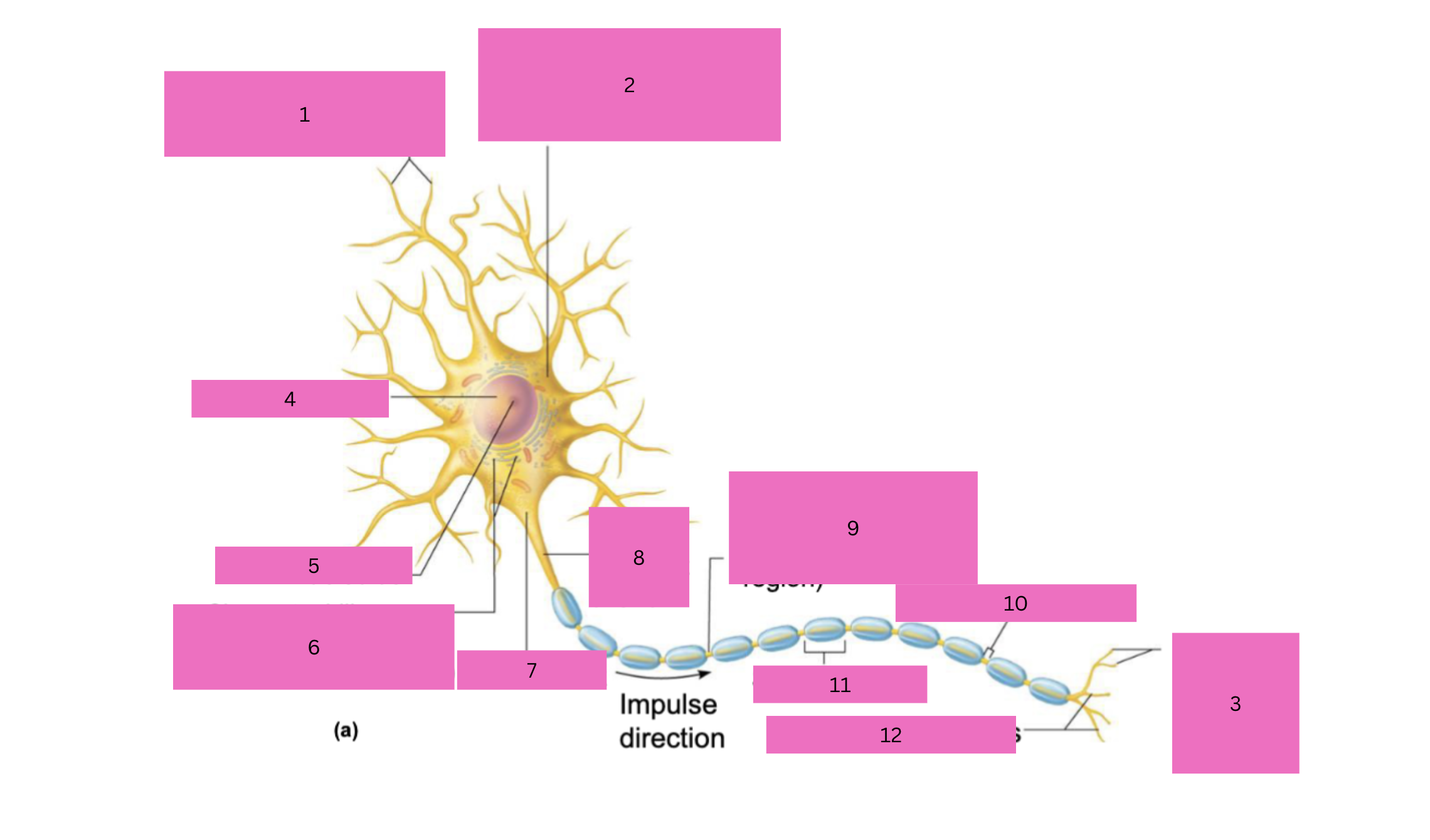
1
dendrites (receptive region)

2
cell body (biosynthetic center and receptive region)

3
axon terminals (secretory region)

4
nucleus

5
nucleolus

6
Chromatophilic substance (rough ER)

7
axon hillock

8
initial segment of axon

9
axon (impulse - generating and conducting region)

10
myelin sheath gap
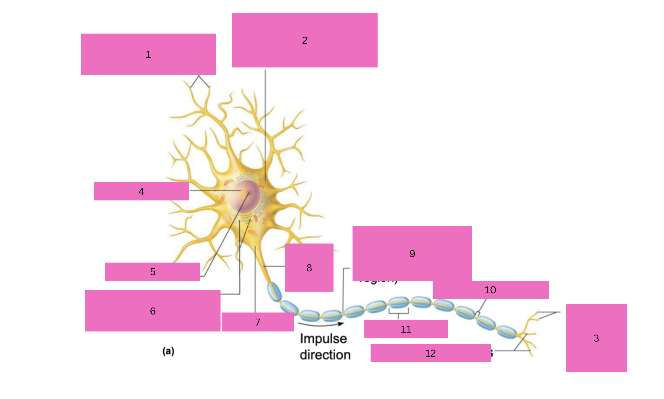
11
schwaan cell

12
terminal branches
phosolipid head
hydrophilic
phosolipid tails
hydrophobic tail
Chemically gated ion channels
open in response to binding of the appropriate neurotransmitter
open and close
depends on if there is a neurotransmitter present
Chemically gated ion channels - state when theres no receptor
closed
K+ stays inside the cell and cant pass through
Na+ stays outside the cell and cant pass through
Chemically gated ion channels - state when theres a receptor
open
Na⁺ moves into the cell
K⁺ may move out of the cell (depending on the channel type)
volage- gated ion channels
open in response to changes in membrane potential
volage- gated ion channels - closed
Closed: when the membrane is at resting potential
Na+ volage- gated ion channels - closed
Na⁺ cannot enter through these channels
However, Na⁺ still has a strong desire to move INTO the cell (because:
higher concentration outside
negative inside attracts it)
K+ volage- gated ion channels - closed
K⁺ can still move through leak channels
So K⁺ slowly diffuses OUT of the cell
volage- gated ion channels - open
Open: when the voltage across the membrane changes enough
Na+ volage- gated ion channels - open
Na⁺ rushing in = depolarization (cell becomes positive)
Voltage-gated Na⁺ channels open first
Na⁺ rushes INTO the cell very quickly
This happens because:
high concentration outside
negative inside attracts Na⁺
👉 So: massive Na⁺ influx → inside becomes more positive
K+ voltage- gated ion channels - open
Shortly after, voltage-gated K⁺ channels open
K⁺ then moves OUT of the cell
👉 So: K⁺ leaves, but slightly later than Na⁺ enters
K⁺ leaving (after) = helps bring the cell back down
resting membrane potential
generating a resting membrane potential depends on:
differences in K+ and Na+ conc. inside and outside cells and,
differences in permeability
Na and K resting membrane potential
Na+ conc is higher outside the cell
K+ conc is higher inside the cell
what maintains the conc. gradient
Na+ and K+ puumps maintain the conc. gradients of Na+ and K+ across the membrane