Day 2 (CNS): Smell and Taste
1/19
There's no tags or description
Looks like no tags are added yet.
Name | Mastery | Learn | Test | Matching | Spaced | Call with Kai |
|---|
No analytics yet
Send a link to your students to track their progress
20 Terms
Smell and taste are forms of chemoreception
Why is it old
What did this evolve into?
Chemoreception is evolutionarily old: bacteria use it to guide their movements; animals without brains use itto find food and mates.
Chemoreception may have evolved into chemical synaptic communication.
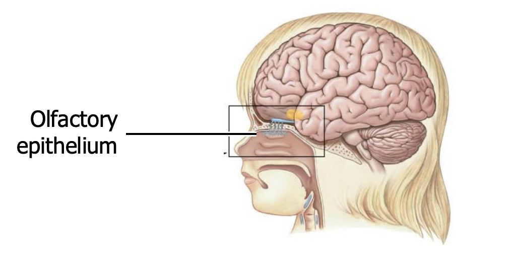
The olfactory receptors are located where?
where in the brain - name of it
This structure itself and its located to elsewhere (comparison)
How many receptor cells in total
How is it related to vision?
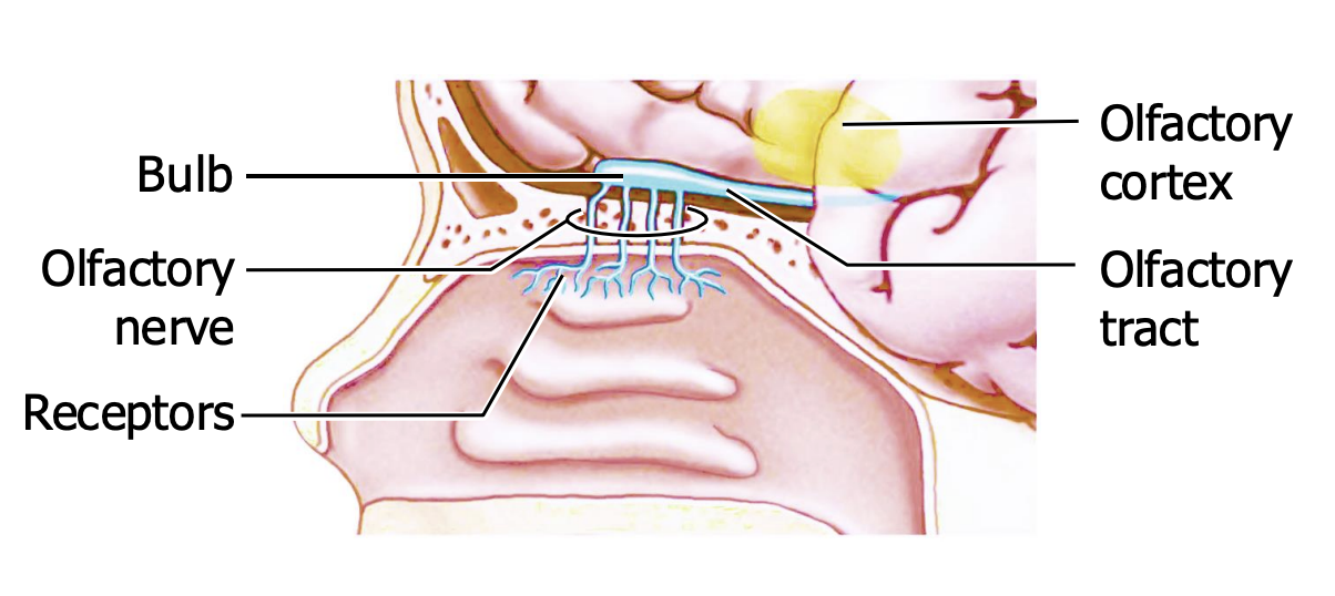
are in the olfactory epithelium
This epithelium lies at the top of the nasal cavity, covering ~3 cm2 in each of the 2 sides. It contains ~10 million receptor cells in total.
The epithelium is pigmented. No one knows why, but the richness of its color correlates with olfactory sensitivity: ipale yellow, it smells like a “dark mustard brown”
The receptor neurons are ciliated neurons
What entends into the olfactory epithelium
Branches to form what?
Why is this important?
Number of total receptor cells = how many of ___
Each cell has a single dendrite that extends into the olfactory epithelium. There it branches to form nonmotile cilia that increase the surface area of the cell, so it has a greater chance of catching odorant molecules.
Each receptor cell has (many copies of) one type of odorant receptor molecule on its membrane. We have ~400 kinds of receptor cell, i.e. ~ 400 “primary odors”.
Type of receptor olfactory receptor cells are
how many genes for these types of receptor molecules in vertebrates
How many genes in humans
Explain the pathway process
What does it activate
What is the process that occurs later
The genes for these receptor molecules form the largest known gene family in vertebrates — 1000 genes, or ~3–5% of the genome — though only ~400 are expressed in humans.
15 genes are expressed in the skin
When an odorant molecule binds its receptor, it activates a G protein, Golf, which increases the local concentration of cAMP.
Olf = olfactory
cAMP-gated cation channels open, depolarizing the receptor neurons and triggering an action potential that travels along the cell’s axon to the olfactory bulb.
How many molecules to activate a cell
how many cells until humans can consciously detect a smell
Some of them can detect a single molecule of their preferred chemical, though ~40 cells must react before we experience a smell.
Olfactory receptor cells have unusual properties
What type of cell are they
How long do they live
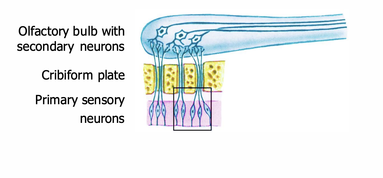
How does the axons reach the brain through the skull
What is this called
Location (approximate to somewhere else)
They are pinocytotic, continually sipping in fluid and sending it along the nerves into the brain. We don’t know why.
They are short-lived, degenerating after a month or 2, to be replaced by new ones from below.
They send their axons into the brain through tiny holes in the cribriform (“sievelike”) plate — the bone at the base of the cranial cavity.
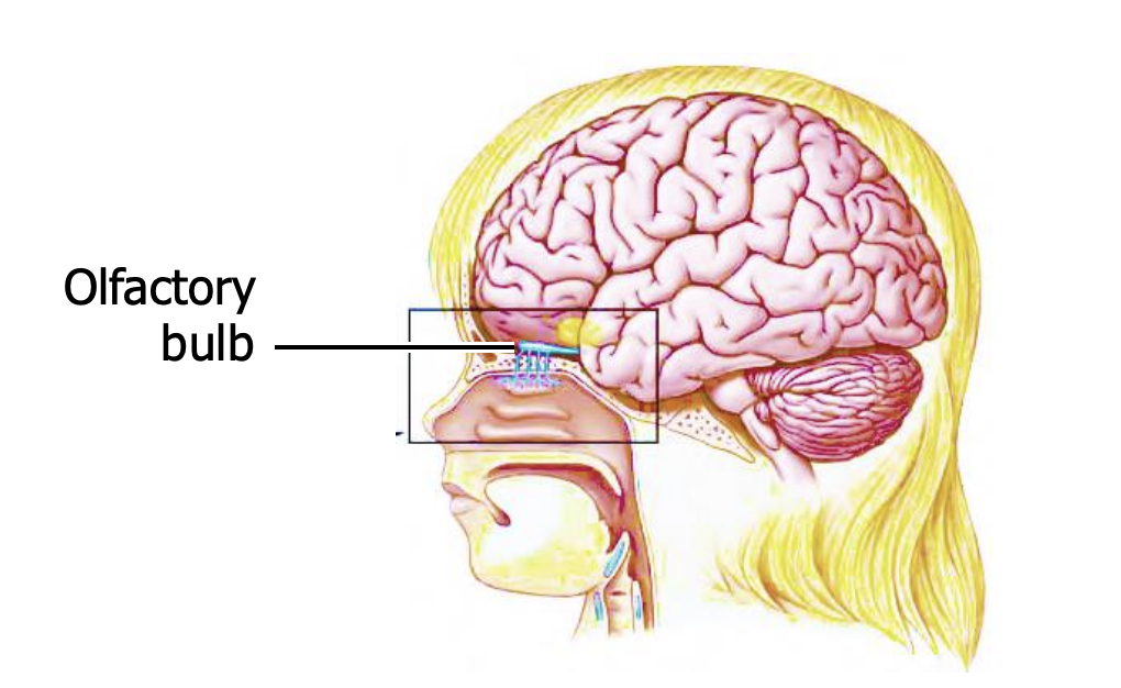
The receptor cells project to the olfactory bulb
what is the bulb
Where is ti located
What connects the receptors to the bulb
The bulb is an extension of the cerebrum, and lies on the underside of the frontal lobes.
The projection from the receptors to the bulb is called the olfactory nerve, or cranial nerve I.
Many receptor cells converge on each bulb neuron
draw the diagram of the sensory neurons to the bulb
As with rods converging on ganglion cells, this arrangement enhances sensitivity but discards spatial information.
Anosmia: loss of smell (due to brain damage)
Converging of cells (lots of primary to secondary) results in the sacrifice of spatial resolution
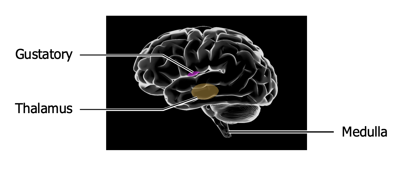
The bulb projects directly to where
what do they skip during the pathway
Draw a diagram
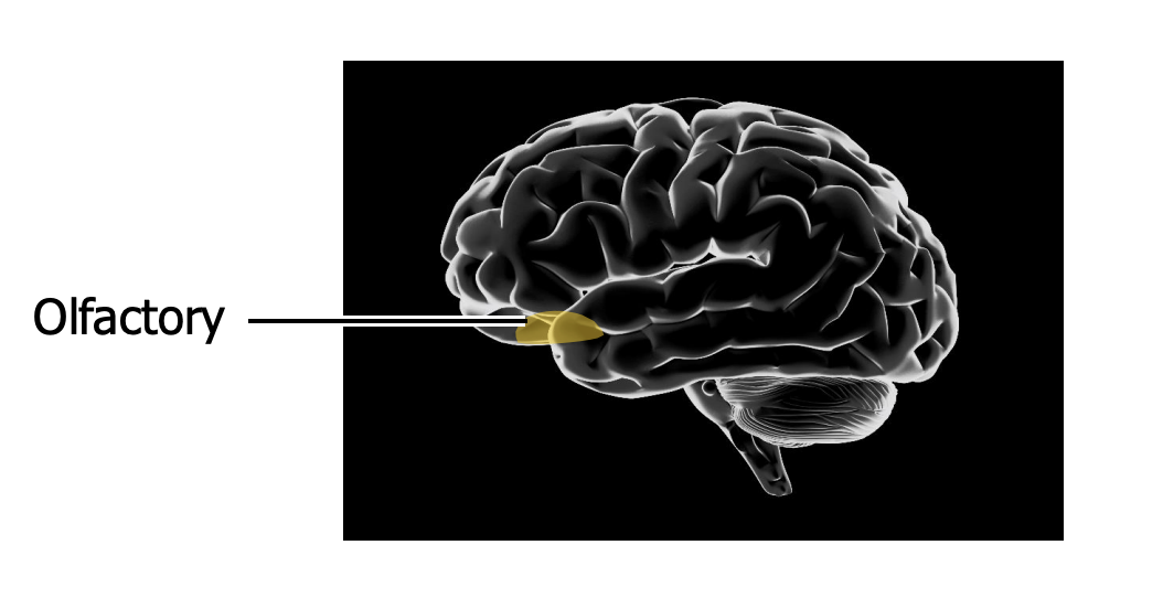
The bulb projects directly to olfactory cortex, bypassing thalamus
In which two lobes is the olfactory located
draw the located (for memorization)
Olfactory cortex is in the frontal and temporal lobes
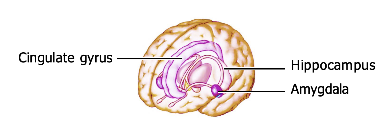
The bulb also projects to the limbic system
explain what this indicates about the old days, mammals (what we did back then, but less now)
This is an old group of brain regions concerned with motivation and emotion. For early animals, motivation was tightly linked to smell: they used their noses to identify food and poisons, mates and predators.
Our emotions are no longer so smell-related (e.g. we like money) but they are still handled by these old olfactory areas. Maybe that is why odors call up emotional memories
Olfaction adapts slowly but completely
Sewer workers don’t notice anything objectionable, and people are often unaware oftheir own body odors.
Food evaluators take steps to avoid adapting, e.g. wine tasters eat biscuits or cheese between sips, and Scottish cheese tasters sip whisky.
Rodents and maybe humans have pheromones
What does pheromones do
In rodents, what organ is involved and what does it do
For humans, explain the difference
Pheromones are chemicals released by an animal into the environment which affect the physiology or behavior of other members of its species.
Rodents have an olfactory structure in the nasal cavity called the vomeronasal organ (VNO), which is involved in their behavioral responses to sex pheromones.
In humans, the VNO disappears during fetal development, but we do respond to airborne chemical signals
Our main taste receptor cells are clustered where?
what is it called
How many do we have
How many does a baby have
Where is it mainly located
How long does last?
How many receptor cells in each one
What type of receptor cell
How are the receptors arranged
What place does the taste receptors contact the oral cavity?
Clustered in taste buds
We have ~5000 taste buds, mainly on the top of the tongue but also on the soft palate, epiglottis and upper esophagus. Babies have 10,0000. A taste bud lives only ~10 days.
Each taste bud contains ~100 receptor cells, which are epithelial cells (not neurons) arranged like petals. They contact the oral cavity through a small opening, the taste pore.
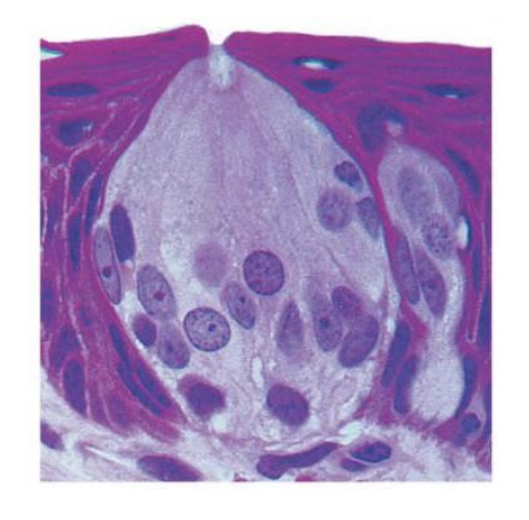
A typical taste bud contains at least 5 kinds of receptor cell
What do we detect in each flavour taste
What is another possible thing we can taste, but not part of the 5 main flavours
Each kind of receptor cell detects one flavor, and all 5 have clear biological roles:
Sweet and umami receptor cells detect sugar (energy) and the amino acid glutamate (indicating protein),respectively.
Bitter receptor cells detect poison.
Salty and sour receptor cells detect Na+ and H +— 2 important ions.
The tongue may also have receptors for fatty acids.
There are receptor cells of all 5 kinds all over the top of the tongue
what is a wrong idea about tasting flavour
But what is a specific part that is true, or that can explain the misunderstanding
For instance, it is not true that sweetness is sensed only by the tip of the tongue.
But different areas of the tongue do vary slightly in their thresholds for different flavors.
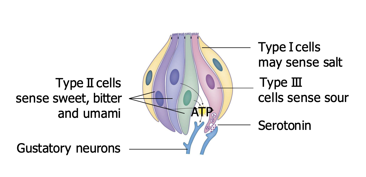
Taste receptor cells are grouped into 3 types
Type I cells
Type II cells
What do they do (2 things)
One about structure
another about function
Type III cells
Functions
What two things do the affect
Type I - detect salt
Only type III cells form direct synapses with sensory neurons, activating them with serotonin.
Detect sour
Type II cells release ATP (signalling molecules or affect the other nearby tissues), which acts on sensory neurons and type IIIs.
Detect sweet, butter and umami
Type I - oldest and therefore sour
Type II - indentity crisis - sweet, sour and umami (always calling for mother)
Can call mother, but expend/give/release energy - ATP
Type III - youngest - type I and II always sour at III because gets direct access to mother (synpase with sensory neurons)
Different kinds of cell employ different membrane proteins
which favours are activated with G-protein
What is this G-protein called
What does it do
Which flavours are activated with ion channels
Cells for sweet, umami, and bitter have receptor molecules coupled to a G protein called gustducin, which activates signal pathways, increasing intracellular [Ca2+ ] and triggering release of ATP.
Detection of salt and sour involves ion channels which are not linked with G proteins.
Recall sour H ions and salty for Na ions = ion channels = no need for g-protein
Our experience of food depends on other sensors besides the taste buds
list some other factors into taste
What does the walls of the mouth have
e.g. vanilloid receptors do what
e.g. TRPM8 receptors do what
It depends on smell, temperature, pain, texture, crunch, appearance, and cognition — if I tell you some lousy food is a delicacy then you like it better.
Nerve endings in the walls of the mouth have TRP channels sensitive to temperature and chemicals, e.g. vanilloid receptors respond to heat and to capsaicin in chilies; TRPM8 channels respond to cold and to menthol.
Pain nociceptors = heat taste
TRP receptors (thermoreceptors) - temperature = taste of colleness when eating mint/menthol
Chemoreceptors in our stomach and intestines monitor their contents; some of these receptors resemble ones on the tongue, e.g. for sweet and umami.
Taste signals take several paths to the brain
what does the receptor cells in the taste buds excite what types of nerve fibers
Number as well as main name
Where do these fibers go to (or pass)
What does the TRP receptors excite what nerve
Main name of the nerve
Receptor cells in the taste buds excite fibers of cranial nerves VII, IX, and X, the facial, glossopharyngeal, and vagus nerves. These pathways synapse in medulla and thalamus en route to the cortex.
TRP receptors in the walls of the mouth excite cranial nerve V, the trigeminal.