BMS 301 Unit 1
1/51
There's no tags or description
Looks like no tags are added yet.
Name | Mastery | Learn | Test | Matching | Spaced | Call with Kai |
|---|
No analytics yet
Send a link to your students to track their progress
52 Terms
What does the transverse/cross-section plane divide the body into?
Divides body into superior and inferior portions
What does the sagittal plane divide the body into?
Divides body into left and right portions
What does the frontal/coronal plane divide the body into?
Divides the body into anterior and posterior portions
What are the four portions of the lower limb? (Work proximal to distal)
Gluteal
Thigh
Leg
Foot
What are the boundaries and bones in the gluteal region?
Boundaries: Iliac crest to inferior gluteal fold
Bones: Ilium, ischium, pubis
What are the boundaries and bones in the thigh region?
Boundaries: Gluteal fold to knee
Bones: Femur
What are the boundaries and bones in the leg region?
Boundaries: Knee to ankle
Bones: Tibia and fibula
What are the boundaries and bones in the foot region?
Boundaries: Everything distal to the ankle
Bones: Tarsals, metatarsals, phalanges
What are the bones of the pelvic girdle?
The left/right hip bones and the sacrum
What is the Acetabulum?
The "socket" of the ball-and-socket hip joint
Describe where the ilium, ischium (spine and tuberosity) and the pubis is
The ilium is the most superior portion
The ischium is the inferior, posterior portion
The ischial spine is more superior
The ischial tuberosity is more inferior
The pubis is the inferior, anterior portion
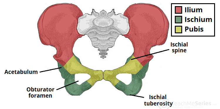
What is the sub-pubic angle and how does the sub-pubic angle differ in males vs females?
Sub-Pubic Angle: Where the two pubic bones meet in the middle
In females it is wider to account for childbirth
Describe a fibrous joint type and give an example
Non-moveable
E.x. Sutures in the skull
Describe a cartilaginous joint type and give an example
Slightly moveable
E.x. Pubic symphysis for childbirth
E.x. Intervertebral discs
Describe a synovial joint type, each subset of a synovial joint, and give an example
Freely moveable
Uniaxial: One direction of movement
Elbow can flex/extend
Biaxial: Two directions of movement
Knuckles can flex/extend and adduct/abduct
Multiaxial: Multiple directions of movement
Hips/shoulders can flex/extend, adduct/abduct and rotate medially/laterally
Nonaxial: Gliding joints
Tarsal bones in foot
What are the four characteristics of every synovial joint?
Articular Cartilage: Line the two points of the bone that come together to articulate
Synovial Membrane: Keeps synovial fluid in joint
Joint/Articular Capsule: Surrounds synovial membrane
Joint Cavity: Filled with synovial fluid
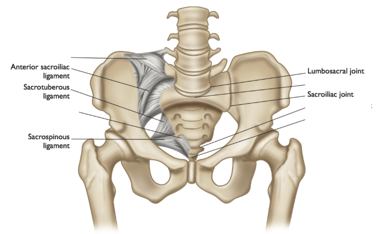
What are the two joints in the pelvic girdle?
Lumbosacral Joint: Connects spine to sacrum, cartilaginous
Sacroiliac Joint: Connects ilium and sacrum, synovial

What are the three ligaments in the pelvic girdle?
Sacroiliac Ligament: Connects sacrum and ilium
Sacrotuberous Ligament: Connects sacrum to ischial tuberosity
Sacrospinous Ligament: Connects sacrum to ischial spine I
What are the two hip ligaments?
Ischiofemoral Ligament: Connects ischium to femur posteriorly
Iliofemoral Ligament: Connects ilium to femur anteriorly (Y-joint)
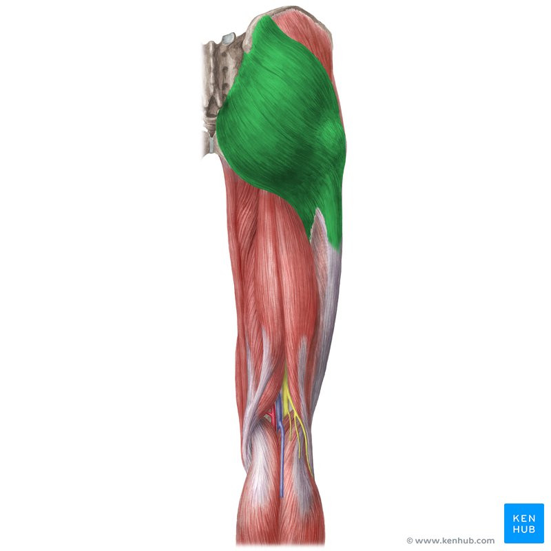
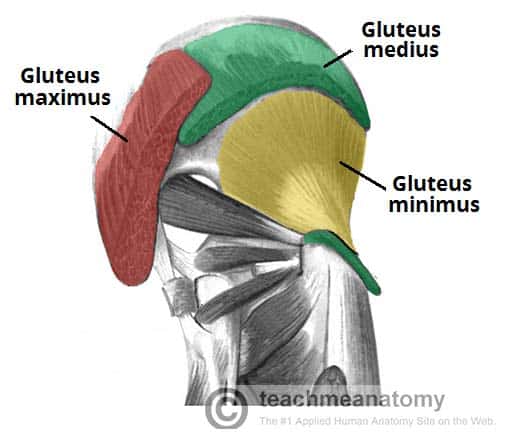
Describe the origin, insertion, action, function and innervation of the gluteus maximus
Origin: Posterior ilium and sacrum
Insertion: Gluteal tuberosity and iliotibial tract
Action: Most powerful hip extensor
Function: Resist/limit hip flexion
Innervation: Inferior gluteal nerve
Describe the origin, insertion, action, function and innervation of the tensor fascia latae
Origin: Iliac crest
Insertion: Iliotibial tract/band (IT band)
Action: Hip flexion, hip abduction (away from midline), medial rotation (towards midline)
Fx: Lateral stabilization of hip (resists lengthening/lateral rotation
Inn: Superior gluteal nerve
Describe the origin, insertion, action, function and innervation of the lesser gluteals
Origin: Posterior ilium
Insertion: Posterior/lateral femur
Action: Hip abduction (pulling femur away from midline) and medial rotation
Fx: Limits hip adduction
Inn: Superior gluteal nerve
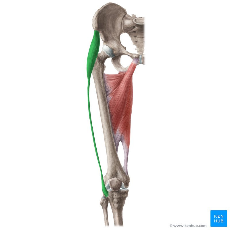
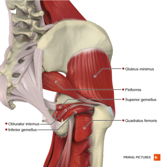
What are the 5 hip intrinsics?
Piriformis
Superior Gemellus
Obturator Internus
Inferior Gemellus
Quadratus Femoris
Describe the origin, insertion, action, function and innervation of the hip intrinsics
A: Posterior sacrum/ilium
B: Posterior/lateral femur
Action: Lateral rotation, abduction
Fx: Proprioception (where limbs/mm. are in space)
Inn: Lumbosacral plexus
What tendon groove runs in-between the calcaneus and the sustentaculum tali?
The groove of flexor hallucis longus tendon
What tendon groove runs in-between the calcaneus and the suboid?
The groove of fibularis longus tendon
Describe where the sustentaculum talis is
On the medial edge of the talus
Which neurons are present in the PNS?
Afferent (sensory) neurons to transmit information from the periphery to the CNS
Which neurons are present in the CNS?
Efferent (Motor) neurons to take motor information from the CNS out to the periphery to have an action
Define Reflex
Stereotypical motor event to sensory stimulus
What are the four components of the reflex arc?
Receptor: Sensory information is coming in to sensory receptors
Afferent (sensory) Neuron: Comes into CNS
Efferent (motor) Neuron
Target Organ: Muscle being innervated
What are the two nerve types in the CNS and what does each nerve type do?
Cranial Nerves
Autonomic function
Somatic function
Spinal Nerves
Autonomic function
Somatic function
Special sense function
Define autonomic function
Motor: Controls smooth muscle, cardiac muscle, and glands (e.g., intestines, heart, sweat glands)
Sensory: Carries visceral sensory information from internal organs (e.g., GI tract)
Control: Unconscious/involuntary
Define somatic function
Motor: Controls skeletal muscles (voluntary movement)
Sensory: Carries information from skin, joints, and skeletal muscles
Control: Conscious/voluntary
Define special sense function
Carries information for the special senses, including hearing, balance (equilibrium), vision, taste, smell
How many bilateral pairs does the cranial nerve have?
12
How many bilateral pairs does the spinal nerve have?
31
What is the location of the cranial nerve?
It exits/enters in the foramina of the base of the skull where they originate from the brain stem
What is the location of the spinal nerve?
It exits/enters the intervertebral foramen
Define Plexus
Ventral/anterior rami come together to create a complex network that innervates skin & skeletal muscle to body wall and limbs
What are the four plexus?
Cervical Plexus: Supplies regions of the neck
Brachial Plexus: Supplies all of the upper limbs
Lumbar Plexus + Sacral Plexus: Join together to form larger lumbosacral plexus to supply the lower limbs
Detail the reflex arc
Sensors in the true muscle and in the skin/limb muscle
The sensors detect a stimulus and send out afferent (sensory) neurons
The signal originating from the true muscle passes through the posterior ramus
The signal originating from the skin/limbs passes through the anterior ramus
The signals meet at the spinal nerve and travel to the posterior root ganglion where their cell bodies are
The signal passes through the posterior root and then synapses onto the posterior horn
Interneurons (not always required) take the signal to the anterior horn where it passes through the anterior root
The signal goes to the spinal nerve and then passes back tthrough the posterior ramus for true muscles or anterior ramus for skeletal/limb muscles as their target organ
The "safe area" in the gluteal region for injections/wounds is _________________________.
A. anywhere because the muscles are so large
B. the inferior medial quadrant
C. just above the gluteal fold
D. the superior lateral quadrant
E. Answers C and D are correct
D. the superior lateral quadrant
The primary function of the foot extrinsics is to move the toes.
TRUE
FALSE
False
Which muscle can extend the hip AND flex the knee?
A. biceps femoris muscle (short head)
B. semitendinosus muscle
C. gastrocnemius muscle
D. adductor magnus muscle
E. answers C and D are correct
B. semitendinosus muscle
The lesser gluteals, (medius & minimus),
A. are innervated by the inferior gluteal nerve
B. prevent hip adduction during locomotion
C. attach distally to the lesser trochanter
D. are innervated by the tibial portion of the sciatic nerve
E. attach distally to the greater trochanter
F. answers B and E are correct
F. answers B and E are correct
Muscle(s) that pass behind the lateral malleolus are the
A. peroneus(fibularis) longus
B. flexor digitorum brevis
C. tibialis posterior
D. all of the above
A. peroneus(fibularis) longus
What is the function of the ilopsoas muscle?
Hip flexion
What nerve innervates the posterior compartment of the leg?
Tibial nerve
What artery supplies the muscles of the thigh?
Deep femoral artery
The hamstring muscles share a common proximal attachment. What is the name of this bony landmark?
Ischial tuberosity
Name the ligament of the ankle that prevents hyper-eversion at this joint.
medial collateral ligament of the ankle