Constrictive Pericarditis
1/18
There's no tags or description
Looks like no tags are added yet.
Name | Mastery | Learn | Test | Matching | Spaced | Call with Kai | Chat |
|---|
No analytics yet
Send a link to your students to track their progress
19 Terms
Constrictive Pericarditis
A fibrotic, thickened and adherent pericardium
restricts diastolic ventricular filling
Constrictive pericarditis is a result of:
chronic pericardial inflammation
fibrosis
calcification
Common causes of constrictive pericarditis include:
TB
extension from lungs / lymph nodes
chronic renal failure
lupus
s/p cardiac surgery
s/p MI
s/p pericardial effusion
idiopathic
The most common cause of constrictive pericarditis in underdeveloped countries:
Tuberculosis (TB)
What is the most common cause of constrictive pericarditis in the US?
Idiopathic
Symptoms of constrictive pericarditis:
exertional dyspnea
JVD
hepatomegaly
peripheral edema
ascites
tachycardia
chronic fatigue
Constrictive Pericarditis Auscultation
diastolic pericadial “knock”
constrictive pericarditis CXR finding:
pericardial calcification
constrictive pericarditis EKG finding:
A-fib
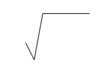
constrictive pericarditis Cath lab findings:
“square root” sign
simultaneous R&L ventricular diastolic pressure recording w/ a “dip and plateau” pattern
early diastolic dip: period of excessively rapid filling
plateau: mod to late diastole where there’s little additional ventricular vol expansion
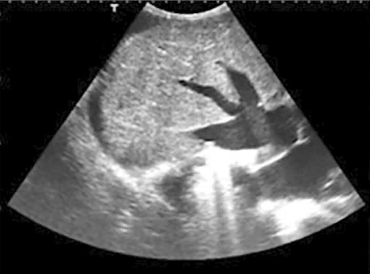
Constrictive pericarditis 2D findings:
thick calcific pericardium
normal size ventricle
enlarged atria
septal (IVS + IAS) bulge**
toward left during inspiration
“bound down” appearance of free walls
best seen A4C
dilated IVC + HV’s
no collapse w/ inspiration
Goose Foot Sign**
Normal IVC
1.2 - 2.3 cm
collapse >50%
What view is constrictive pericardial septal bulging best seen in?
A4C & Subcostal
Constrictive pericarditis m-mode findings:
thick calcific pericardium
rapid, early ventricular filling may cause:
flattening of the LV free wall during mid-late diastole
sharp downward motion of the posterior Ao root in early diastole
increased / steep E-F slope of the MV
exaggerated anterior motion of the IVS
“Spanish notch”
abnormal systolic SM
PSM, hypokinetic, akinetic
premature PV opening
seen mid-diastole w/ an increase in RVDP
Why does the IVS have exaggeration motion w/ constrictive pericarditis in m-mode?
b/c the posterior free wall of the LV is unable to expand properly, an increase in LV volume w/ atrial systole produces an anterior displacement of the septum toward the lower pressure RV, following by a brisk posterior rebound (“Spanish notch”)
What helps explain the doppler findings of constrictive pericarditis?
Cardiac Tamponade
Treatment of constrictive pericarditis:
Pericardectomy
partial / complete surgical removal of the pericardium
often performed when pericardium is scarred
may stabilize heart location
hrt shifts left
more RV seen in PLAX
will not affect hrt function
may be congenital
absent pericardium
What may a pericardectomy cause?
excessive LVPW motion & chamber dilation
Absent Pericardium
may be congenital
partial → usually lt sided
complete → rare