PEDIATRIC ECHO I
1/93
There's no tags or description
Looks like no tags are added yet.
Name | Mastery | Learn | Test | Matching | Spaced | Call with Kai |
|---|
No analytics yet
Send a link to your students to track their progress
94 Terms
____in _____children are born with Congenital Heart Defects *****
8 in 1000 children are born with Congenital Heart Defect
Some forms of CHD cause how big of a problems with growth and development of a child
Some forms of CHD cause little to no problems with growth and development of a child
Others may cause significant what?
if detected late, the child may suffer from what defcts
Others may cause significant morbidity and mortality
•If detected late, the child may suffer from neurologic defects
Performing a pediatric echo will help to identify what?
Performing a pediatric echo will help to identify any malformations of the heart
Pediatric echo includes age ranges from what?
Pediatric echo includes age ranges from newborn to 18 yrs
INDICATIONS FOR PEDIATRIC ECHO,
what is the #1 reason why?
Suggested heart disease through symptoms or family history of heart disease
(#1 reason family history)
INDICATIONS FOR PEDIATRIC ECHO
name the rest other reasons why? 4 others to name
known heart disease
symptoms that suggest heart disease
presence of structural heart disease
acquired heart disease and non - cardiac disease
INDICATIONS FOR PEDIATRIC ECHO
known heart disease name 5
check on progression of disease
valve function
growth of cardiovascular structures
LV function
interrogation of surgical intervention and treatments
INDICATIONS FOR PEDIATRIC ECHO:
3. Symptoms that suggest heart disease, name 11
whats the #1 most common (???) symptom?
Cyanosis (low O2 sats) #1 most common symptom
Failure to thrive (no weight gain) ****
Chest pain
Syncope
Respiratory disease
Murmurs
Cardiomegaly
Arrhythmias
Congestive Heart Failure
Abnormal arterial pulse - BP’s on all 4 limbs to rule out CHD ****
(different BP of the Limps point to CHD)
INDICATIONS FOR PEDIATRIC ECHO
Presence of Structural Heart Disease name 7
Shunting lesions •
Obstructive lesions •
Regurgitant lesions •
Abnormal systemic or pulmonary venous connections •
Conotruncal anomalies •
Coronary artery anomalies
• arrhythmias
INDICATIONS FOR PEDIATRIC ECHO
Acquired Heart Disease and Non-Cardiac Disease name 8
Cardiomyopathies •
Infective endocarditis •
Kawasaki disease •
Rheumatic fever •
Systemic lupus erythematousus •
Myocarditis •
Cardiotoxic drug exposure •
Teratogen influences (maternal diabetes, rubella, etc.)
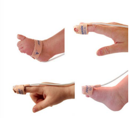
what is O2 SATS
pulse oximetry is a ______,_______,_______ what test performed on ____ infants
pulse oximetry is a painless, simple, timely, noninvasive test performed on all infants
O2 SATS
what is it used to detect?
Used to detect the amount of oxygen in arterial blood
O2 SATS is a ______ nerborm critical ______ screnning endorsed by the AAP, AHA, ACC
its a universal newborm critical CHD screening
O2 SATS, screening should be performed when?
Screening should be performed after 24 hours of life or as late as possible if early discharge is planned
(when babies are born the Rt - side pressures drop - so babies O2 sats are checked before the leave to make sure they are good and not blue)
Oxygen saturation (SpO2) is measured in the which hand (____) and on either ____(____).
Oxygen saturation (SpO2) is measured in the right hand (preductal) and on either foot (postductal).
O2 SATS
what normal?
what % will have CHD if they fail an O2
Normal is above 95%
20-25% will have CHD if they fail an O2
Done prior to discharge there by minimizing complications associated with delayed diagnosis. name 3
May not look cyanotic initially
Anemia may hide it
May only be cyanotic (blue) at 60- 70% pulse ox
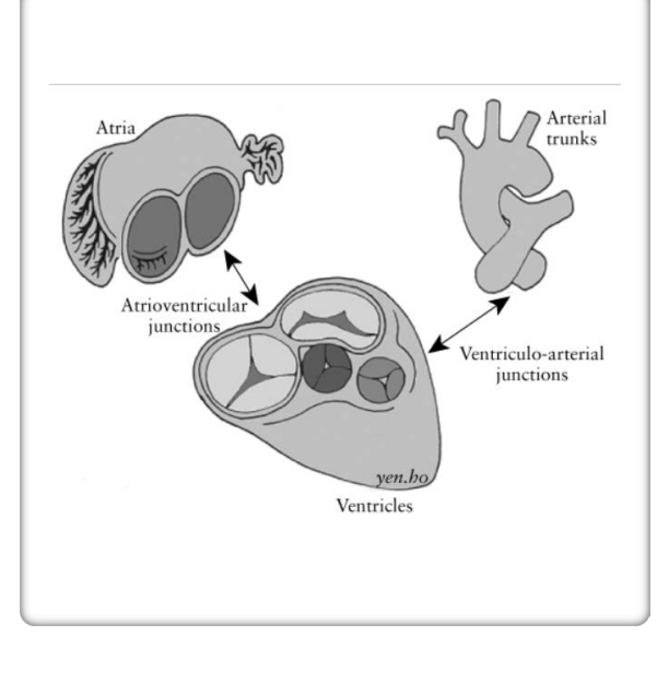
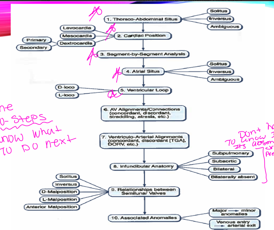
The what approach divides the heart into three basic segments and then the junctions between them
The segmental approach (or can be called the sequential approach) divides the heart into three basic segments and then the junctions between them
what are the Basic Segment:
Basic Segment: •
Atria •
Ventricles •
Great arteries
Junctions between the segments and Venous and arterial connections
name 4
Venoatrial • (SVC, IVS, Pul vein, and how they attach to the atria )
Atrioventricular and Inflow valves (chamber connection and inflow valves, the valve always follow the ventricle)
Infundibulum • (RV is more muscular and LVOT has the fibrus mitral continity)
Semilunar valves and Arterial trunks
SEGMENTAL APPROACH explain the first and secound steps
Start with Subcostal view and determine abdominal thoracic situs
Second position of apex and cardiac position
SEGMENTAL APPROACH
determines what? name 2
define the relationship of the what?
Determine atrial situs •
Determine Ventricular looping •
Define the relationship of the arterial trunks
SEGMENTAL APPROACH
whats the 3rd step (look at what 3 things and this defines the relationships of what?
whats the finally steps
Look for the venous to atrium connections
Look at the atrioventricular connection and inflow valves
Look at the outflow tract morphology
Define the relationships of the semilunar valves and connections
Next look for shunting lesions, abnormal connections, and any anomalies
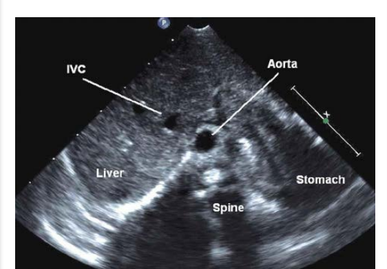
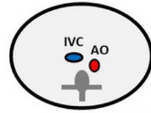
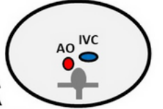
ABDOMINAL VISCERAL SITUS SOLITUS
explain where each lay
RA, AO, Liver, IVC, trilobed lung, bi lobed lung, stomach, spleen
RA is on the right •
Aorta on the left •
Liver on the right
• IVC anterior and to the right of the aorta
• Right sided trilobed lung
• Left sided bi lobed lung •
Left sided stomach
• Left sided spleen
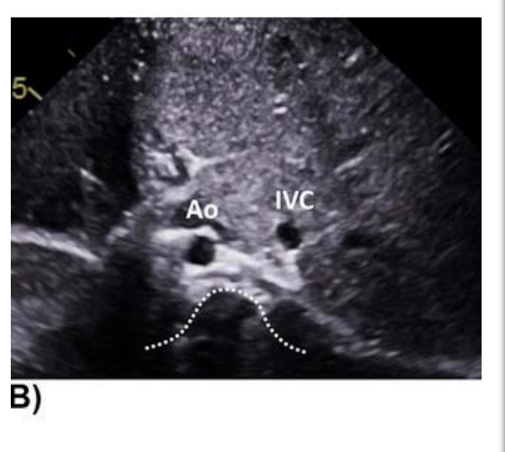
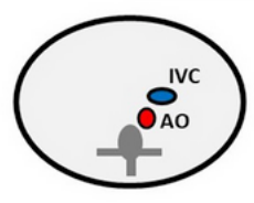
SITUS INVERSUS
describe where the following lay
AO, IVC, liver, RA, trilobed lung, bi lobed lung, stomach, spleen
Situs Inversus •
Aorta on the right •
IVC anterior and to the left of the aorta •
Liver on the left • RA is on the left •
Left sided trilobed lung •
Right sided bi lobed lung •
Right sided stomach •
Right sided spleen
what does SITUS AMBIGUOUS includes
Includes Left isomerism and right isomerism

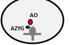
Situs Ambiguous Left Isomerism
what sign ?
has interrupted what?
where is the liver?
what is wrong with the LA
whats wrong with the ling?
what is the stomach position
how many spleen
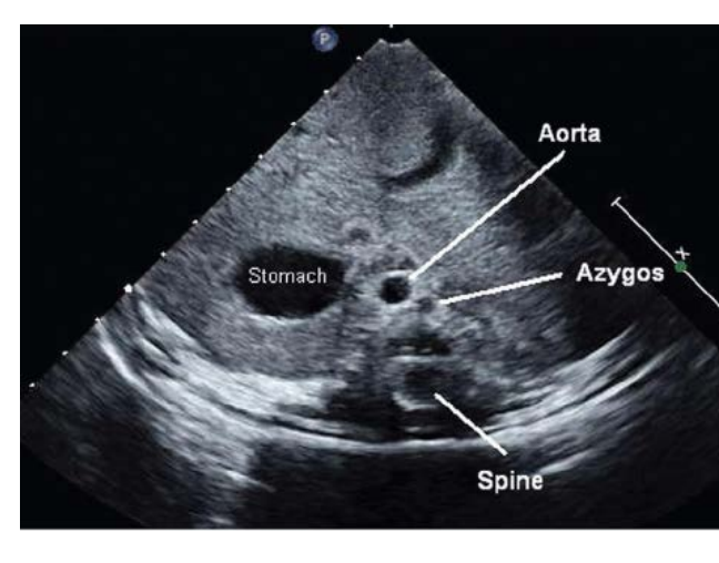
Double vessel sign with Aorta and “vein stacked” •
Interrupted IVC with Azygous continuation •
Liver is midline •
Will have two left atrium •
Bilateral bilobed lung •
Variable stomach position •
Multiple spleen
SITUS AMBIGUOUS:
Asplenia – (what syndrome?)
has what sidedness?
which atrial isomerism
Asplenia – (Ivemark’s syndrome)
bilateral right sidedness, right atrial isomerism
Asplenia – (Ivemark’s syndrome)
what wrong with the lungs?
where is the liver?
how many gallbladders?
how many splees?
Two right lungs, midline liver, two gallbladders, no spleen
which SITUS AMBIGUOUS is Almost always associated with CHD of the severe spectrum?
Asplenia – (Ivemark’s syndrome)
Polysplenia
what sidedness
what atrial isomerism
Polysplenia – bilateral left sidedness, left atrial isomerism.
Polysplenia
what is wrong with the lungs
where is the liver?
how many spleens?
75% have what forms of what?
Two left lungs, midline liver, multiple spleens
•75% have mild forms of CHD
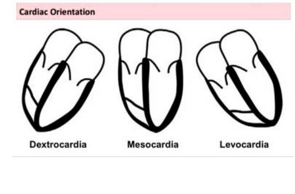
APEX AND THORACIC POSITION! • Apex Position
name the 3 different postion?
Levocardia • Dextrocardia • Mesocardia
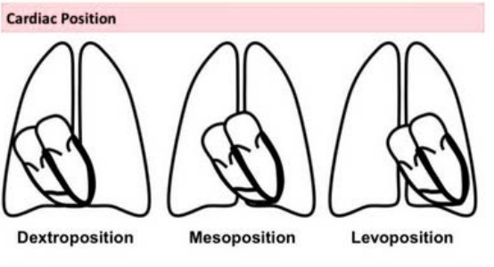
Thoracic cavity position
name 3 different position
Levoposition • Dextroposition • mesoposition
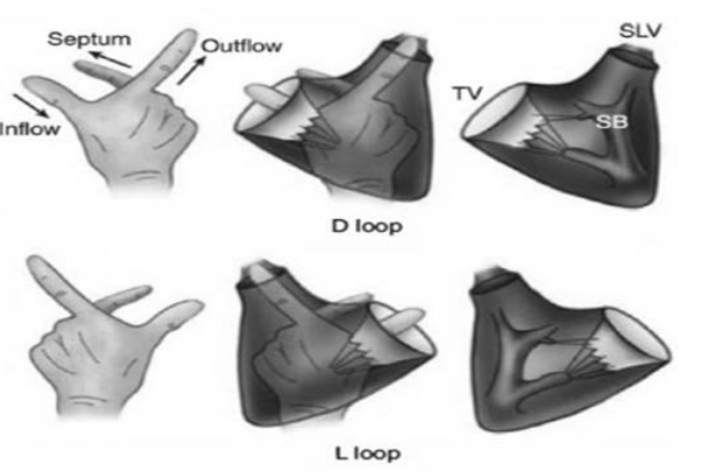
TOPOLOGY (HAND RULE) •
the thumb is for which flow?
the pointer is for which flow?
when we use the right hand what kind of looping is that?
when we use the left hand what kind of looping is that?
Thumb is for the inflow
pointer is for the outflow
Right hand D-Loop
Left hand L -loop
The point of this rule is to determine the looping of the ventricle
ATRIAL SEG MENTAL SITUS
what does S mean
what does I mean
what does A mean and what is it associated with?
S - situs solitus - (normal heart)
•I - situs inversus - all anatomy is flipped •
A - situs ambiguous - Associated with isomerism of atria (two left or two right)
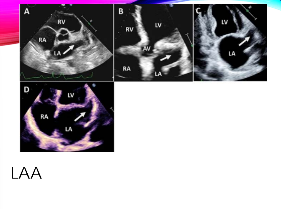
The LAA has a what shape?
what are the two best views we can see the LAA? and name another two that we can use
pinky shape
SAX - great artery level and 2 chamber are the best two views
5 chamber sweeping to AV, and 4 chamber
what is the shape of the RAA
what is the first view we see the RAA
and name 3 more
1st view is RVIF view
Bicaval
subcostal 4 chamber
subcostal bicaval view (both vena cava attached to the RA)
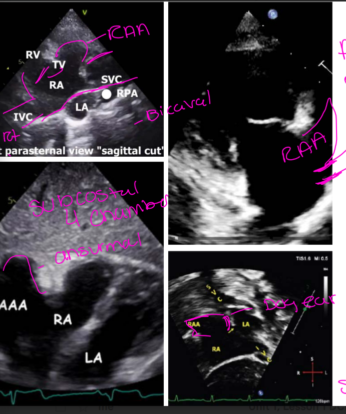
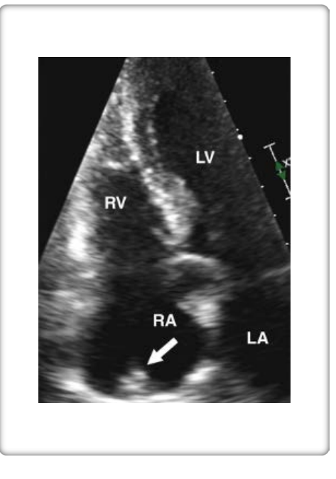
what is ANOTHER IDENTIFIER OF RA?
CRISTA TERMINALIS IS ANOTHER IDENTIFIER OF RA
VENTRICULAR SEGMENTAL SITUS
D-Loop= explain the twist
D-Loop (normal heart) Bulboventricular loop twists to the right
VENTRICULAR SEGMENTAL SITUS
L-Loop= explain the twist
L-Loop (dextrocardia) Bulboventricular loop twists to the left
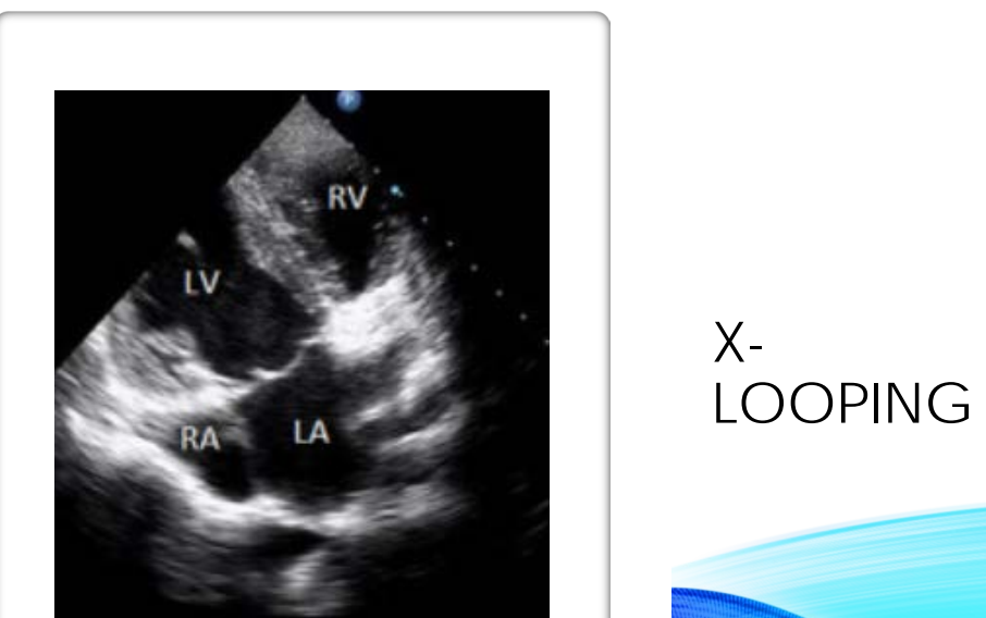
VENTRICULAR SEGMENTAL SITUS
X-Loop= explain the twist
Single ventricle is present. Unable to determine looping
VENTRICULAR LOOPING
The usual what looping of the heart tube (D—loop) leads to the morphological right ventricle being to the what of the morphological left ventricle
The usual rightward looping of the heart tube (D—loop) leads to the morphological right ventricle being to the right of the morphological left ventricle

VENTRICULAR LOOPING
what looping (L— loop) results in the morphological right ventricle being to the what of the morphological left ventricle
Leftward looping (L— loop) results in the morphological right ventricle being to the left of the morphological left ventricle
VENTRICULAR LOOPING
(x-Loop) what about the looping?
• (X-Loop) Cannot determine looping

GREAT ARTERY SITUS
S- Solitus; explain the relatipn between the AO and PV
S- Solitus; (normal) aorta is rightward and posterior to pulmonic valve

GREAT ARTERY SITUS
I – Inversus; = explain the relatipn between the AO and PV
I – Inversus; aortic valve is leftward and posterior to pulmonic valve

GREAT ARTERY SITUS
D – D-malposed;= explain the relatipn between the AO and PV
•D – D-malposed; aortic valve is rightward and anterior to pulmonic valve
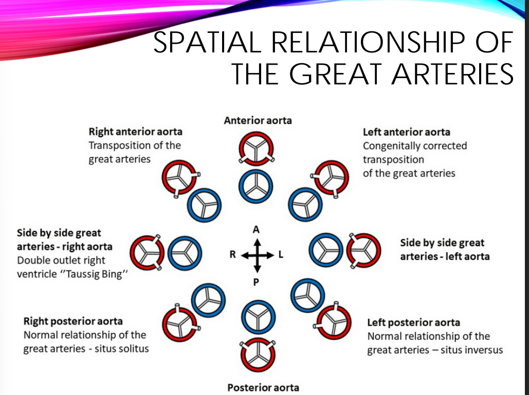
GREAT ARTERY SITUS
L – L-malposed;= explain the relatipn between the AO and PV
L – L-malposed; aortic valve is leftward and anterior to pulmonic valve
GREAT ARTERY SITUS
A – Anterior= explain the relatipn between the AO and PV
•A – Anterior; aortic valve directly anterior to pulmonic valve.

Normal heart with situs solitus:
Normal heart with situs inversus (mirror image of normal): •
D-TGA:
•L-TGA with situs solitus:
VENO-ATRIAL CONNECTION – SYSTEMIC VEINS
Systemic vein connections
Subcostal long axis of IVC connecting into
Subcostal long axis of IVC connecting into RA
VENO-ATRIAL CONNECTION – SYSTEMIC VEINS
Subcostal bicaval view connecting the SVC into the
• Subcostal bicaval view connecting the SVC into the RA
VENO-ATRIAL CONNECTION – SYSTEMIC VEINS
Hepatic veins draining into the
• Hepatic veins draining into the IVC
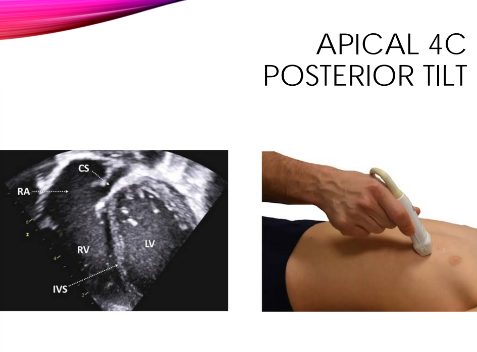
VENO-ATRIAL CONNECTION – SYSTEMIC VEINS
Apical 4 Chamber with posterior tilt to show what into what?
Apical 4 Chamber with posterior tilt to show Cononary sinus ostia into RA
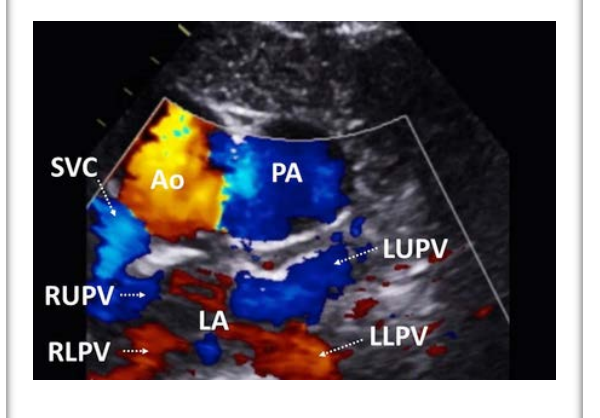
VENO-ATRIAL CONNECTION - PVEINS • Pulmonary vein connections
follow the veins entering what and use what to show drainage in what views
and do you need to do with the scale?
Follow the veins entering atrium and use color flow to show drainage in SSN Short axis •
Lower color scale
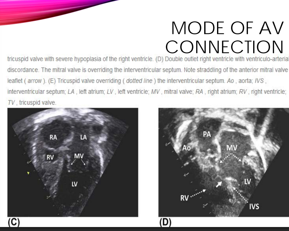
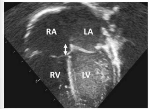
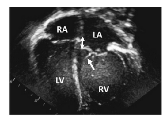
ATRIOVENTRICULAR CONNECTION
Time to assess whether the heart is biventricular, univentricular, and the mode of AV connection
Remember the inflow valves follow the what?
Remember the inflow valves follow the ventricle •
Features of TV?
• Features of MV?
• What are the different features of the ventricles?
START WITH ____TO VENTRICLE CONNECTION
START WITH ATRIA TO VENTRICLE CONNECTION
UNIVENTRICULAR CONNECTIONS
The word “univentricular” refers just to the type of what and not to the number of ventricles?
The word “univentricular” refers just to the type of connection and not to the number of ventricles
UNIVENTRICULAR CONNECTIONS
The term univentricular AV connection describes the connection of one or two atrial chambers to?
The term univentricular AV connection describes the connection of one or two atrial chambers to only one (dominant) ventricle
UNIVENTRICULAR CONNECTIONS
Univentricular hearts commonly have a one what ventricle and a second what ventricle
Univentricular hearts commonly have a one functional dominant ventricle and a second (rudimentary) ventricle
UNIVENTRICULAR CONNECTIONS •
When describing the type of AV connection…we should determine whether it’s what?
When describing the type of AV connection…we should determine whether it’s biventricular or univentricular
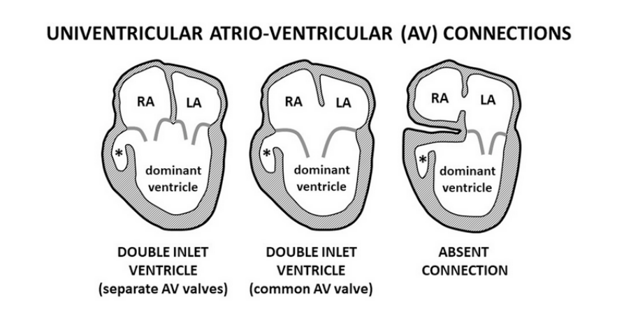
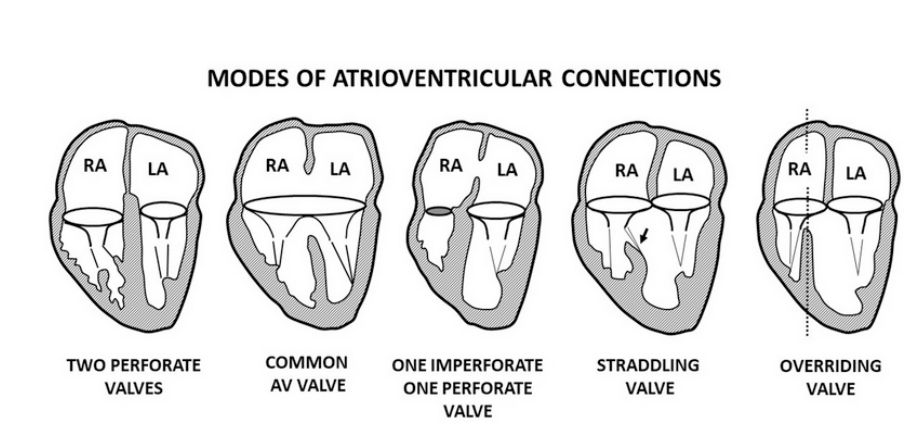
MODES OF CONNECTION INCLUDE name 5
MODES OF CONNECTION INCLUDE TWO PERFORATE VALVES, COMMON AV VALVE, ONE IMPERFORATE AND ONE PERFORATE VALVE AND AV VALVE STRADDLING AND OVERRIDING
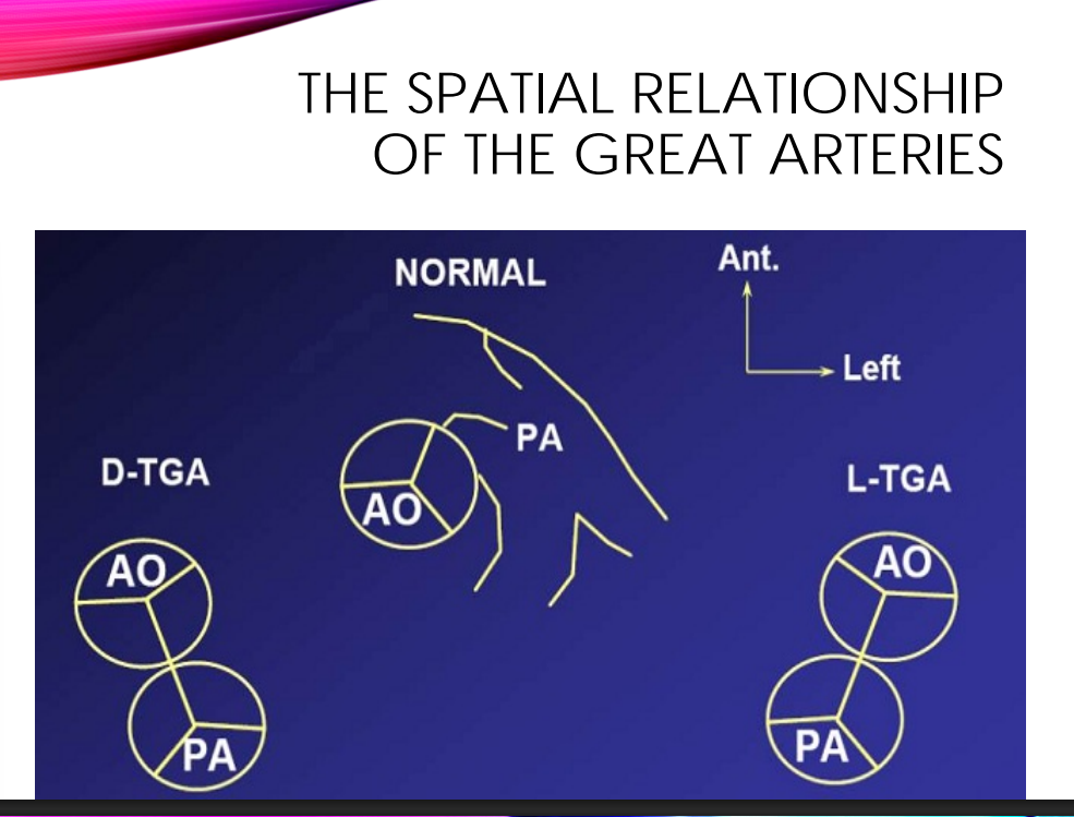
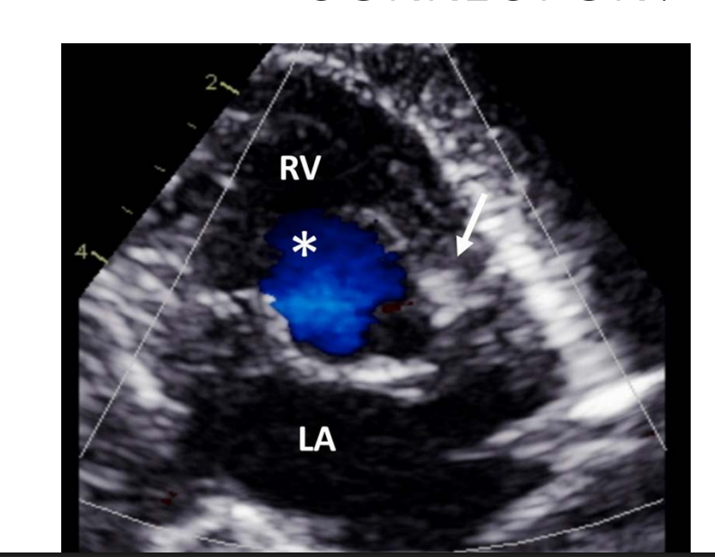
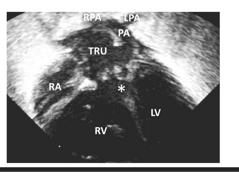
VENTRICULO-ARTERIAL CONNECTION AND RELATIONSHIP BETWEEN THE GREAT ARTERIES
how is the PA anatomic orination
Pulmonary artery is Anterior and leftward
pulmonary artery is characterized by what?
pulmonary artery is characterized by bifurcation into the right and left pulmonary arteries
Aorta gives rise to what?
and whats its anatomic orination
Aorta gives rise to coronary arteries and systemic arteries (Brachiocephalic arteries) •
Posterior and rightward
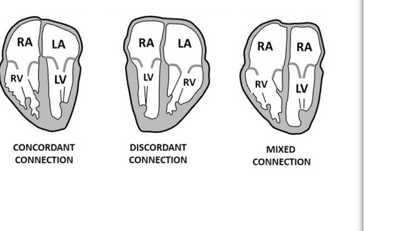
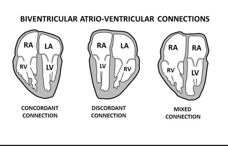
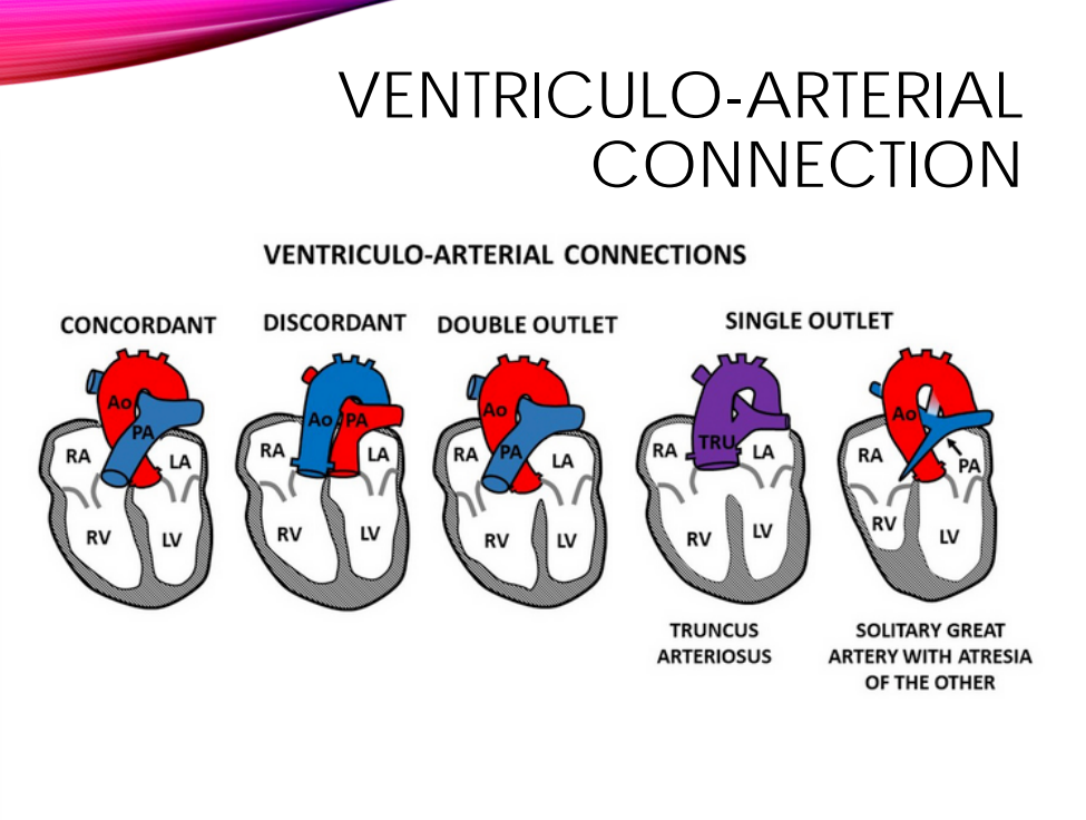
VENTRICULO-ARTERIAL CONNECTION
Which great artery is connected to which ventricle
• For types of connections name 4
Concordant •
Discordant •
Double outlet
• Single outlet
•Normal heart with situs solitus: S, D, S
•Normal heart with situs inversus (mirror image of normal): I, L, I •
D-TGA: S, D, D
•L-TGA with situs solitus: S, L, L
CONCORDANT/DISCOR DANT
which one is the two wrongs make a right?
Atrial – ventricular discordance; ventricular – arterial discordance. Two wrongs make a right!
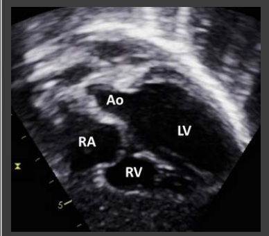
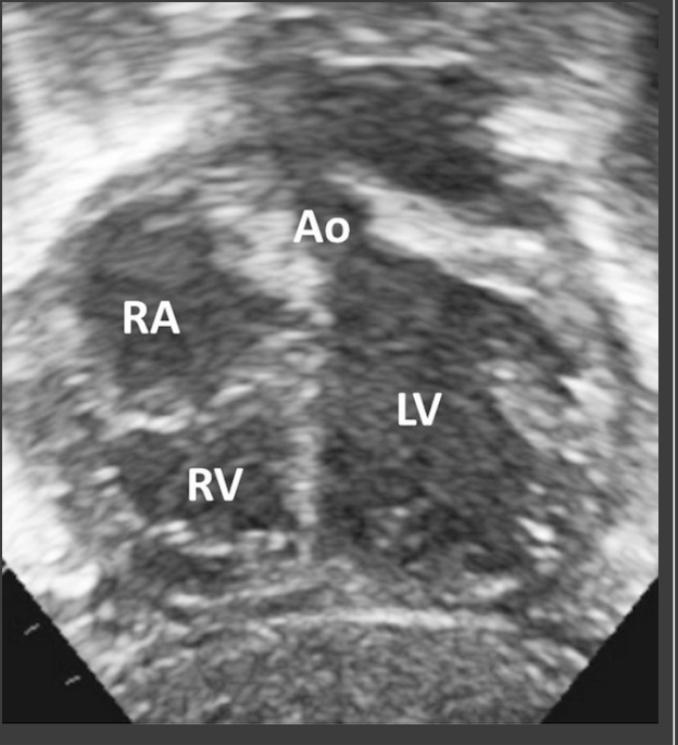
WHAT IS THE CONNECTION?
WHAT IS THE CONNECTION?
WHAT IS THE CONNECTION?
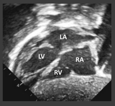
what are the morphological features of the ventricle
AO
PA
RA and LA
RV - MB, muscular
LV - bullet shaped, smooth, false tendon
AO 3 head and neck branched / vessels
PA branches
RA and LA appendages
for the SEGMENTAL APPROACH, what are all the steps
what situs is this
situs solitus
what situs is this
situs inversus
what situs is this
situs ambiguous, rt atrial isomerism
what situs is this
situs ambiguous lt atrial isomerism
which situs ambiguous has 100% association very sever congential heart defects ? more severe !!!
situs ambiguous, rt atrial isomerism
what APEX AND THORACIC POSITION is this
Dextrocardia
what APEX AND THORACIC POSITION is this
Levocardia
what APEX AND THORACIC POSITION is this
Mesocardia & mesoposition (equally divded by the RT and LT side of the chest)
Use the atrial what to determine right situs
Use the atrial appendages to determine right situs
what kind of looping is this? concordance vs discordance?
D- Looping and concordance
what kind of looping is this? concordance vs discordance?
L-Looping and discordance
Normal heart with situs solitus
SDS
Normal heart with situs inversus (mirror image of normal):
ILI
D-TGA
SDD
L-TGA with situs solitus
SLL
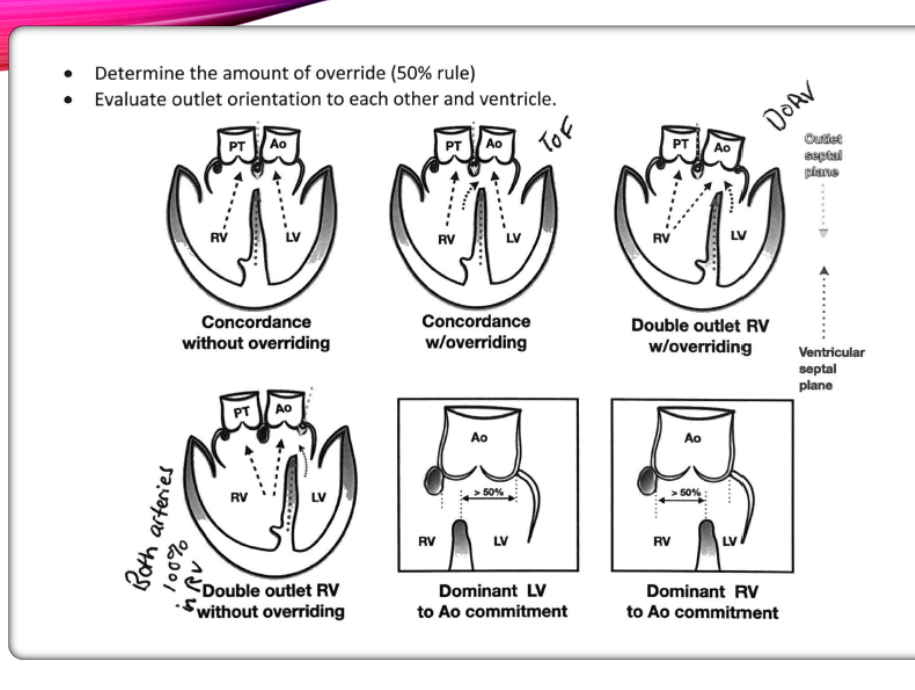
DORV is determine the amount of override is what %?
> greather than or equal to 50%