MRI lecture 5:
1/21
There's no tags or description
Looks like no tags are added yet.
Name | Mastery | Learn | Test | Matching | Spaced | Call with Kai |
|---|
No analytics yet
Send a link to your students to track their progress
22 Terms
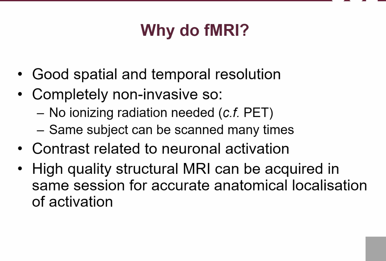
why should you use fMRIs?
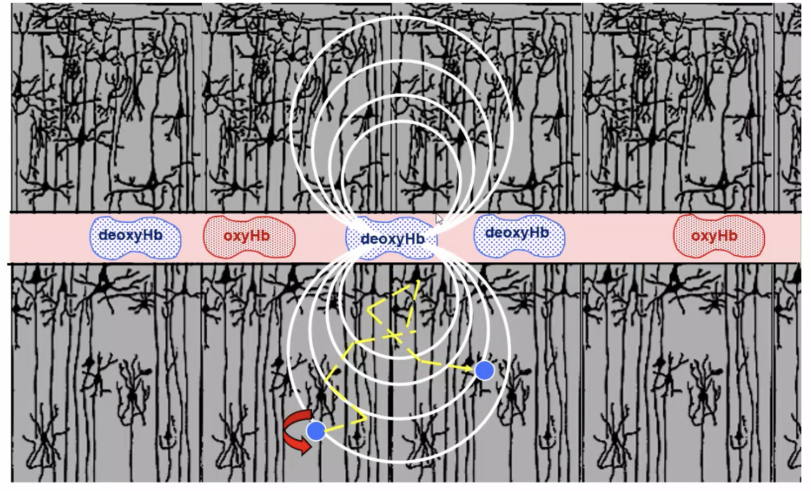
How does oxygenation affect haemoglobin’s magnetic properties?
Oxyhaemoglobin = weakly magnetic
deoxyhaemoglobin = paramagnetic because removal of oxygen exposes the iron core.
What effect does deoxyhaemoglobin have on the MRI magnetic field?
It creates local magnetic field inhomogeneities around blood vessels, causing nearby water molecules to experience different magnetic field strengths.
How does deoxyhaemoglobin affect the MRI magnetic field+ proton behaviour?
It creates local field inhomogeneities, causing nearby protons to experience different magnetic fields, precess at different frequencies, and rapidly dephase.
What is the effect of dephasing on MRI signal and T2*?
Dephasing causes signal cancellation → faster signal decay → shorter T2* → lower signal intensity.
What is the relationship between blood oxygenation and T2*-weighted MRI signal?
Higher oxygenation → less deoxyhaemoglobin → less field disturbance → longer T2* → higher signal;
lower oxygenation → lower signal.
What is the key principle behind BOLD fMRI?
MRI signal changes reflect blood oxygenation levels because deoxyhaemoglobin alters magnetic field uniformity and signal decay.
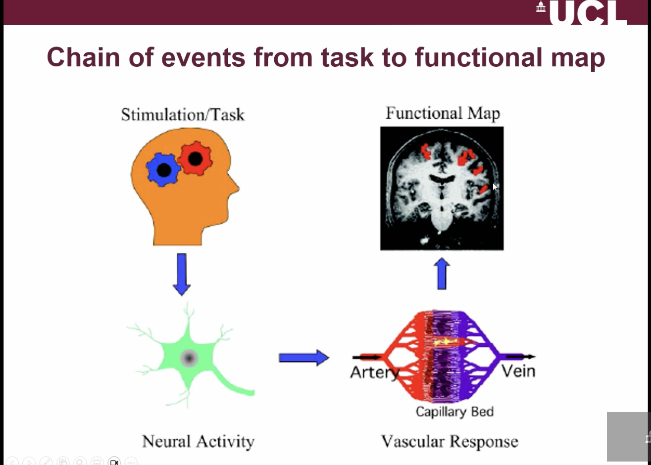
What is the sequence of events in fMRI (BOLD imaging)?
A task or stimulus → triggers neuronal activity
Neurons signal nearby blood vessels (neurovascular coupling)
This causes changes in blood flow, volume, and oxygenation
These changes alter deoxyhaemoglobin levels, producing the MRI (BOLD) signal
Result → functional brain maps showing active regions
What is neurovascular coupling?
The process by which neuronal activity signals blood vessels to increase local blood flow to meet metabolic demand.
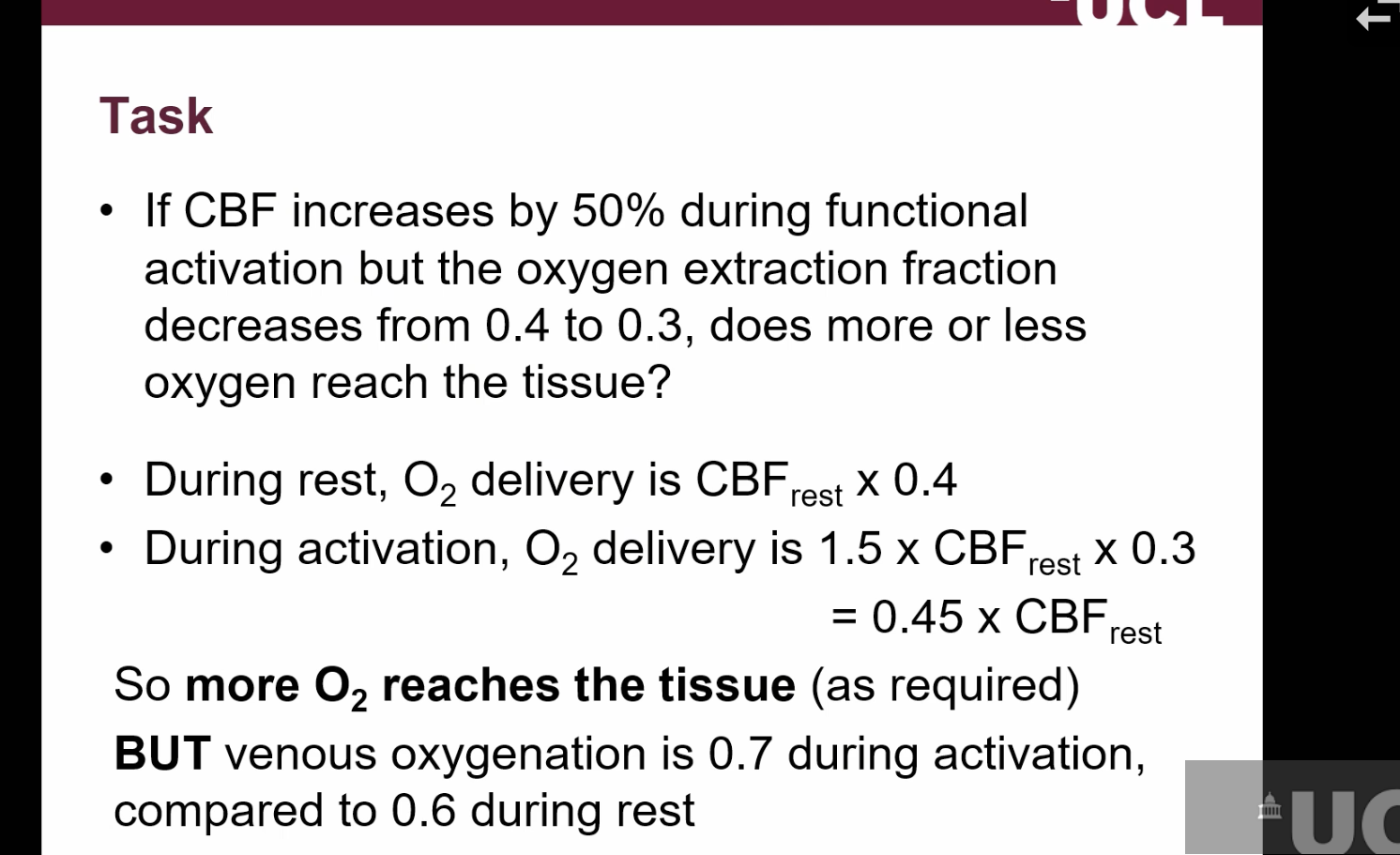
what happens during neuronal activation?
cerebral blood flow increases to accommodate for the increased metabolic demand/glucose + oxygen
BUT because blood flow is passing through the capillaries more quickly, the efficacy of oxygen being extracted from the blood is reduced(extraction fraction)
this results in concentration of deoxyhaemoglobin in the veins decreasing on activation.
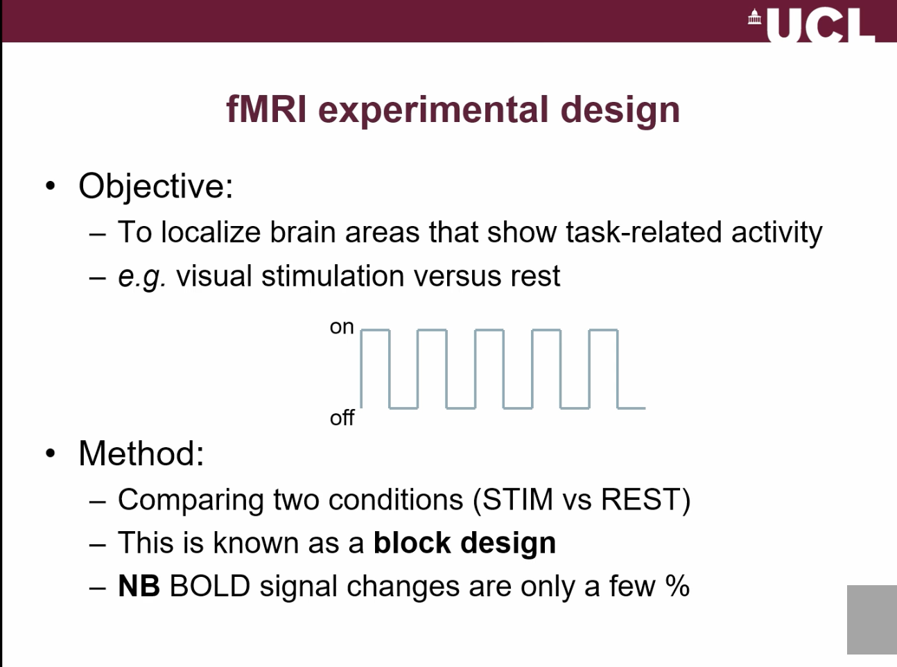
How is an fMRI experiment typically designed?
Using a block design: alternate stimulus ON and OFF periods while continuously acquiring T2*-weighted images to detect signal changes.
what is the objective of fMRI experimental design?
What is the hemodynamic delay and why is it important?
A few-second lag between neural activity and blood response; must be accounted for to correctly detect activation.
What are the two main analysis methods in fMRI?
1. Simple subtraction (ON vs OFF)
Idea: Compare “stimulus” vs “rest”
Take all the brain images when the stimulus is ON
Take all the images when it is OFF
Average each group
Subtract them
👉 What’s left = areas that are more active during the stimulus
2. Correlation analysis (pattern matching)
Idea: Look for signals that follow the same pattern as the stimulus over time
You know when the stimulus was ON and OFF (a pattern like: ON–OFF–ON–OFF)
For each pixel in the brain:
Check if its signal goes up and down in the same pattern
If it matches well → that area is active
👉 It’s like asking: “Does this pixel behave like the stimulus?”
Simple analogy:
Like listening to music and trying to find:
Which lights are flashing in time with the beat
If a light flashes in sync → it's “connected” to the music
for every pixel do statistical test to see if there is correlation between measured signal + expected Time course of an activated pixel.
How are fMRI results typically displayed?
As activation maps with coloured hotspots showing regions where signal correlates with the stimulus.
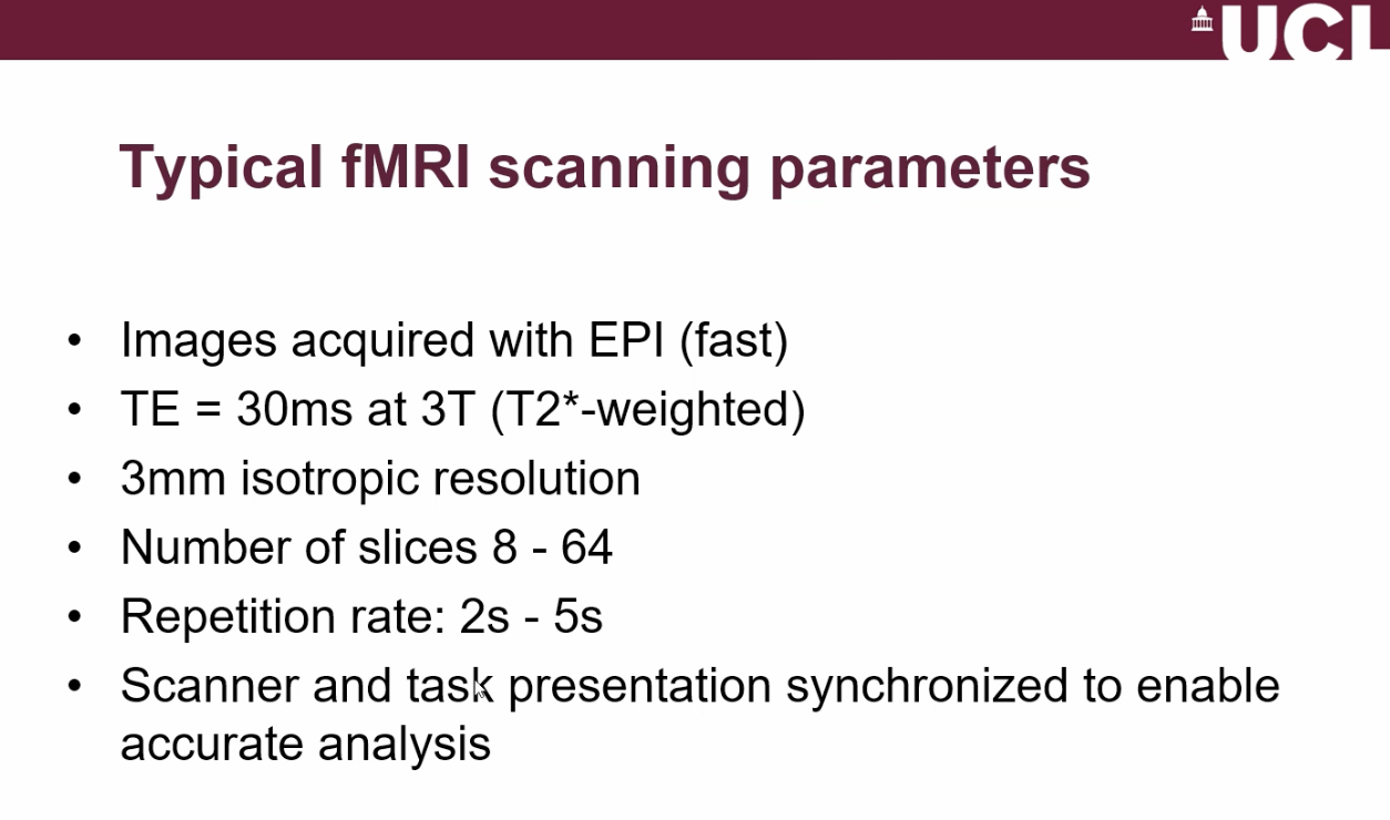
What technology enables fast fMRI acquisition?
Echo Planar Imaging (EPI), allowing whole-brain scans in seconds. - Peter Mansfield 1977.
What are the limitations of EPI in fMRI?
Lower spatial resolution and possible image distortions in exchange for very fast imaging
What are the main MRI scanner requirements for fMRI?
Needs high stability to detect very small signal changes (a few %)
Requires large data storage due to rapid, continuous image acquisition
Uses T2*-weighted imaging with moderate spatial resolution (~3 mm)
What imaging method is used in fMRI and why?
T2*-weighted imaging, because it is sensitive to blood oxygenation (BOLD contrast).
What are the fMRI scanning parameters/settings?
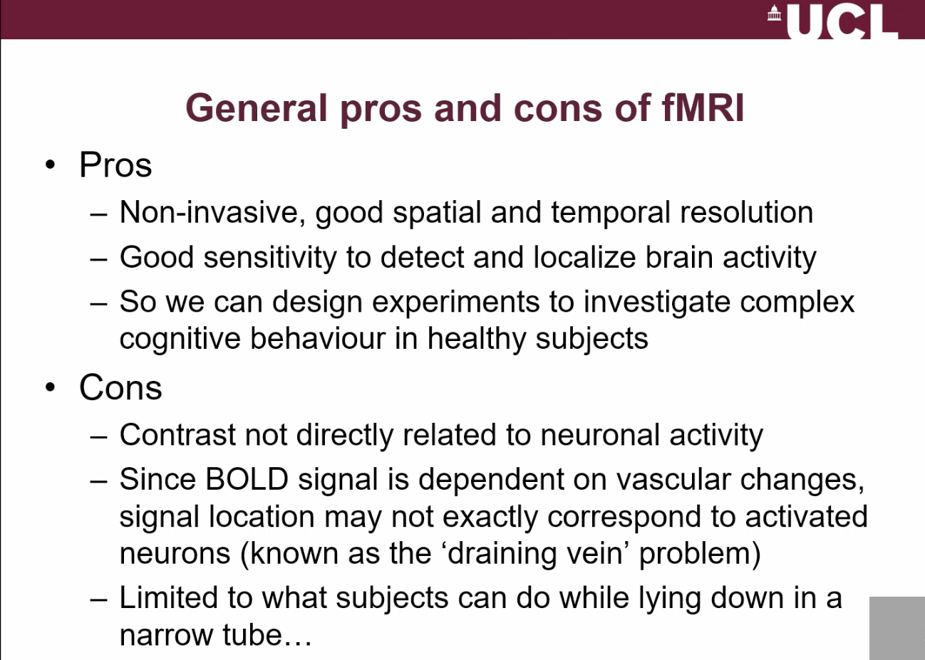
What are the advantages + limitations of fMRI?