cardiac cycle
1/21
There's no tags or description
Looks like no tags are added yet.
Name | Mastery | Learn | Test | Matching | Spaced | Call with Kai |
|---|
No analytics yet
Send a link to your students to track their progress
22 Terms
what is the cardiac cycle
the 3 stages in which the myogenic cardiac muscle (in walls of the heart) contracts and relaxes to pump blood - one complete cycle creates a heart beat
what is diastole
relaxation
what is systole
contraction
what are the 2 types of systole
atrial systole
ventricular systole
what is AP
atrial pressure = blood pressure inside the atria
what is VP
ventricular pressure = blood pressure inside the ventricles
outline the 3 stages of the cardiac cycle
1.atrial systole
2.ventricular systole
3.diastole
what is the AoP
aortic pressure = blood pressure in the aorta, as blood leaves the heart from the ventricles
what occurs during atrial systole
1. atria contract causing AP increase (as atria contracts the space gets smaller but since there is still the same volume of blood so pressure incr.)
2. AV valves open causing blood to be pushed into ventricles (AP > VP, a pressure gradient is generated forcing AV valves open)
what occurs during ventricular systole
ventricles contract causing VP increase, while atria relax causing AP decrease
AV valves close (VP > AP blood tries to move backwards into atria therefore valves close to prevent backflow of blood)
SL valves open causing blood to be pushed into arteries (VP > arteries e.g. AoP, a pressure gradient is generated forcing SL valves open)
what occurs during diastole
ventricles and atria relax causing VP decrease
2. SL valves close (AoP > VP blood tries to move backwards into ventricles therefore valves close to prevent backflow of blood)
3. blood flows passively into atria (through vena cava and pulmonary artery) and ventricles so cycle restarts
How many seconds roughly does one cardiac cycle take
0.7 - 0.8s
draw diagram for cardiac cycle + label
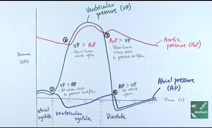
What side of the heart does this graph apply to
left side of the heart
» graphs will always present left side of heart because pressure changes are much greater
why is VP significantly bigger/ higher than AP
because the ventricle walls are much thicker than atria walls meaning they can contract more strongly
…
both ventricles and both atria contract together
why would there never be whole heart systole
-blood moves through the heart via pressure gradient:
1. atrial systole = atria > ventricles because ventricles are at lower pressure
2. ventricular systole = ventricles > arteries because arteries are at lower pressure
» if whole heart systole occurred the atria and ventricles would contract at the same meaning
…
why is there whole heart diastole
entire cardiac muscle is relaxed meaning pressure in all four chambers is lower than pressure of incoming veins = allows heart chambers to passively fill up with blood
what is cardiac output
volume of blood pumped by 1 ventricle in 1 minute/ per minute
how do you calculate cardiac output
Cardiac Output = Heart Rate X Stroke Volume
define what is Heart Rate and Stroke Volume
Heart Rate - number of beats per min
Stroke Volume - volume of blood pumped out the ventricles with each beat
why is cardiac output useful
to calculate how efficiently the heart is supplying body with oxygenated blood