KU BIOL 480 Exam 1
1/83
There's no tags or description
Looks like no tags are added yet.
Name | Mastery | Learn | Test | Matching | Spaced | Call with Kai |
|---|
No analytics yet
Send a link to your students to track their progress
84 Terms
definition of parasitism
An organism that associates with a host at least once throughout their life with interspecific association
Pathogen
When harm is expressed (not all parasites)
Pathogenicity
The property of causing a disease
Ectoparasite
Lives outside the host
Endoparasite
Live inside the host
Mesoparasite
Lives with a portion of their body inside/outside
Zoonosis
Can transfer from animals to human
reservoir host
infected organism (usually an animal) which makes the parasite available for transmission to other hosts
Definitive Host
Parasite undergoes sexual reproduction
Intermediate Host
Parasite develops but NO sexual reproduction
Paratenic
Parasite has no development
Prevalence
# of individuals in a pop infected (50 pigs, 10 infected, Prevalence = 20%)
Intensity
# of individuals of a PARASITE per infected host (10 pig, 250 worm, mean intensity = 25 worm) (1 pig, 25 worm, intensity = 25 worm)
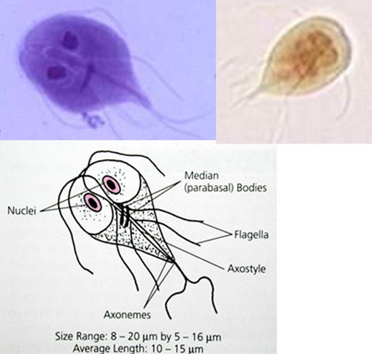
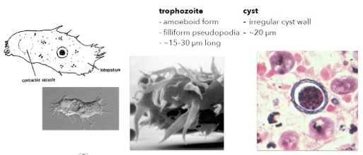
Trophozoite
Membrane-bound, motile, active feeding stage, pathogenic stage
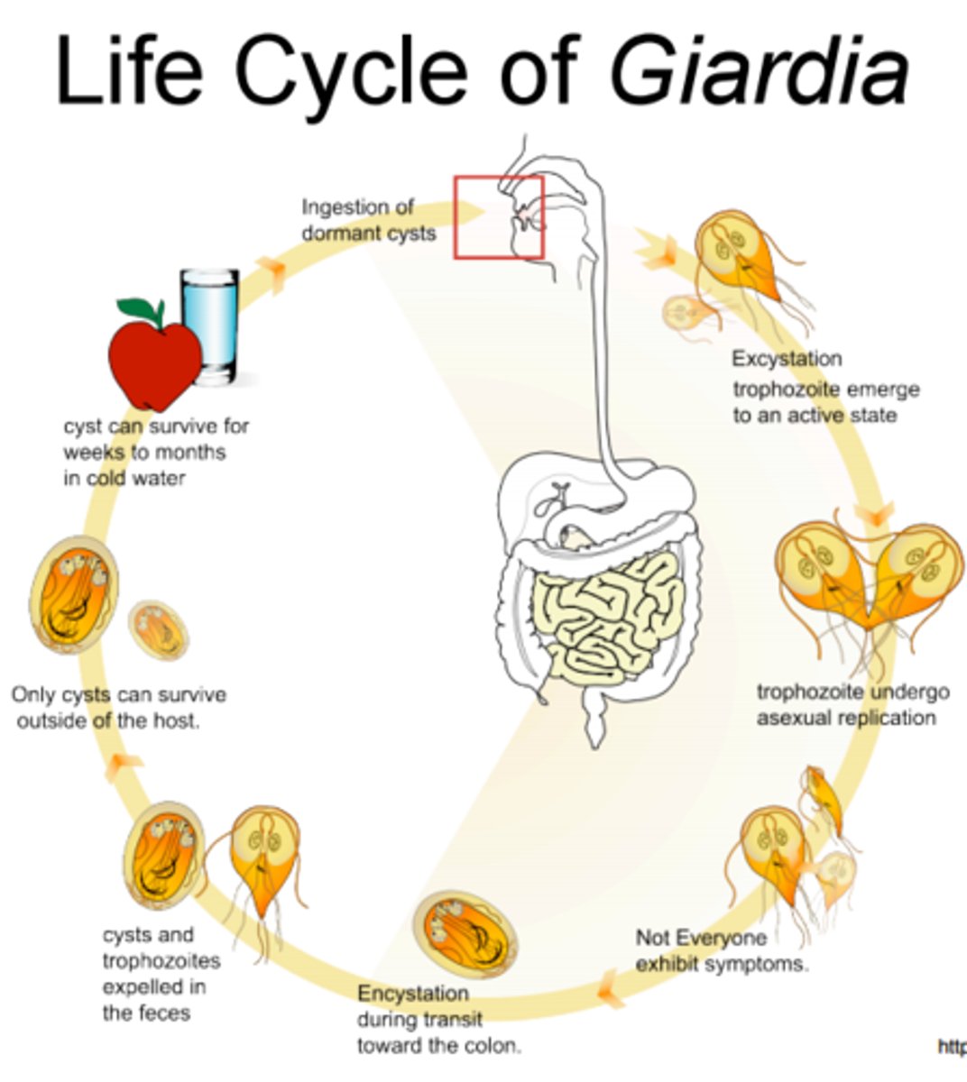
Cyst
Outer protective wall, able to survive outside of host, resting stage, critical for diagnosis
Giardia duodenalis
host(s), zoonotic? site in host,
Zoonotic, multiple species serve as host, asexual reproduction, gastrointestinal tract (Small Intestine)
disease caused, sources of pathogenicity
Giardiasis, Direct contact/ Toxic substance produced by parasite (antigenic)/ Loss of absorptive surface
diagnosis (life stage and type of sample), symptoms, treatment, transmission.
Cysts in stool, Fat in stool/ Explosive Diarrhea/ Dehydration/weight loss/ intestinal discomfort, Antibiotics
Transmission = 6 Fs (Fingers, food, feces, flies, fluids, fomites (object capable of carrying infection))
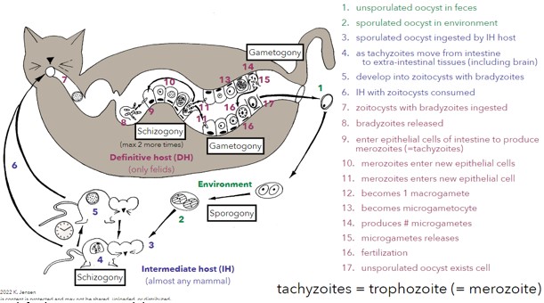
Giardia duodenalis Life Cycle
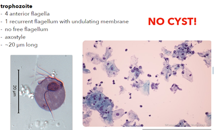
Phylum Parabasalia: Order Trichomonadida: general features
undulating membrane, recurrent flagellum, axostyle
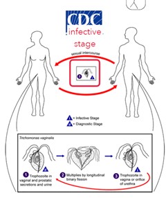
Trichomonas vaginalis
host(s), site in host,
Monoxenous, Upper vagina and urethra (female)/ urethra and prostate (males)
disease caused, sources of pathogenicity,
Trichomoniasis, genitals, spreads though sexual intercourse
diagnosis (life stage and type of sample), symptoms, treatment (if any), transmission;
Vaginal smear for trophozoites (Nucleic acid amplification test); intense itching, possible tissue damage, asymptomatic in males; antibiotics
Trichomonas vaginalis Life Cycle
Trichomonas gallinae
host(s), monoxenous or heteroxenous?,
Birds, monoxenous -
site in host, transmission, disease caused;
Mouth & throat; feeding/contamination; Avian trichomoniasis
Tritrichomonas foetus
host(s), monoxenous or heteroxenous?,
Cattle; monoxenous
site in host, pathology, transmission, disease caused
Vagina & uterus in cows, preputial cavity in bulls; abortion in cattle, infertility in cows, bulls asymptomatic; Venereal disease cattle (intercourse)
problem with cryopreservation
Trophozoite can survive cryopreservation
Histomonas meleagridis
cyst?, monoxenous or heteroxenous?, host(s),
No cyst; Heteroxenous (intestinal nematode of birds as intermediate host); Gallinaceous birds
site in definitive host, disease caused.
Lesions in caecum, liver; Histomonas (Blackhead disease)
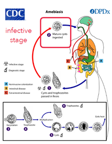
Entamoeba histolytica
host(s), site occupied within host (1º & 2º [i.e., ectopic] infections)
Monoxenous (Humans); 1º: large intestine, 2º: ectopic sites if the conditions are met
disease caused, pathogenicity, symptoms, diagnosis of infection, treatment, transmission.
Amoebiasis (Amoebic Dysentery); Cyst both infective and diagnostic stage; dysentery, 4-6 loose stools per day, cramps, vomiting, blood in stool; Diagnosis: Large intestine = cysts or trophs in feces, ectopic = x-ray; Antibiotics; fecal contamination of water/food
![<ul><li><p>host(s), site occupied within host (1º & 2º [i.e., ectopic] infections)</p><ul><li><p>Monoxenous (Humans); 1º: large intestine, 2º: ectopic sites if the conditions are met</p></li></ul></li><li><p>disease caused, pathogenicity, symptoms, diagnosis of infection, treatment, transmission.</p><ul><li><p>Amoebiasis (Amoebic Dysentery); Cyst both infective and diagnostic stage; dysentery, 4-6 loose stools per day, cramps, vomiting, blood in stool; Diagnosis: Large intestine = cysts or trophs in feces, ectopic = x-ray; Antibiotics; fecal contamination of water/food</p></li></ul></li></ul><p></p>](https://knowt-user-attachments.s3.amazonaws.com/5e4f394c-24b4-4201-8241-995e9d8b1231.png)
Entamoeba histolytica Life Cycle
Non-pathogenic species that parasitize humans
Entamoeba coli, E. dispar, E. hartmanni, E. moshkoviski, Endolimax nana, lodamoeba buetschlii all in large intestine
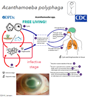
Acanthamoeba polyphaga
disease caused, pathogenicity, transmission (multiple possible sources of infection), normal habitat.
Acanthamoeba keratitis (AK); ulceration of cornea; hot tubs and contaminated contact lens fluid; topical antibiotics; commonly found in lakes, swimming pools, tap water, and heating and air conditioning units
Acanthamoeba polyphaga Life Cycle
ALVEOLATA: Phylum Ciliophora
general features (macro and micronuclei, etc.), mode of locomotion
Multiple Cilia, Infraciliature, Macro/Micronucleus
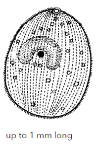
Balantidoides coli
host(s), site, disease caused, pathogenicity, diagnosis of infection, treatment, transmission;
Zoonotic (pigs as reservoir), monoxenous; only ciliated and the largest protozoon known to infect humans; Balantidiosis (asymptomatic in pigs); produces enzyme lyse host cell, ulcer of colon (could perforate -> ectopic); cecum & colon in cyst form (feces); antibiotic; water as vehicle or human-human transmission+
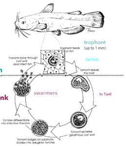
Ichthyopthirius multifilis
host(s), site, disease caused, pathogenicity, diagnosis of infection.
Freshwater fish, monoxenous; small white spots on fish; look for trophonts
Ichthyopthirius multifilis life cycle
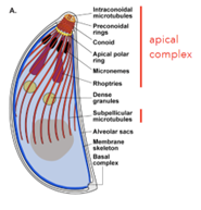
Phylum Apicomplexa
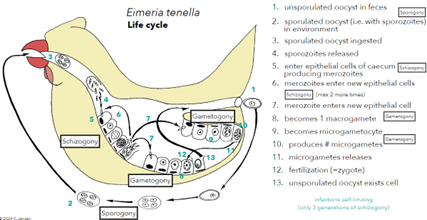
basic features, key organelles of apical complex;
Apical complex (penetration of host cells (conoid), made of secretory organelles and specific cytoskeletal elements), all intracellular parasitic, single nucleus, no pseudopods, cilia, or flagella ext. microgametes)
Phylum Apicomplexa three distinct reproductive processes
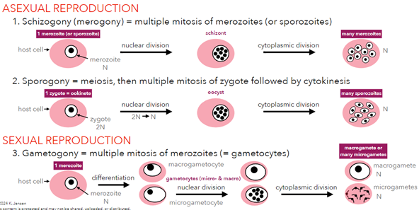
Phylum Apicomplexa initial life-cycle stages
Subphylum Aconoidasida: distinguishing feature
Conoid (tip) only in ookinete stage
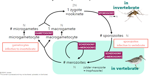
Order Haemospororida
general features (sporozoites naked, etc.)
Life in blood cells at sometime in life, zygote motile, sporozoites naked, heteroxenous
diversity (three main genera with ~475 species)
Plasmodium, Hemoproteins, Leucocytozoonosis
Plasmodium
types of vertebrate hosts, types of invertebrate hosts;
Lizards, birds, monkeys
1 zoonotic species and its reservoir hosts
Plasmodium knolesil; Reservoir host: macaques (monkey)
Plasmodium Life Cycle
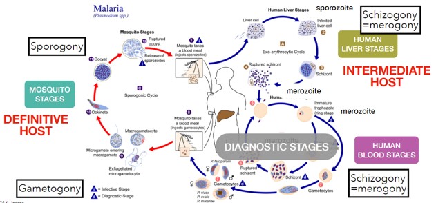
Plasmodium falciparum
(50%) MOST DEADLY
Malignant Tertian Malaria, infects all ages, relapse 1-3 years
Gametocytes banana-shaped, 65% of RBC, blood becomes “sticky”
Symptoms: Cytoadherence (sticky), rosetting (binding of RBCS), RBC Cancer
Plasmodium vivax
(43%)
Benign Tertian Malaria, infects young RBC, only 1 merozoite/RBC, relapse 8 years
Gametocyte circular-shaped, lots of Schüffner’s dots
Symptoms: Rosetting (binding of infect/uninfected RBC, causes clots)
Plasmodium malariae
(7%)
Quartan Malaria, Paroxysms 72 hr, infects aging RBC, 1 parasite/20,000 cells, relapse 53 year
Band-shaped trophozoite
Plasmodium ovale
(1%)
Mild Tertian Malaria, Paroxysms 48 hr, infects young RBC, relapse months
Fewer merozoites per schizont
Gametocyte circular-shaped w/ red, more little dots
Plasmodium Symptoms
Jaundice, Anemia, Enlarged spleen
Paroxysm
Periodic fever/chills caused by release of erthocytic merozoites into bloodstream
P. vivax: every 48 hrs.
P. ovale: every 48 hrs.
P. malariae: every 72 hrs.
P. falciparum: every 48 hrs, not as synchronous
Relapse
When paroxysms reappears after a number of paroxysm-free months (P. ovale) or years (P. vivax)
From 2nd population of dormant sporozoites in the liver becoming active
Recrudescence
When paroxysms reappears after a number of paroxysm free years (P. falciparum and P. malariae)
From merozoites remaining low until rapid schizogony
Strategies of malaria control
elimination of mosquito hosts (= vector control; destroy mosquito breeding sites, etc.), treat infected people (antimalarials, ACT, mass drug administration), prevent uninfected people from becoming infected (mosquito nets, vaccines, spraying), individual resistance to malaria
Babesia bigemina
intermediate and definitive hosts
Def: boophilus (tick), Inter: cattle, deer, zebu, water buffalo
disease caused, pathology
Babesiosis (Texas red-water fever); destroys RBCs, blood in urine
Babesia microti
intermediate, definitive, and reservoir hosts
Def: Ixodes scapularis (deer tick), Inter: humans, Reservoir: meadow moles ZOONOTIC!!
geography, pathogenicity
Babesiosis (Nantucket fever), fever, chills, fatigue, headache
Subphylum Conoidasida: distinguishing features
Conoid in all life cycle stages, zygote not motile, sporozoites in cysts, monoxenous, sporogony (asexual) occurs outside of host
Order Coccidia: general features
Parasites of vertebrates (incl. humans), Mature gametocytes intracellular, Undergoes schizogony
Eimeria morphology and lifecycle
4 sporocysts, 8 sporozoites, monoxenous
Cystoisospora morphology and lifecycle
2 sporocysts, 8 sporozoites, monoxenous

Cyclospora morphology and lifecycle
2 sporocysts, 4 sporozoites, monoxenous

Toxoplasma morphology and lifecycle
2 sporocysts, 8 sporozoites, heteroxenous

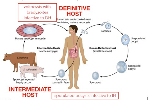
Sarcocystis morphology and lifecycle
2 sporocysts, 8 sporozoites, heteroxenous

Cryptosporidium morphology and lifecycle
0 sporocysts, 4 sporozoites, monoxenous

Eimeria
incredible diversity of species (1,500+ species),
Host and Tissue specific, each species has a specific host
infections self-limiting (what does this mean, contrast to Haemospororida?), general name for diseases caused
Self-limiting, infection will run its course and resolve on its own without external intervention
Only 3 cycles of schizogony, while Haemospororida will infinitely reproduce
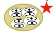
Eimeria tenella
pathogenicity, why is parasitemia of cells of mucosa of caecum so high?
Specific to epithelial cells of domestic chickens
Parasitemia of cells high due to constant generation cycling.
Eimeria tenella Life Cycle
Cystoisopspora belli
host(s), individuals most at risk, disease caused, pathologenicity
Humans; immunocompromised individuals; Cystoisoproiasis; epithelial cells of jejunum of intestine, can cause death
Cyclospora cayatensis
symptoms, pathology, individuals most at risk;
Cyclosporiasis; fatigue, diarrhea, weight loss; immunocompromised, self-liming for only immunocompetent
Toxoplasma gondii
infective stages to each host
Sporozoite (C): in oocysts (environment), infective to intermediate hosts
Tachyzoite (A): highly active form (cats and human), in cells of any organs or blood, disease stage
Bradyzoite (B): still in cats and humans, in zoitocyst (tissue cyst)
Serious in pregnant women (fetus), immunocompromised; transmits through food-bone, zoonotic, and congenital
Toxoplasma gondii Life cycle
Sarcocystis Life Cycle
distinctive rice grained zoitocyts
Cryptosporidium parvum
hosts: Zoonotic, +150 spp. Of vertebreates as reservoir hosts, monoxenous
disease caused, pathogenicity, diagnosis, common transmission scenario
Cryptosporidiosis; infects intestinal cells; oocysts in feces (lacks sporocytes); fecal contamination surfaces or water;
potential issue with phylogenetic affinities (Order Cryptosporidia)
Each treatment is extremely different
Order Gregarinasina
association with mandibulate arthropods only
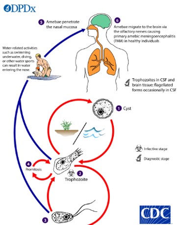
Naegleria fowleri
Facultative Parasite
disease caused, pathogenicity, diagnosis (life stage and type of sample), treatment(?), transmission
PAM (Primary Amoebic Meningoencephalitis); Trophozoites into nasal passages, enters brain via cribriform plate, lyse brain tissue with amoebocytes; diagnosis and treatment difficult (PCR)
Naegleria fowleri Life Cycle
Monoxenous, Facultative, migrates to brain via olfactory nerves
Class Kinetoplastea
general features
Reproduces asexually via binary fission, vertebrate considered definitive host, invertebrate intermediate
Kinetoplast: a specialized region of the mitochondria of trypanosomatids that harbors the most complex and unusual mitochondrial DNA found in nature
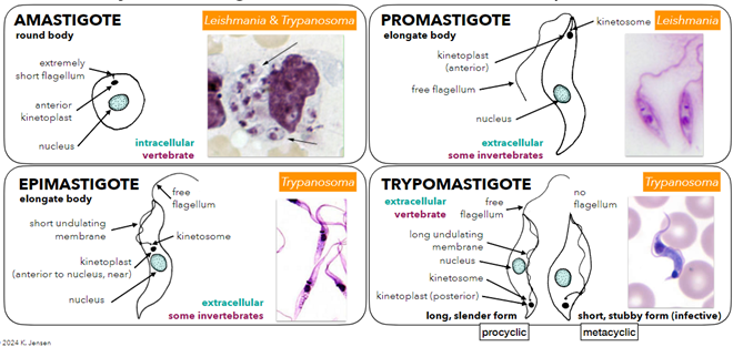
4 major trophozoite forms
Amastigote:
Round body, extremely short flagellum, antieror kinetoplast, intracellular vertebrate
Promastigote:
Elongated body, normal kinetosome, free flagellum, extracellular invertebrates
Epimastigote
Elongated body, kinetoplast near nucleus, short undulating membrane, extracellular invertebrates
Trypomastigote: Posterior kinetoplast, Extracellular vertebrate
Pericyclic: long slender form
Free flagellum
Metacyclic: Short stubby form (infective)
No flagellum
Anterior station species
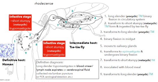
Trypanosoma brucei
Posterior station species
Trypanosoma cruzi, Leishmania
Trypanosoma brucei
Rhodesiense (8%)
Definite host: Humans
Reservoir hosts: Zoonotic (rhinos, hyenas)
Intermediate: Glossina morsitans (savannah)
Disease: Acute African sleeping sickness (East)
Fatal in 6 months,
Gambiense (92%)
Definitive host: Humans
Reservoir hosts: Zoonotic (pigs, bulls)
Intermediate: Glossina palpalis (forest)
Disease: Chronic African sleeping sickness (West)
Brucei
Definite host: Bulls, Horse, Donkey
Reservoir hosts: None
Intermediate: Glossina paldipes
Disease: Nagana (sleeping sickness in cattle)
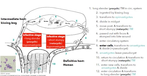
Trypanosoma cruzi
morphology, life cycle, hosts (intermediate, definitive and reservoir)
Definitive host: Humans
Reservoir hosts: Zoonotic o Intermediate: Triatoma Rhodnius (kissing bug)
Disease: South American sleeping sickness
site occupied within hosts
Cardiac and skeletal muscle
disease caused, pathogenicity (acute and chronic phases of Chaga’s disease), diagnosis,
Changas Disease
Acute; affects children, romana sign (swollen eye at bite)
Chronic; adults, 70% are asymptomatic, could affect the heart or digestive organs
explanation for why treatment is difficult, transmission, control, geographic distribution, recognize diversity of species in other hosts
Transmits through vector and blood transfusions; does not respond well to drugs
Trypanosoma cruzi Life Cycle
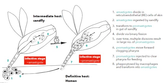
Leishmania
general features (only promastigotes and amastigotes, etc.);
No trypomastigote form, All species with amastigote form in vertebrate, all species with promastigote form in invertebrate, Generally heteroxenous (human/sandfly)
Leishmania Life Cycle
Cutaneous leishmaniasis tropica
Host: Human (Def), Phelebotomus (Inter, Old World), Rodents (vector, zoonotic)
Site: At bite, in reticuloendothelial system (lymph)
Disease/ Pathogenicity: Cutaneous Leishmaniasis (Oriental Sore); ulcerating dry lesions, painless, frequently multiple, self healing
Diagnosis: Amastigotes in skin lesions
Geographic Dist.: Middle East, India
Treatment: Protective immunity
Cutaneous leishmaniasis major
Host: Human (Def), Phelebotomus (Inter, Old World), Rodents (vector, zoonotic)
Site: At bite, in reticuloendothelial system (lymph)
Disease/ Pathogenicity: Cutaneous Leishmaniasis (Oriental Sore); Rapid necrosis, multiple wet sores, inflammation, slow to heal, severe scarring
Diagnosis: Amastigotes in skin lesions
Geographic Dist.: N/W Africa, Middle East, Central Asia
Treatment: Protective immunity
Cutaneous leishmaniasis mexicana
Host: Human (Def), Lutzomyia (Inter, New World), Rodents and Oppossum (vector, zoonotic)
Site: cutaneous; nasopharyngeal and visceral (rare)
Disease/ Pathogenicity: Chicloro ulcer (Bay sore); Ulcerating lesions, single or multiple, often self-healing
Diagnosis: Amastigotes in skin lesions
Geographic Dist.: Central and South America
Treatment: Protective immunity
Mucocutaneous leishmaniasis
Host: Human (Def), Lutzomyia (Vector, New World)
Site: At bite to mucocutaneous tissue
Disease/ Pathogenicity: Espundia, ulcerating lesions that’s disfiguring, degenerates’ cartilage
Diagnosis: amastigotes from mucosal lesions and lymph node
Geographic Dist.: Central and South America
Treatment: Pentavalent antimonial
Visceral leishmaniasis
Host: Human (Def), Phlebotomus (Vector, Old World)
Site: In cells of reticuloendothelial system (spleen, bone marrow, lymph nodes)
Disease/ Pathogenicity: Visceral Leishmaniasis, Persistent fever, splenomegaly, weight loss, rash, fatal within two years
Diagnosis: amastigotes in smear, PCR detection
Geographic Dist.: Ethiopia, Sudan, Kenya, India, China, Bangladesh, Burma
Treatment: Miltefosine