(MINE) Interpret EKG Rhythms
1/65
There's no tags or description
Looks like no tags are added yet.
Name | Mastery | Learn | Test | Matching | Spaced | Call with Kai |
|---|
No analytics yet
Send a link to your students to track their progress
66 Terms
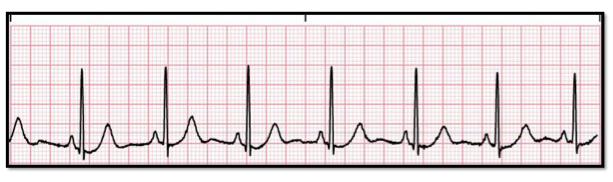
Identify:
a. normal sinus rhythm
b. sinus bradycardia
c. 1st degree heart block
d. sinus tachycadia
a. normal sinus rhythm
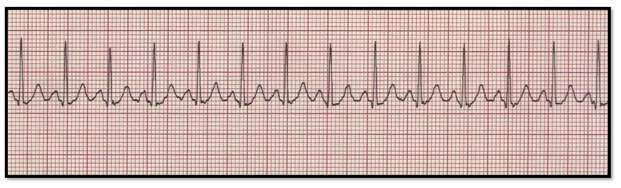
Identify:
a. normal sinus rhythm
b. sinus arrhythmia
c. atrial fibrillation
d. sinus tachycadia
d. sinus tachycadia
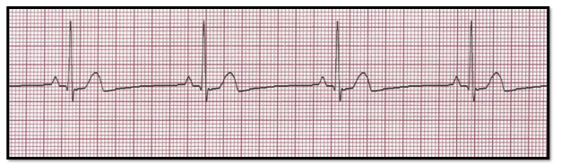
Identify:
a. normal sinus rhythm
b. sinus bradycardia
c. 1st degree heart block
d. sinus arrhythmia
b. sinus bradycardia
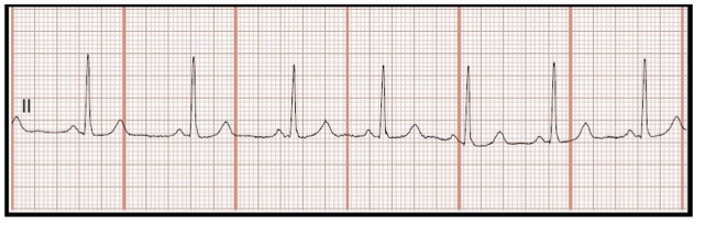
Identify:
a. normal sinus rhythm
b. sinus bradycardia
c. 1st degree heart block
d. sinus arrhythmia
d. sinus arrhythmia
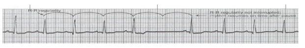
Identify:
a. sinus block
b. sinus arrest
c. 1st degree heart block
d. sinus arrhythmia
a. sinus block
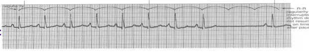
Identify:
a. sinus block
b. sinus arrest
c. 1st degree heart block
d. sinus arrhythmia
b. sinus arrest.
Note is sinus arrest and not a block. It is a sinus arrest because the rhythm is regular, but missing more than 2 QRS’s?
(if it was missing only one and regular then it is sinus block?)
Identify the above waveform:
a. Atrial flutter
b. Atrial fib
c. Ventricular fib
d. Ventricular tach
b. Atrial fib
Identify the above waveform:
a. Atrial flutter
b. Atrial fib
c. Ventricular fib
d. Ventricular tach
a. Atrial flutter
PVCs are most dangerous if they:
a. Are multiformed and increase in frequency.
b. Appear wide and bizarre.
c. Occur after the T wave.
d. Are uniform and wide.
a. Are multiformed and increase in frequency.
The term pulseless electrical activity refers to a condition in which there's:
a. An extremely slow heart rate but no pulse.
b. Asystole on a monitor or rhythm strip.
c. Electrical activity in the heart but no actual contraction.
d. Asystole and a palpable pulse and blood pressure.
c. Electrical activity in the heart but no actual contraction.
Identify the above waveform:
a. Atrial flutter
b. Atrial fib
c. Ventricular fib
d. Ventricular tach
d. Ventricular tach
Identify the above waveform:
a. Atrial flutter
b. sinus bradycardia
c. PVC (premature vent contraction)
d. PAC (premature atrial contraction)
d. PAC (premature atrial contraction)
Identify the above waveform:
a. SVT or atrial tachycardia
b. VTACH
c. PAC
d. AFIB
a. SVT or atrial tachycardia
Identify the above waveform:
a. Atrial flutter
b. Atrial fibrillation
c. PVC (premature vent contraction)
d. PAC (premature atrial contraction)
a. Atrial flutter
Identify the above waveform:
a. Atrial flutter
b. Atrial fibrillation
c. SVT or atrial tachycardia
d. sinus tachycardia
b. Atrial fibrillation
Identify the above waveform:
a. Atrial flutter
b. Atrial fibrillation
c. PVC (premature vent contraction)
d. PAC (premature atrial contraction)
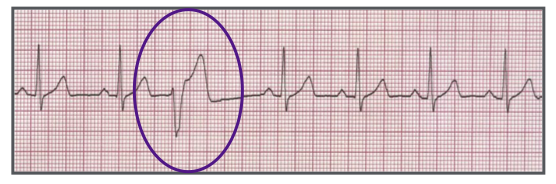
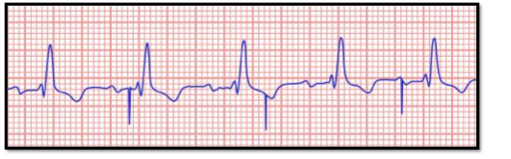
c. PVC (premature vent contraction)
Identify the above waveform:
a. Paired PVCs
b. Trigeminy
c. Bigeminy
d. PAC (premature atrial contraction)
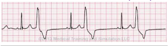
c. Bigeminy (PVC every other QRS, one yes and one no)
Identify the above waveform:
a. Paired PVCs
b. Trigeminy
c. Bigeminy
d. PAC (premature atrial contraction)
b. Trigeminy
ONE PVC every 2 QRS…
PVC — QRS — QRS — PVC
Identify the above waveform:
a. Paired PVCs
b. Trigeminy
c. Bigeminy
d. PAC (premature atrial contraction)
a. Paired PVCs (2 PVCs in a row)
Identify the above waveform:
a. unifocal PVCs
b. Trigeminy
c. Torsades
d. multifocal PVCs
d. multifocal PVCs
Identify the above waveform:
a. unifocal PVCs
b. Trigeminy
c. First degree heart block
d. multifocal PVCs
a. unifocal PVCs
Identify the above waveform:
a. unifocal PVCs
b. Trigeminy
c. Torsades
d. multifocal PVCs
c. Torsades
Identify the above waveform:
a. SVT or atrial tachycardia
b. VTACH
c. PAC
d. AFIB
b. VTACH
Identify the above waveform:
a. Torsades
b. V fib
c. A fib
d. V fib
a. Torsades (a type of V tach)
Identify the above waveform:
a. asystole
b. monitor malfunction
c. A fib
d. V fib
d. V fib = fine ventricular fibrillation
Identify the above waveform:
a. asystole
b. monitor malfunction
c. A fib
d. V fib
d. V fib = course ventricular fibrillation
Identify the above waveform:
a. asystole
b. monitor malfunction
c. A fib
d. fine V fib
a. asystole
In 2nd Degree Heart Block Type I, the PR interval:
a. Varies according to the ventricular response rate.
b. Progressively lengthens until a QRS complex is dropped.
c. Remains constant despite an irregular ventricular rhythm.
d. Is unmeasurable.
b. Progressively lengthens until a QRS complex is dropped.
Identify the above waveform:
a. First degree heart block
b. Second degree heart block Type I
c. Second degree heart block Type I|
d. Normal sinus rhythm
c. Second degree heart block Type I|
Identify the above waveform:
a. First degree heart block
b. Third degree heart block
c. Second degree heart block Type I|
d. Sinus bradycardia
b. Third degree heart block
Identify the above waveform:
a. First degree heart block
b. Third degree heart block
c. normal sinus rhythm
d. Second degree type I
a. First degree heart block
Identify the above waveform:
a. First degree heart block
b. Third degree heart block
c. normal sinus rhythm
d. Second degree heart block type I
a. First degree heart block
Identify the above waveform:
a. First degree heart block
b. Third degree heart block
c. normal sinus rhythm
d. Second degree heart block type I
d. Second degree heart block type I
Identify the above waveform:
a. First degree heart block
b. Third degree heart block
c. Second degree heart block type II
d. Second degree heart block type I
c. Second degree heart block type II
Identify the above waveform:
a. First degree heart block
b. Third degree heart block
c. Second degree heart block type II
d. Second degree heart block type I
b. Third degree heart block
Note: some P waves do not have a QRS, and the ones that do happen at different frequencies.
P and QRSs do not agree so it is a third degree
The identification of the above ECG is:
a. Atrial pacing 1:1
b. Ventricular pacing 1:1
c. Failure to capture
d. Failure to sense
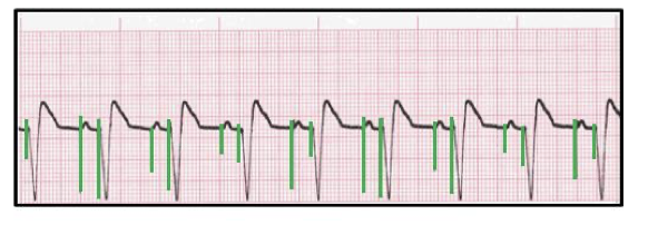
b. Ventricular pacing 1:1 → all the spikes are before the QRS indicating that the device generated a contraction
Failure to capture is represented on the ECG as:
a. No pacemaker activity.
b. Spikes where they shouldn't be.
c. A spike on a T wave.
d. A spike without a complex.
d. A spike without a complex. → Capturing– is when the heart has responded to the stimulus. Electrical capture is the pacing spike, followed by a P or QRS wave. A spike without a complex means the heart muscle did not capture the impulse (spike).
The identification of the above ECG is:
a. Atrial pacing 1:1
b. Ventricular pacing 1:1
c. Failure to capture
d. Failure to sense
d. Failure to sense → the device is not sensing when the contractions happened so it puts random spikes in random places. Sensing– ability of pacemaker to recognize the patient’s intrinsic beats.
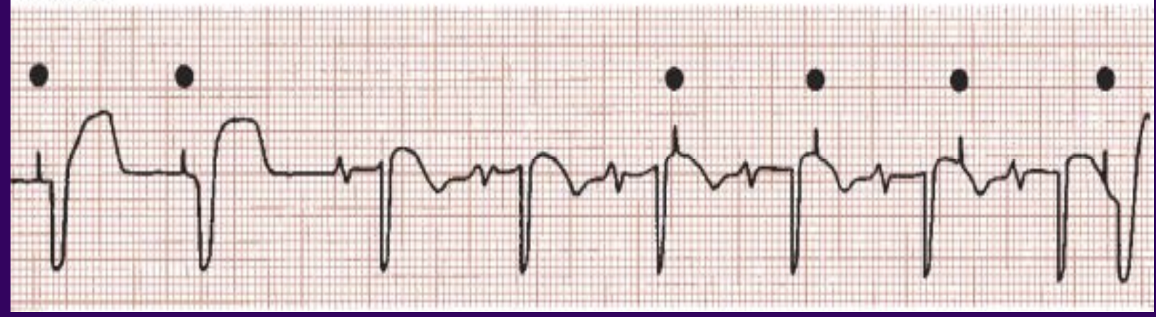
The identification of the above ECG is:
a. Atrial pacing
b. Ventricular pacing
c. Failure to capture
d. Failure to sense
a. Atrial pacing
The identification of the above ECG is:
a. Atrial pacing
b. Ventricular pacing
c. AV sequential pacing
d. Failure to sense
b. Ventricular pacing
The identification of the above ECG is:
a. Pacemaker malfunction
b. Ventricular pacing
c. AV sequential pacing
d. Failure to sense
c. AV sequential pacing
The identification of the above ECG is:
a. Failure to capture
b. Ventricular pacing
c. AV sequential pacing
d. Failure to sense
a. Failure to capture

The identification of the above ECG is:
a. Failure to capture
b. Ventricular pacing
c. AV sequential pacing
d. Failure to sense
d. Failure to sense
a. First degree heart block
b. Third degree heart block
c. atrial fibrillation
d. atrial tachycardia
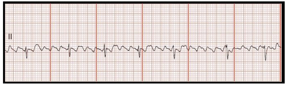
C. atrial fibrillation
This is atrial fibrillation. For atrial fibrillation, the rhythm will be irregular, and the P-waves will be characteristically absent and replaced with fine, chaotic waves. The ventricular rate is usually variable and may be slow, normal or rapid. Patients with symptoms will usually have a rapid ventricular rate. The PR interval will be absent, and the QRS complex is typically normal but can be widened in certain cases.
a. Atrial flutter
b. Third degree heart block
c. atrial fibrillation
d. atrial tachycardia
a. Atrial flutter
This is atrial flutter. Atrial flutter can be regular or irregular. For atrial flutter, the atrial rate is typically extremely fast. (250-350 bpm). The ventricular rate is typically slower than the atrial rate. Saw-toothed flutter waves are usually seen instead of a single P-wave. The PR interval will not be measurable and the QRS is typically normal (0.06-0.10 sec).
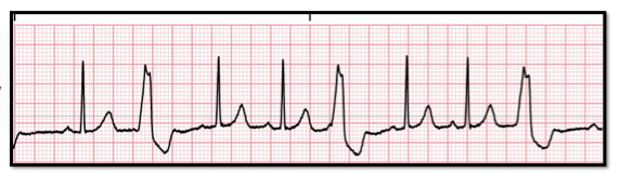
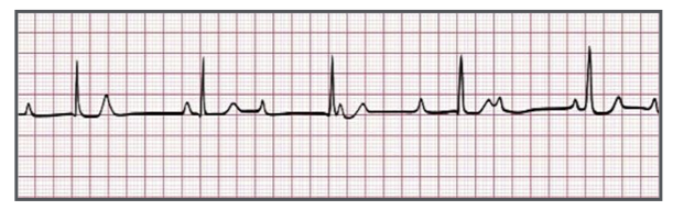
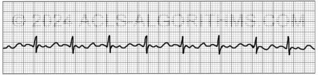
Identify:
a. normal sinus rhythm
b. sinus bradycardia
c. 1st degree heart block
d. sinus arrhythmia
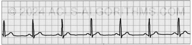
C. 1st degree heart block
I bet you picked NSR and did not count the stupid little boxes. ..
This is first-degree heart block. For first-degree heart block, the rhythm is regular. The PR interval will be prolonged at > 0.2. The QRS complex will usually be normal. First-degree heart block indicates slowed but not blocked conduction through the AV node.
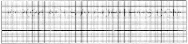
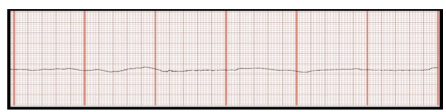
Identify the above waveform:
a. asystole
b. monitor malfunction
c. A fib
d. fine V fib
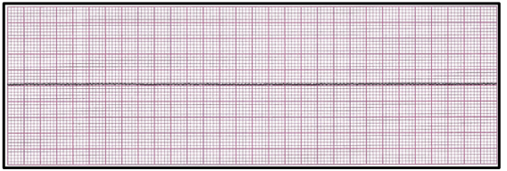
a. asystole
This is asystole. For asystole, there is no discernible electrical activity on the ECG monitor. The isoelectric line may also have very light and intermittent deviation away from the baseline. When asystole is present, there may be intermittent agonal rhythms which will usually have a wide and bizarre form.
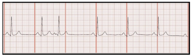
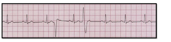
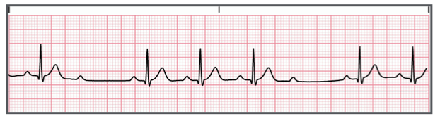
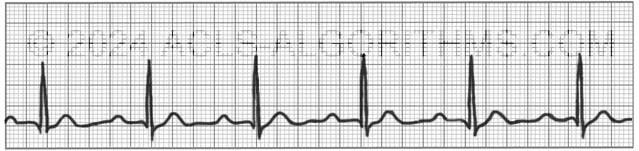
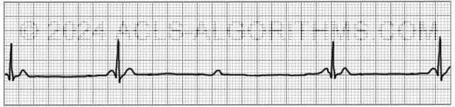
Identify:
a. normal sinus rhythm
b. sinus bradycardia
c. 1st degree heart block
d. sinus arrhythmia
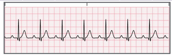
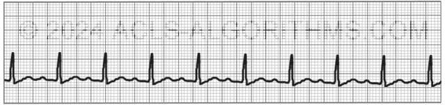
a. normal sinus rhythm
ahhh…you counted the stupid little boxes and picked NSR this time I hope!
This is normal sinus rhythm. For normal sinus rhythm, the rhythm will be regular, and the rate will be 60-100/min. A P-wave will come before each QRS complex. The PR interval will be normal (0.12-0.20 sec), and the width of the QRS complex will typically be normal (0.06-0.10 sec).
Identify the above waveform:
a. First degree heart block
b. Third degree heart block
c. Second degree heart block type II
d. Second degree heart block type I
c. Second degree heart block type II
This is second-degree heart block type II. For second-degree heart block type II, the ventricular rhythm will be irregular due to intermittent dropped QRS complexes. The atrial rhythm will be regular. The P-waves will look normal, and there will be more P-waves than QRS complexes. The PR interval will be normal or prolonged when a P-wave is followed by a QRS complex. The QRS complex will usually be normal or wide.
Identify the above waveform:
a. First degree heart block
b. Third degree heart block
c. Second degree heart block type II
d. Second degree heart block type I
d. Second degree heart block type I
This is second-degree heart block type I. For second-degree heart block type 1, the PR interval becomes progressively longer until finally there will be a dropped QRS complex. Then the cycle will repeat. The P-waves will be regular. The width of the QRS complex is usually normal (0.06-0.10 sec). (**Remember: longer, longer, longer, drop.)
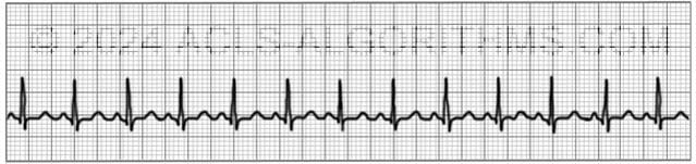
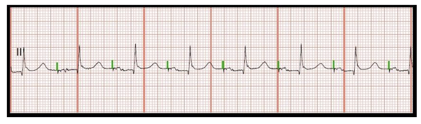
Identify the above waveform:
a. Atrial flutter
b. Atrial fibrillation
c. Sinus tachycardia
d. PAC (premature atrial contraction)
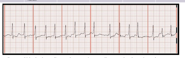
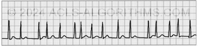
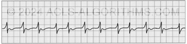
c. Sinus tachycardia
RHYTHM REGULAR (sinus) = 130 bpm
This is sinus tachycardia. For sinus tachycardia, the rhythm will be regular, with heart rate > 100 bpm. The P-waves will be normal but may be buried in the T-wave if the rate is very fast. The PR interval will be normal (0.12-0.20 sec), and the QRS complex will usually be normal. (0.06-0.10 sec). For adults, sinus rhythm rates typically will not exceed 150/min.

Identify the above waveform:
a. First degree heart block
b. Third degree heart block
c. Second degree heart block type II
d. Normal sinus rhythm
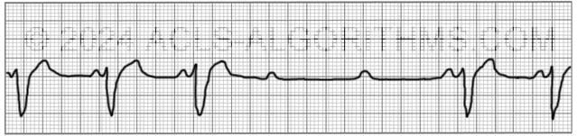
b. Third degree heart block
This is third-degree heart block. For third-degree heart block, the rhythm will be regular, but atrial and ventricular rhythms are disassociated from one another. The atrial rate will usually be normal and faster than the ventricular rate. The P wave will be normal but may be missing if the atrial impulse occurs at the same time as ventricular firing. The P-wave will be buried in the QRS complex. The PR interval is not measurable since the atria and ventricles are completely disassociated. The QRS complex is typically normal, but can be wide.

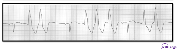
Identify the above waveform:
a. asystole
b. monitor malfunction
c. A fib
d. V fib
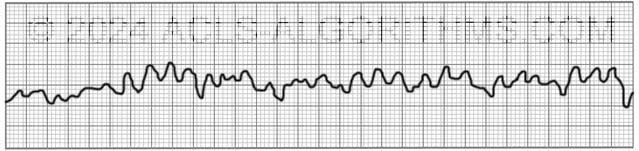
d. V fib
This is fine ventricular fibrillation. For ventricular fibrillation, the rhythm will be chaotic and wavy, with an unmeasurable heart rate. P-waves and QRS complexes are absent. Typically, this chaotic wavy line with varying amplitude will be the defining characteristic of ventricular fibrillation. It is possible that this could be asystole. When in doubt treat as ventricular fibrillation.
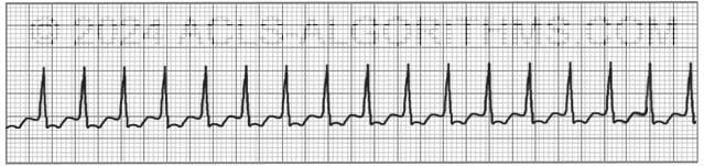
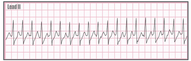
Identify the above waveform:
a. Atrial flutter
b. Atrial fibrillation
c. Sinus tachycardia
d. Supraventricular tachycardia
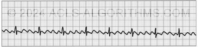
d. Supraventricular tachycardia
COUNT THE RATE IS WAYYY OVER TACHY (which is 101-160)
This is supraventricular tachycardia (SVT). For SVT, the rhythm will be regular, and the heart rate will be fast (150-250 bpm). The P wave will typically be merged with t wave. If measurable, the PR interval will be normal (0.12 sec). The QRS complex will usually be normal (.10 sec).
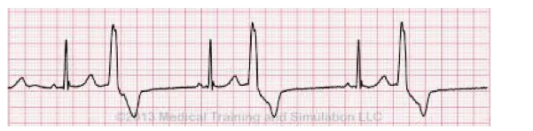
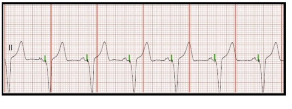
Identify the above waveform:
a. First degree heart block
b. Third degree heart block
c. Second degree heart block type II
d. Second degree heart block type I
a. First degree heart block
This is first-degree heart block. For first-degree heart block, the rhythm is regular. The PR interval will be prolonged at > 0.2. The QRS complex will usually be normal. First-degree heart block indicates slowed but not blocked conduction through the AV node.
Identify the above waveform:
a. First degree heart block
b. Third degree heart block
c. Normal sinus rhythm
d. A fib
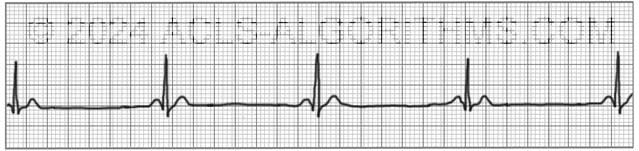
c. Normal sinus rhythm
This is normal sinus rhythm. For normal sinus rhythm, the rhythm will be regular, and the rate will be 60-100/min. A P-wave will come before each QRS complex. The PR interval will be normal (0.12-0.20 sec), and the width of the QRS complex will typically be normal (0.06-0.10 sec).
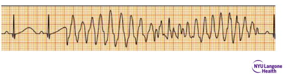
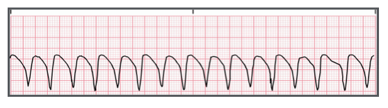
a. ventricular tachycardia
b. ventricular fibrillation
c. atrial fibrillation
d. heart block type I
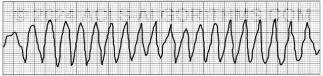
a. ventricular tachycardia
This is ventricular tachycardia. For ventricular tachycardia, the rhythm is regular, and the heart rate will typically be >150/min. The P waves will be absent and therefore, there will be no PR interval. The QRS will usually be wide (>0.10 sec). This particular rhythm happens to be polymorphic VT.

third degree heart block
1st degree heart block type 1
1st degree heart block type 2
first degree heart block
third degree heart block
This is third-degree heart block. For third-degree heart block, the rhythm will be regular, but atrial and ventricular rhythms are disassociated from one another. The atrial rate will usually be normal and faster than the ventricular rate. The P wave will be normal but may be missing if the atrial impulse occurs at the same time as ventricular firing. The P-wave will be buried in the QRS complex. The PR interval is not measurable since the atria and ventricles are completely disassociated. The QRS complex is typically normal, but can be wide.
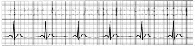
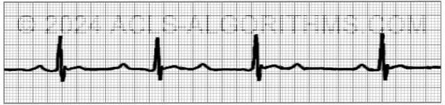
Identify:
a. normal sinus rhythm
b. sinus bradycardia
c. 1st degree heart block
d. sinus arrhythmia
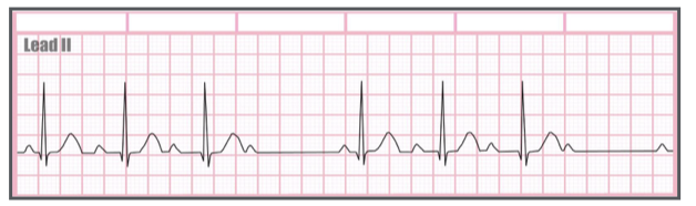
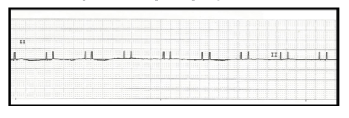
b. sinus bradycardia
This is sinus bradycardia. For sinus bradycardia, the rhythm is regular, with a heart rate that will be < 60/min. The P-waves will be normal. The PR interval will be normal (0.12-0.20 sec), and the QRS complex is typically normal (0.06-0.10 sec).
a. third degree heart block
b. sinus tachycardia
c. sinus arrhythmia
d. atrial fibrillation
d. atrial fibrillation
This is atrial fibrillation. For atrial fibrillation, the rhythm will be irregular, and the P-waves will be characteristically absent and replaced with fine, chaotic waves. The ventricular rate is usually variable and may be slow, normal or rapid. Patients with symptoms will usually have a rapid ventricular rate. The PR interval will be absent, and the QRS complex is typically normal but can be widened in certain cases.

a. third degree heart block
b. sinus tachycardia
c. trigeminy
d. atrial fibrillation
a. third degree heart block
This is third-degree heart block. For third-degree heart block, the rhythm will be regular, but atrial and ventricular rhythms are disassociated from one another. The atrial rate will usually be normal and faster than the ventricular rate. The P wave will be normal but may be missing if the atrial impulse occurs at the same time as ventricular firing. The P-wave will be buried in the QRS complex. The PR interval is not measurable since the atria and ventricles are completely disassociated. The QRS complex is typically normal, but can be wide.
third degree heart block
1st degree heart block type 1
1st degree heart block type 2
first degree heart block
1st degree heart block type 2
This is second-degree heart block type II. For second-degree heart block type II, the ventricular rhythm will be irregular due to intermittent dropped QRS complexes. The atrial rhythm will be regular. The P-waves will look normal, and there will be more P-waves than QRS complexes. The PR interval will be normal or prolonged when a P-wave is followed by a QRS complex. The QRS complex will usually be normal or wide.
a. atrial flutter
b. atrial fib
c. atrail tachycardia
d. sinus tachycardia
a. atrial flutter
This is atrial flutter. Atrial flutter can be regular or irregular. For atrial flutter, the atrial rate is typically extremely fast. (250-350 bpm). The ventricular rate is typically slower than the atrial rate. Saw-toothed flutter waves are usually seen instead of a single P-wave. The PR interval will not be measurable and the QRS is typically normal (0.06-0.10 sec).
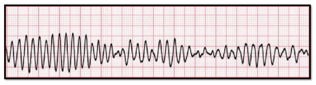
a. asystole
b. ventricular fibrillation
c. ventricular tachycardia
d. atrial fibrillation
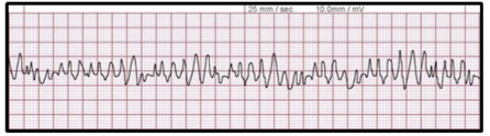
b. ventricular fibrillation
This is ventricular fibrillation. For ventricular fibrillation, the rhythm will be chaotic and wavy, with an unmeasurable heart rate. P-waves and QRS complexes are absent. Typically, this chaotic wavy line with varying amplitude will be the defining characteristic of ventricular fibrillation.
Analyze:
a. failure to capture
b. failure to sense
c. bigeminy
d. trigeminy
b. failure to sense
