Lab Practical 2
1/97
There's no tags or description
Looks like no tags are added yet.
Name | Mastery | Learn | Test | Matching | Spaced | Call with Kai |
|---|
No analytics yet
Send a link to your students to track their progress
98 Terms
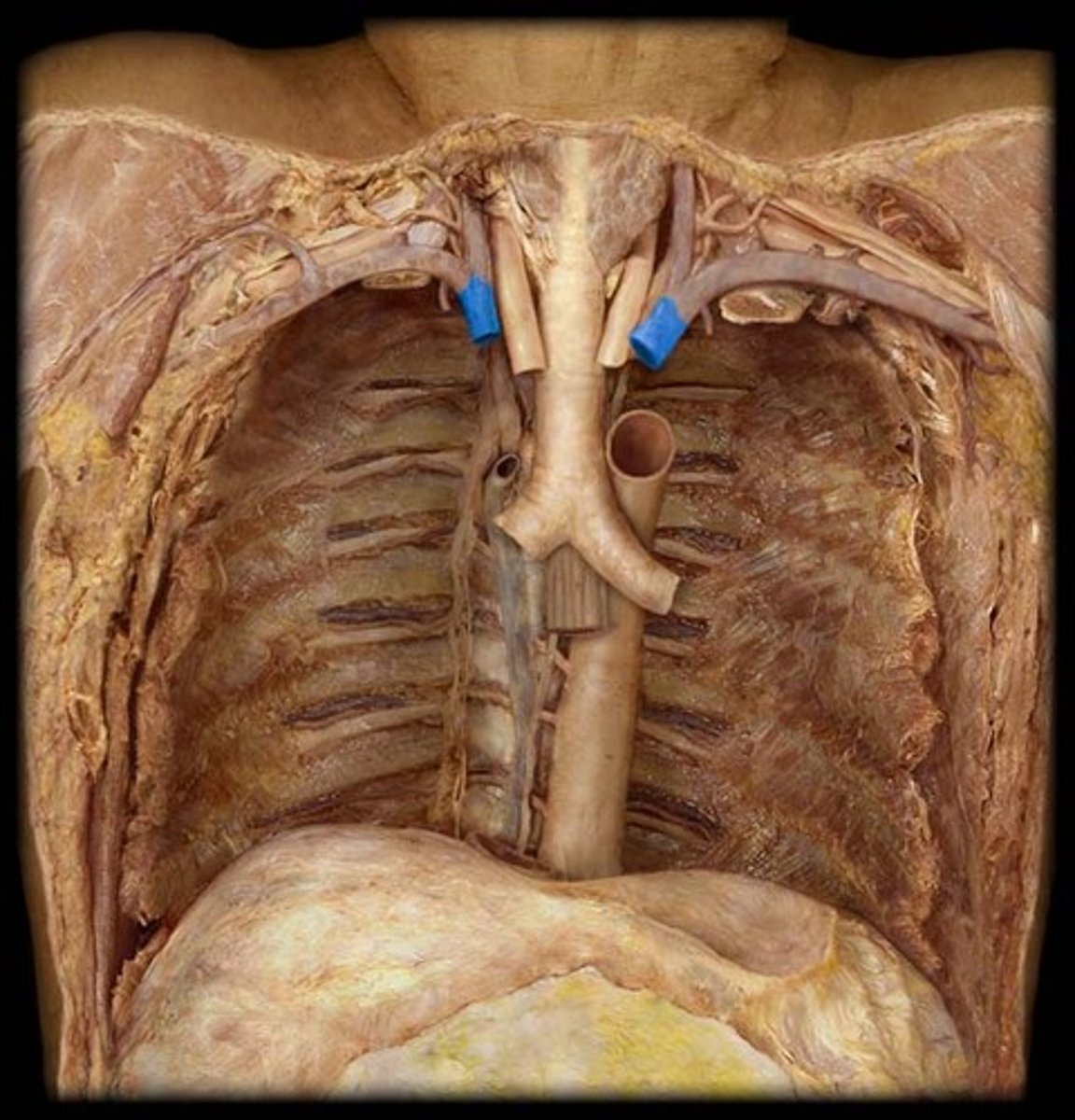
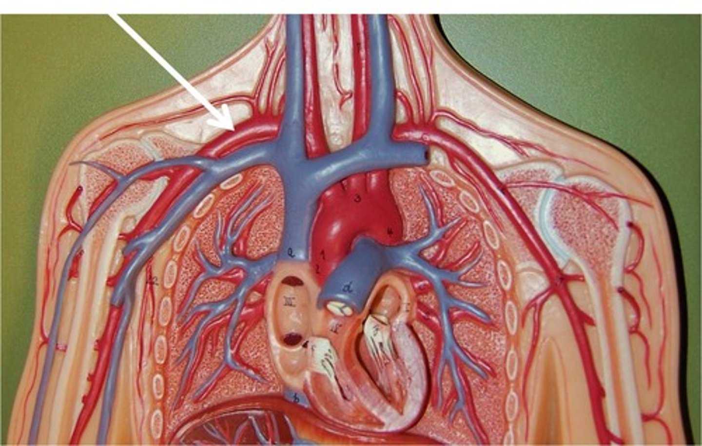
Brachiocephalic trunk
Supplies blood to the right side of the head, neck and upper limb.
common carotid arteries
Supplies structures of the head and neck.
external carotid artery
Generally supplies blood to the face, mouth, sinuses and structures of the lateral head
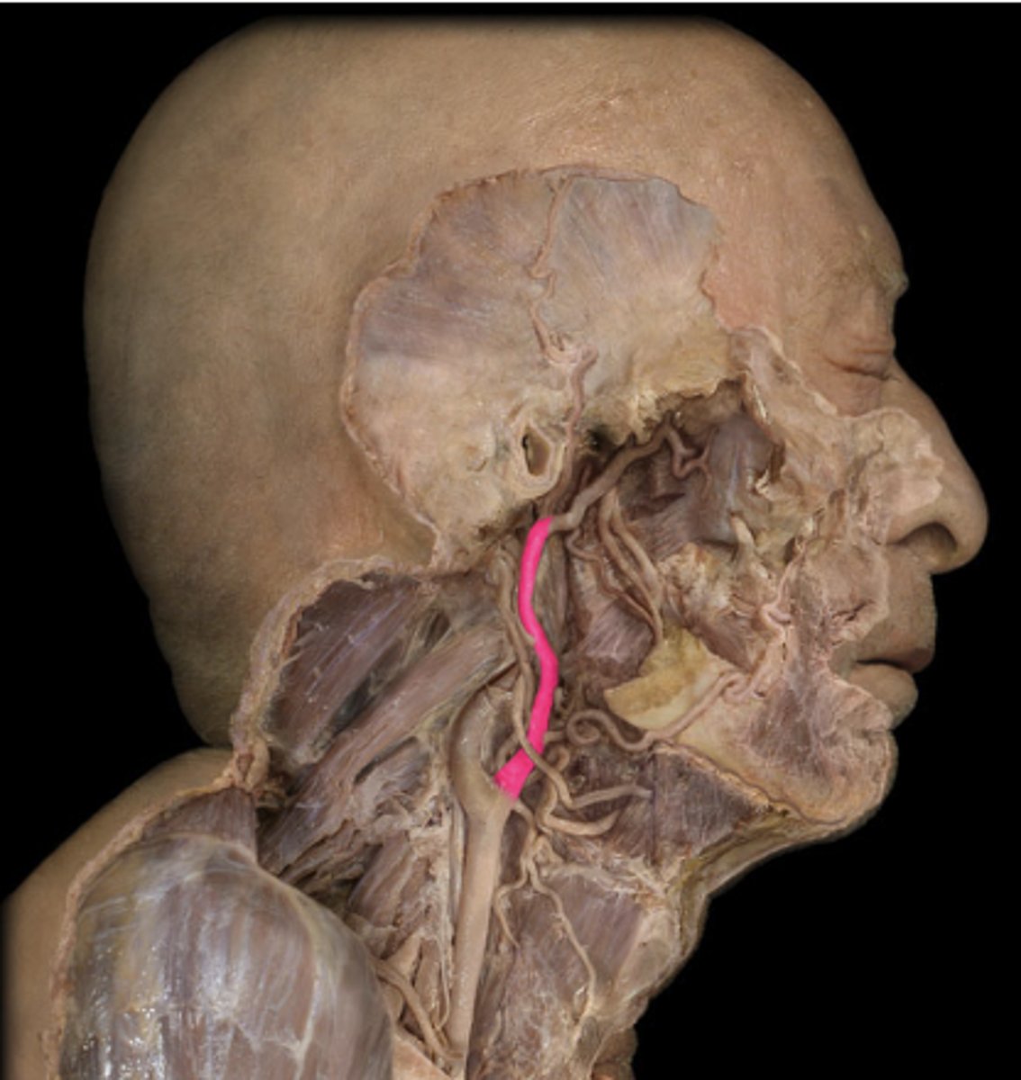
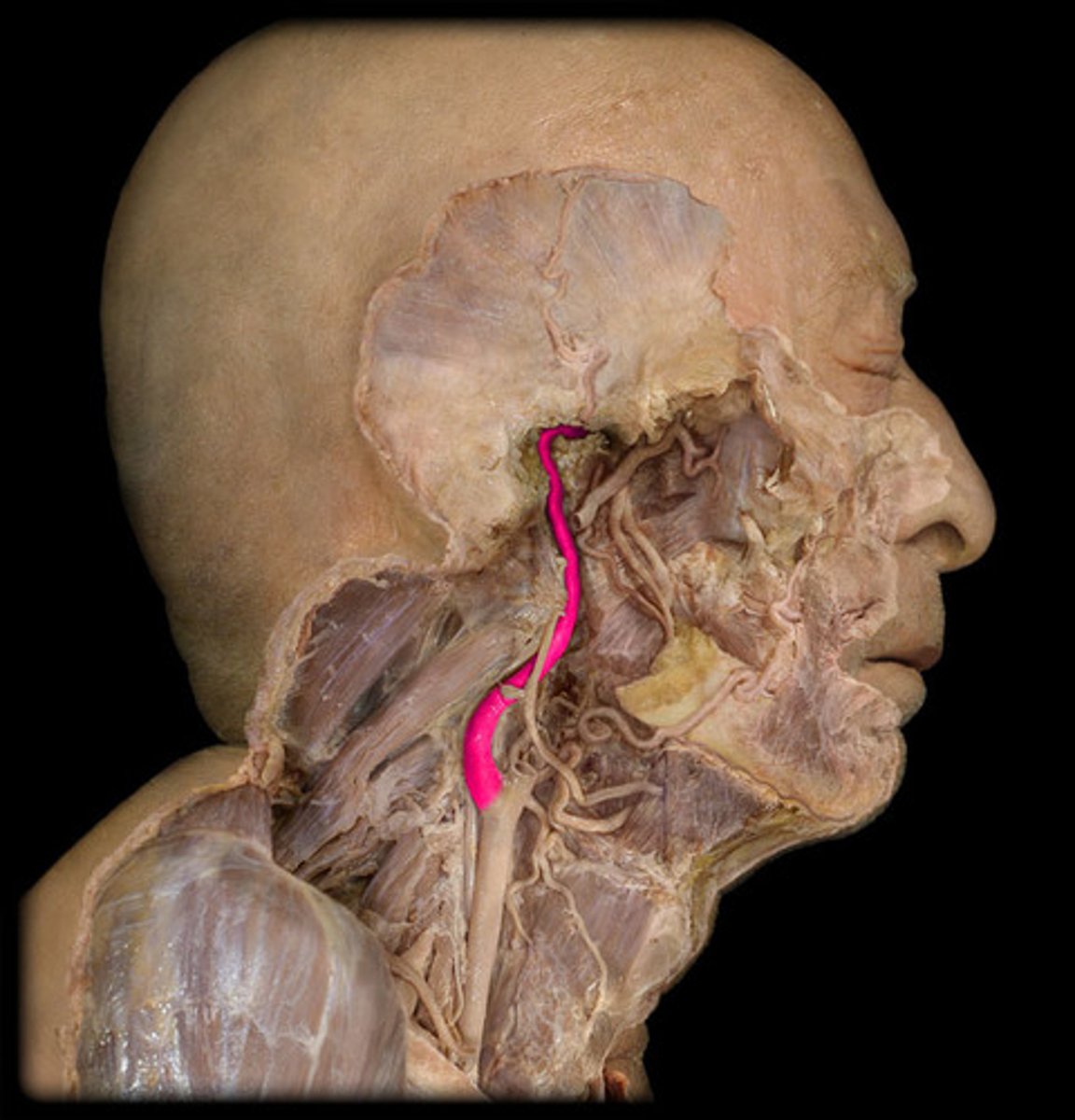
internal carotid artery
Generally supplies blood to structures of the interior of the cranium including the brain, pituitary gland and orbit.
carotid sinus
baroreceptor that senses changes in arterial blood pressure
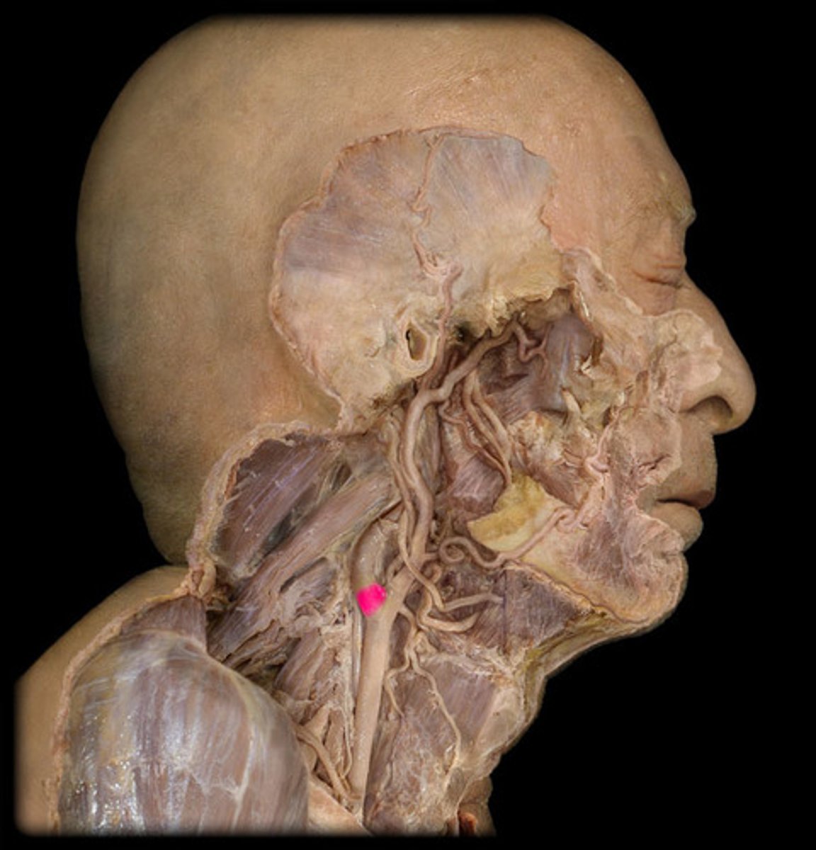
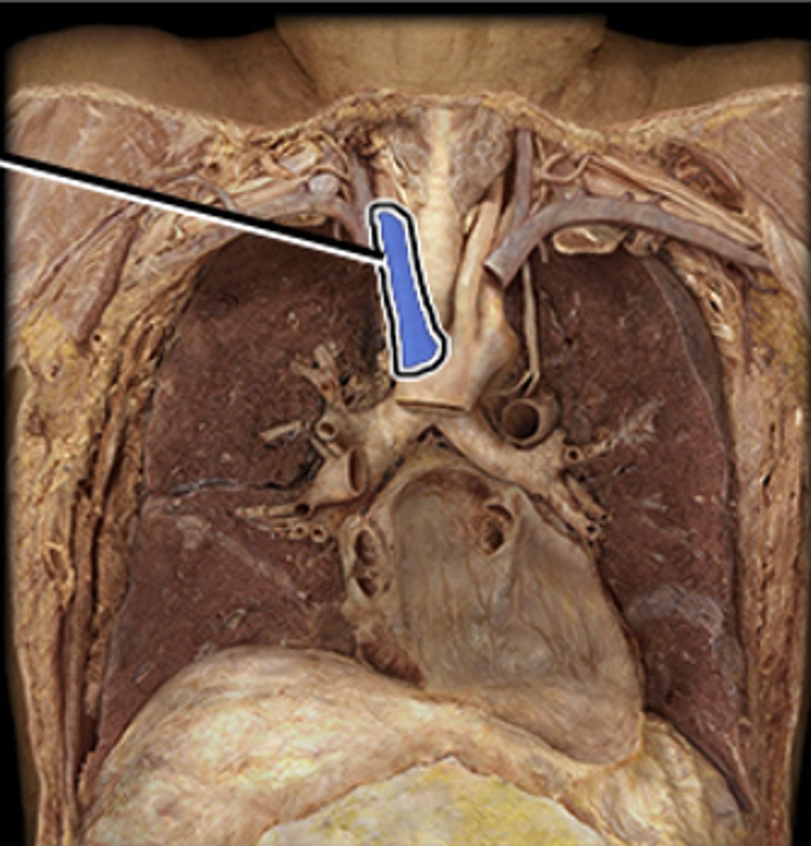
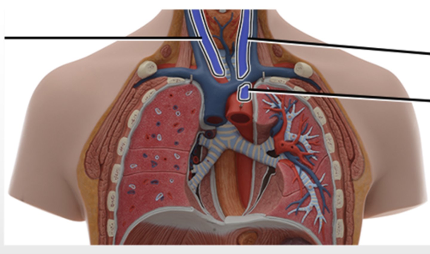
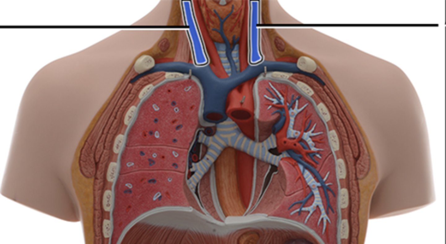
internal jugular veins
drains blood from the skull, brain, superficial face and most of the neck
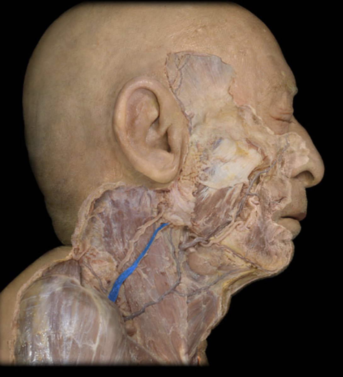
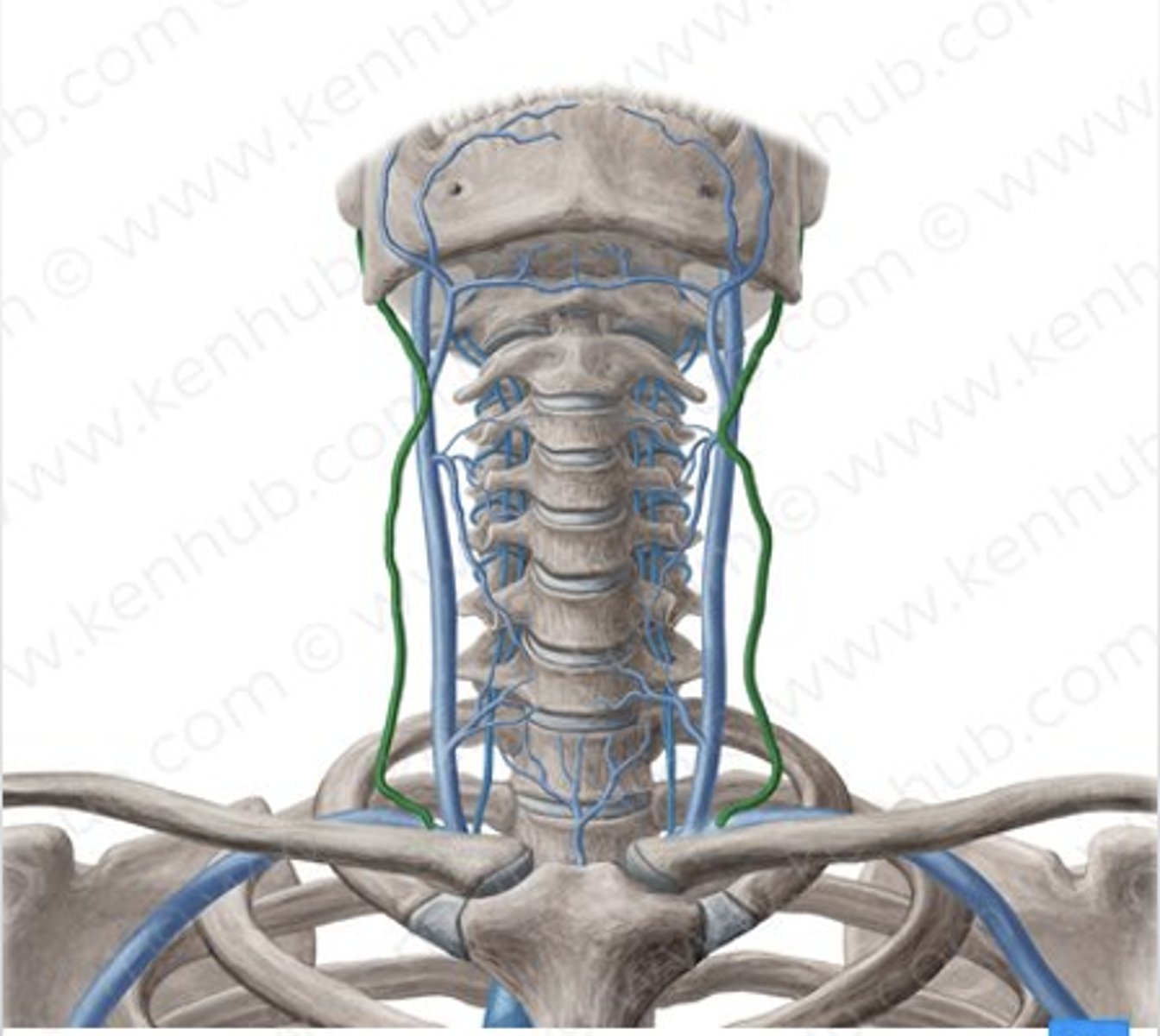
external jugular veins
drains blood from superficial skull/head structures and the deep face
anterior jugular veins
Drains blood from the anterior neck
on the front side
brachiocephalic veins
Drains head, neck and upper limb.
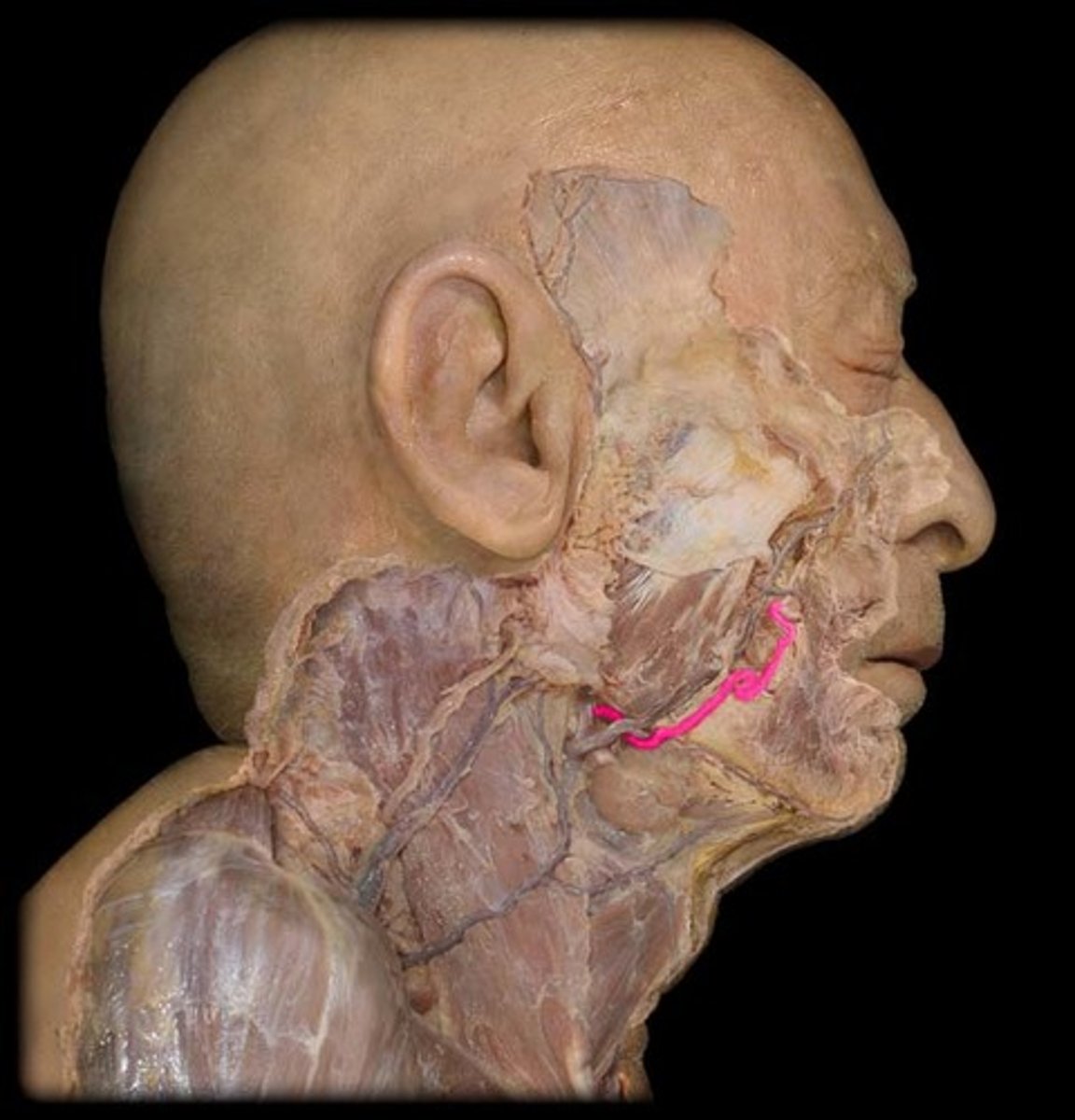
facial artery
Supplies blood to the glandular tissue and musculature of the face as well as the overlying skin and fascia.
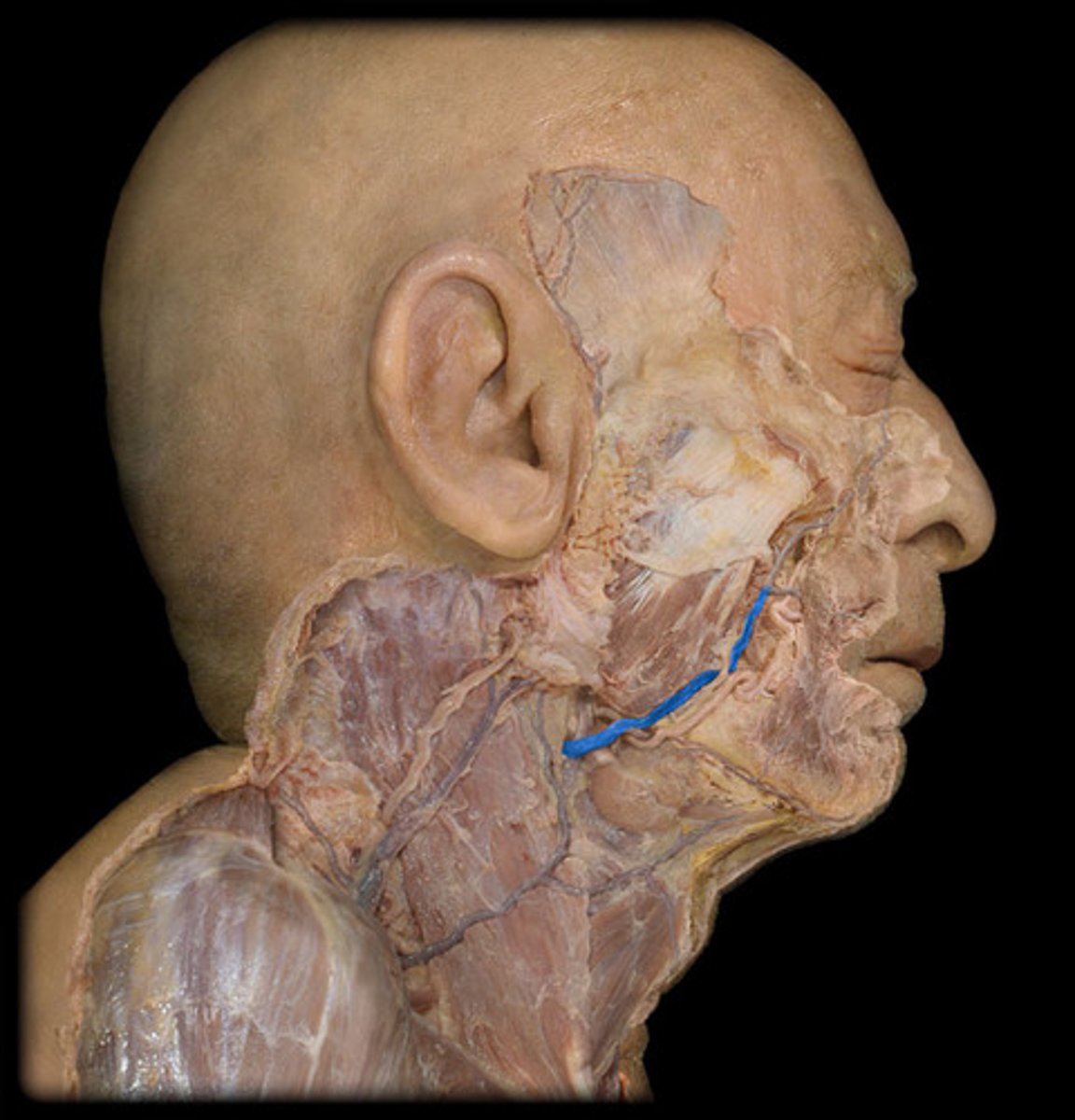
facial vein
drains blood from structures of the face
superficial temporal artery
Supplies structures within the temporal region - skin, muscles and scalp.

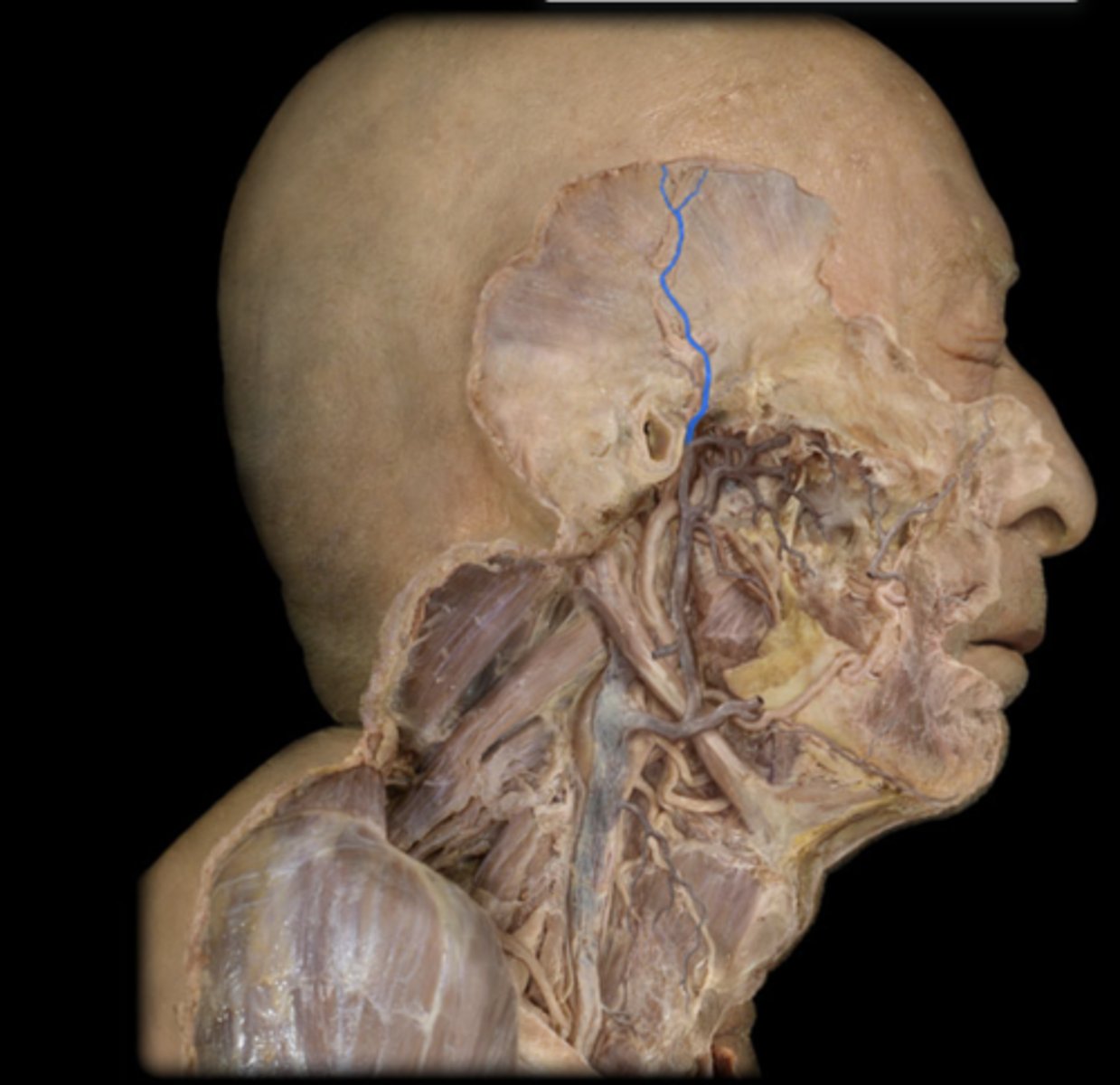
superficial temporal vein
drains blood from the forehead, parietal, temporal regions
subclavian arteries
Supplies structures of the shoulder and upper limb
axillary artery
Supplies blood to structures within the axillary, shoulder and pectoral regions.
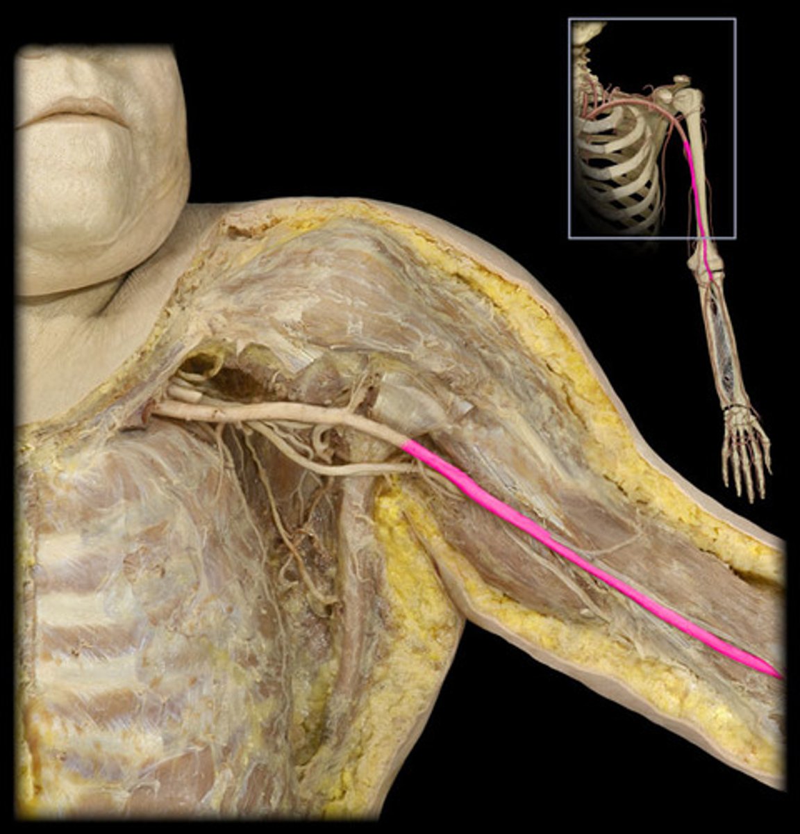
brachial artery
Supplies blood to the biceps brachii, coracobrachialis, brachialis, humerus and elbow joint
ulnar collateral arteries
Supplies the medial head of the triceps brachii, cutaneous tissue and elbow joint.
radial collateral arteries
Supplies the brachialis and the brachioradialis muscles, as well as the radial nerve
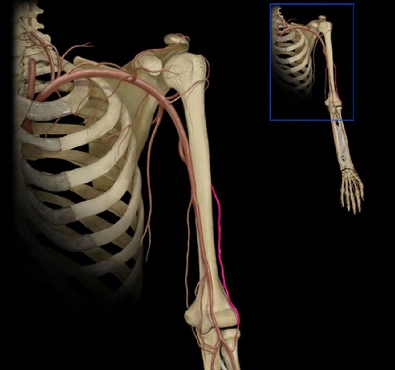
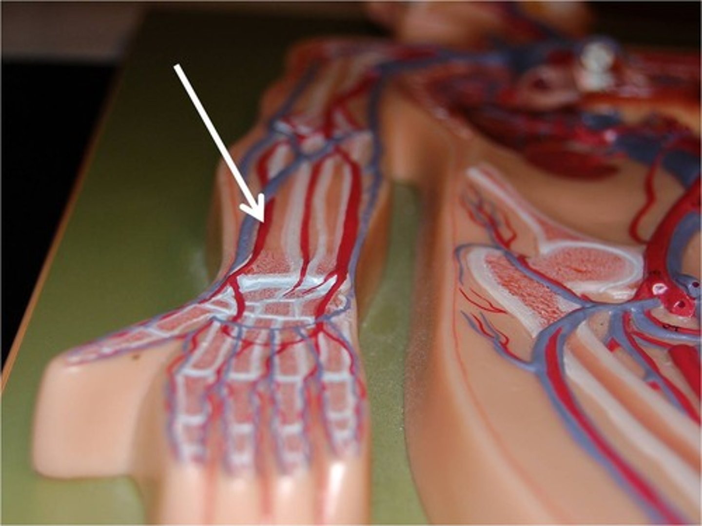
ulnar artery
Supplies blood to the elbow joint and forearm muscles
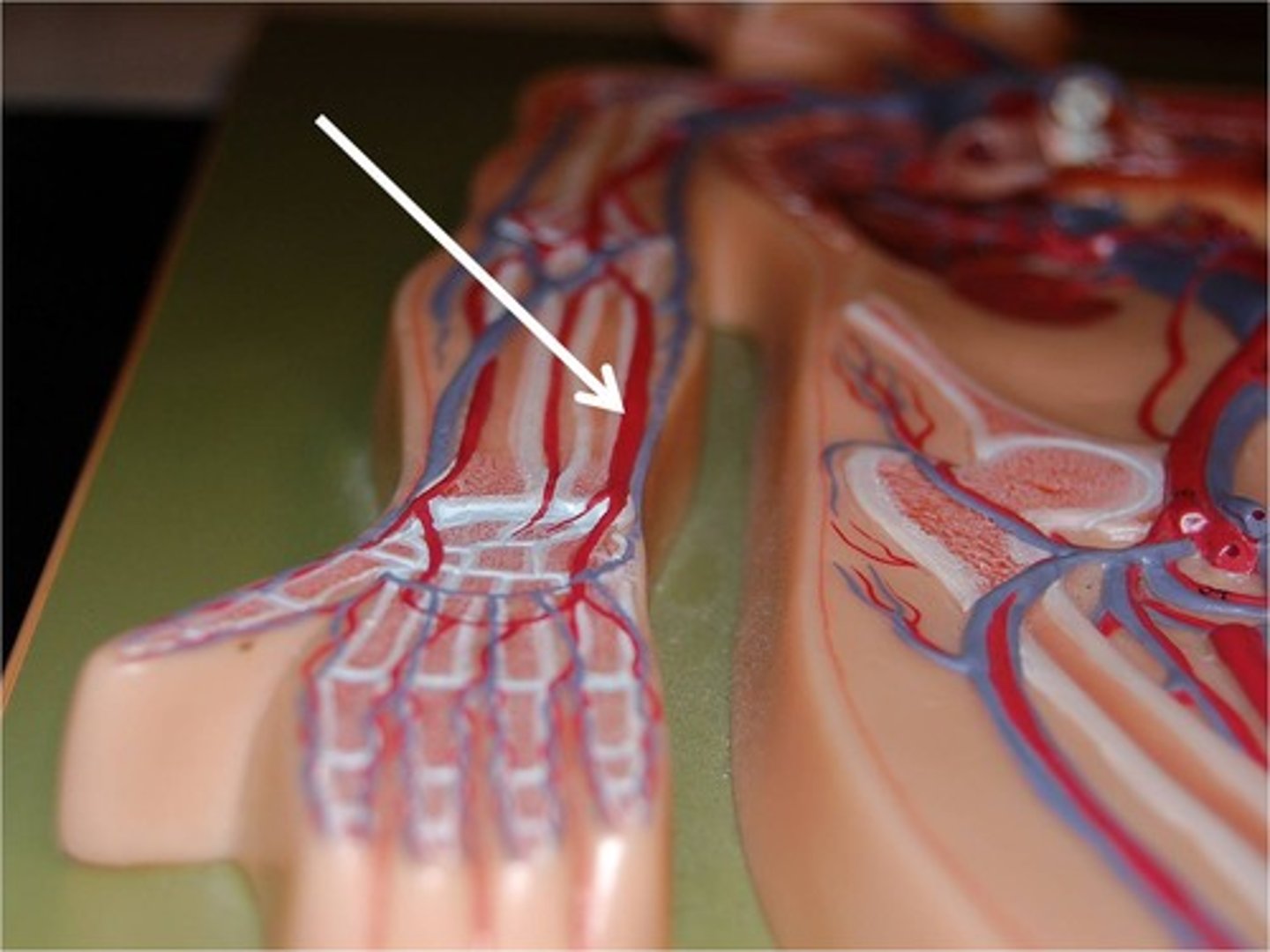
radial artery
Supplies muscles on the lateral aspect of the anterior forearm
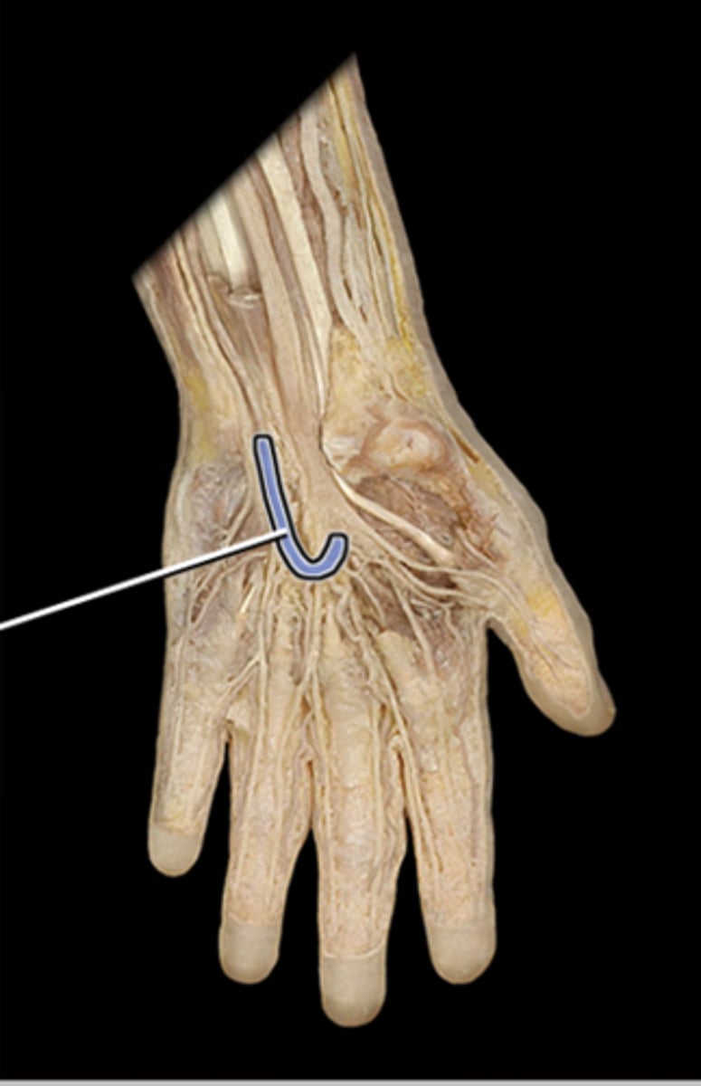
palmar arch arteries
Supplies blood to the palmar aspect of metacarpals and digits
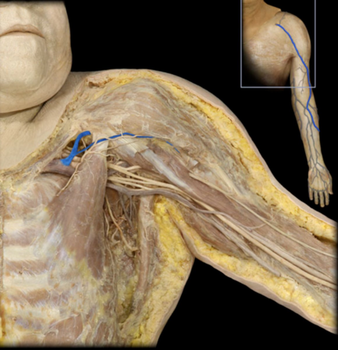
cephalic vein
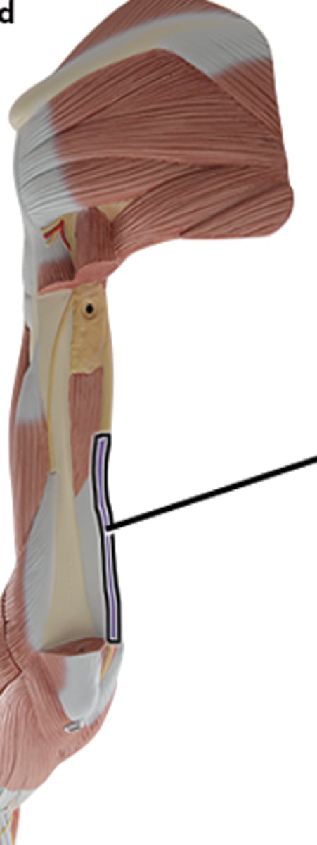
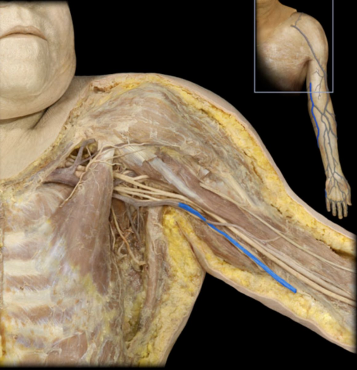
basilic vein
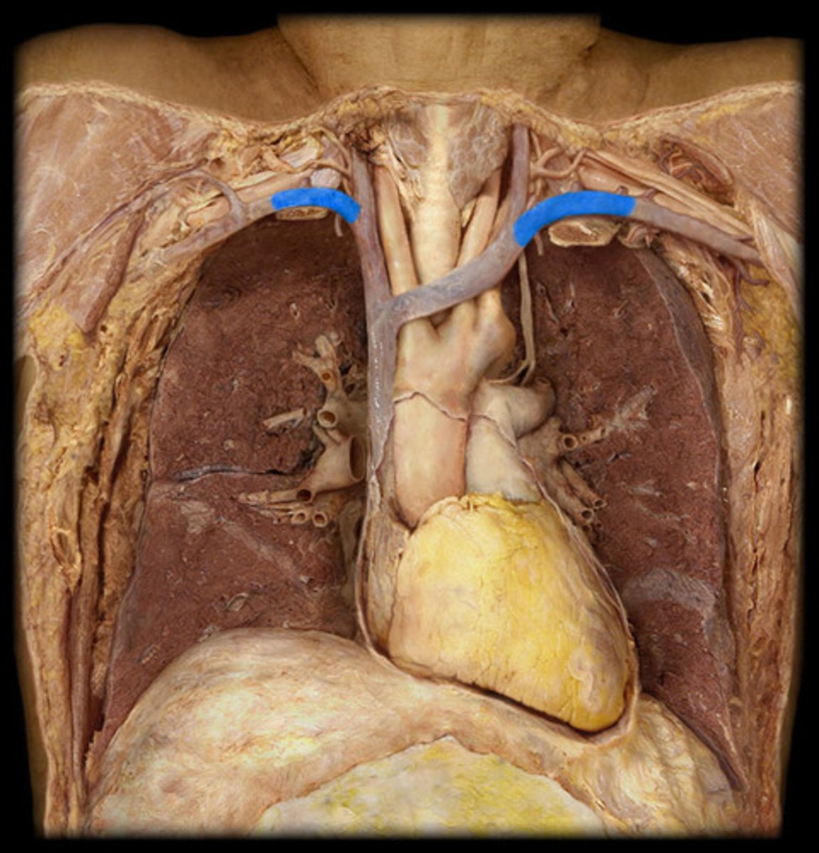
axillary veins
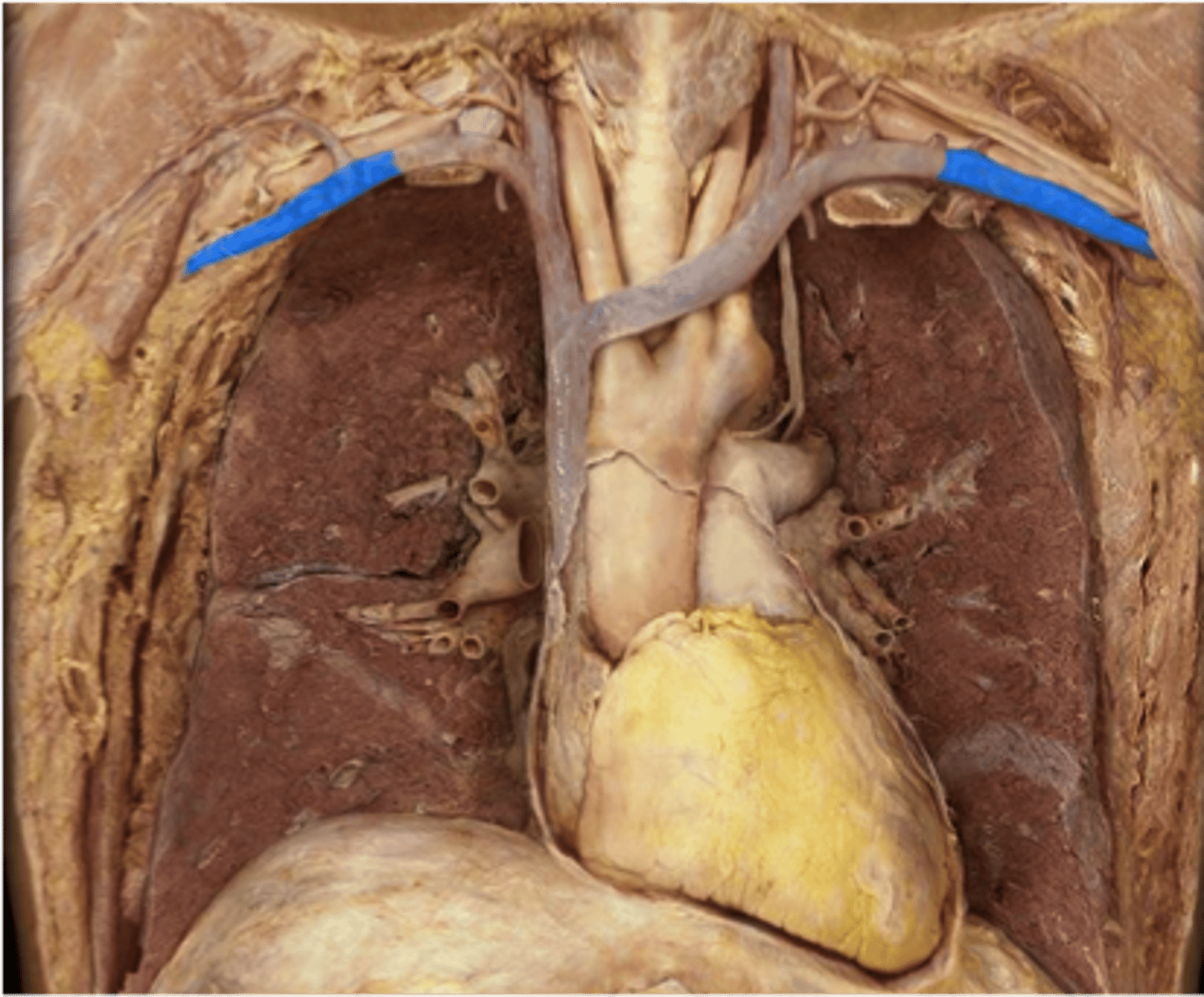
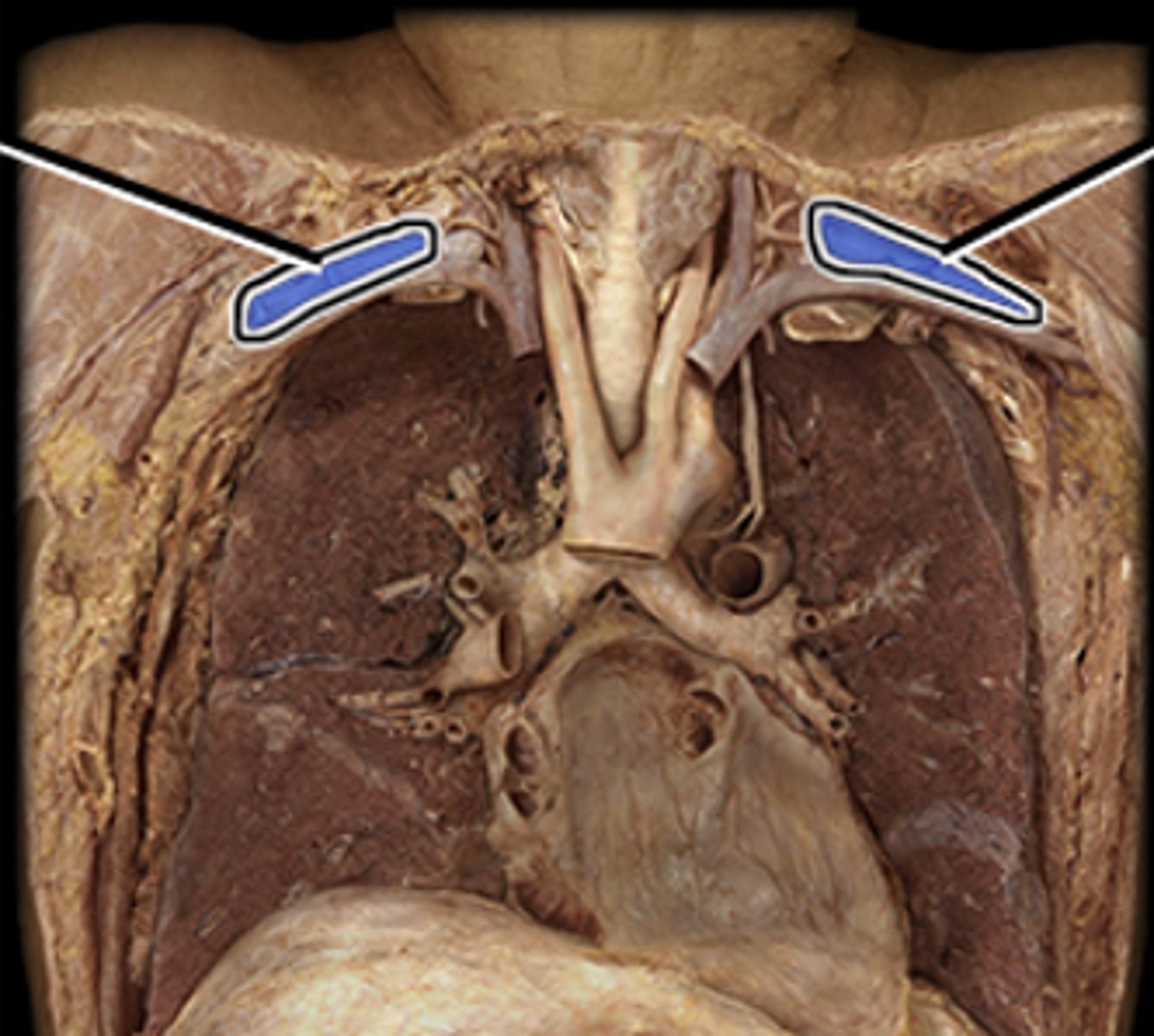
subclavian veins
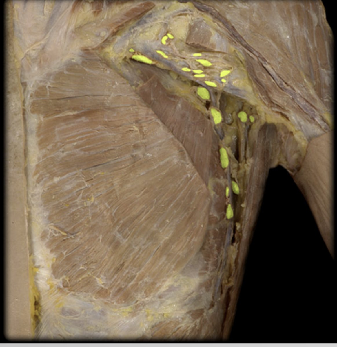
axillary lymph nodes
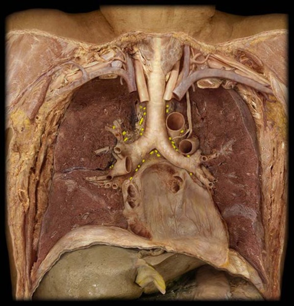
mediastinal lymph nodes
lingual tonsil
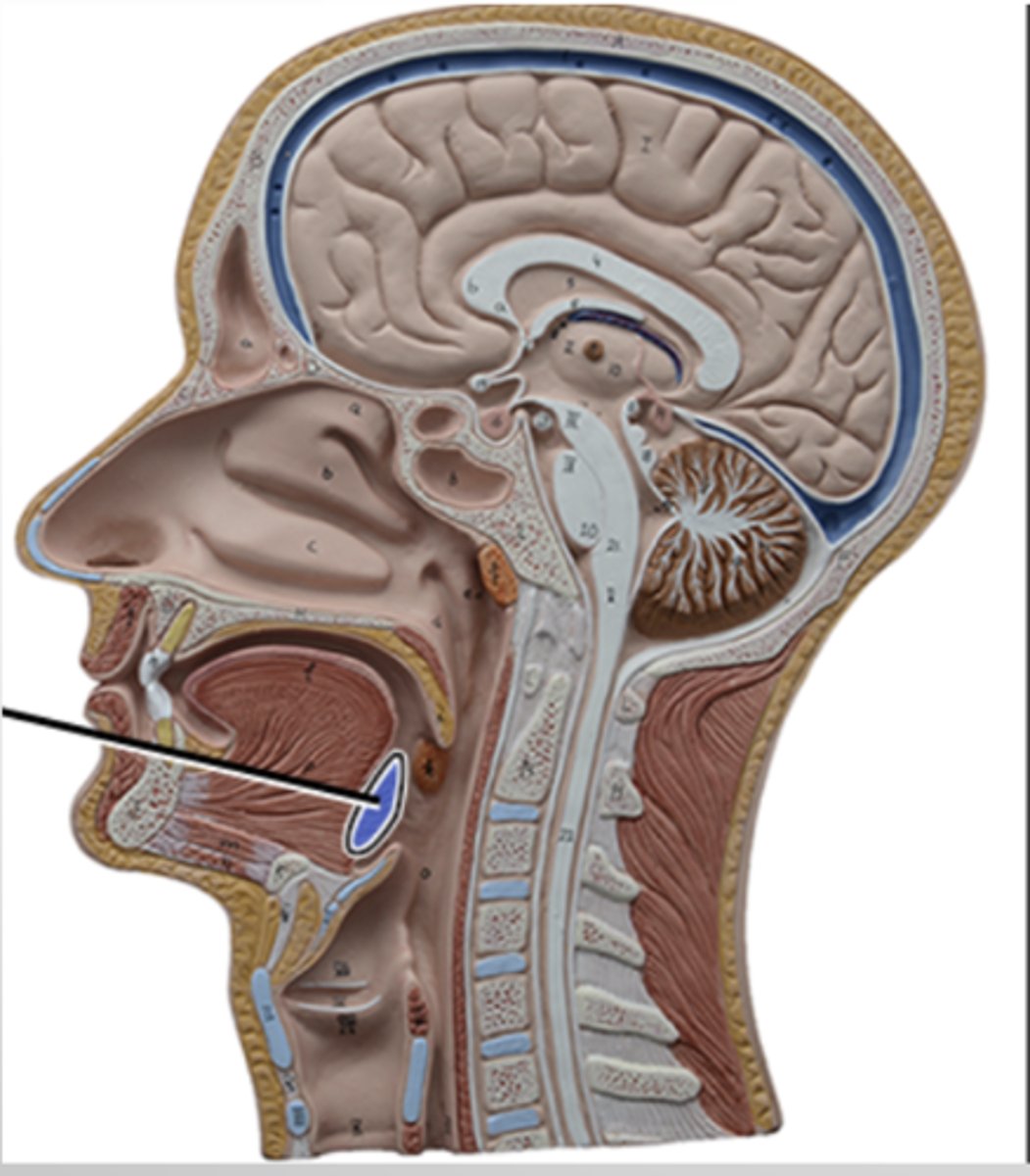
palatine tonsil
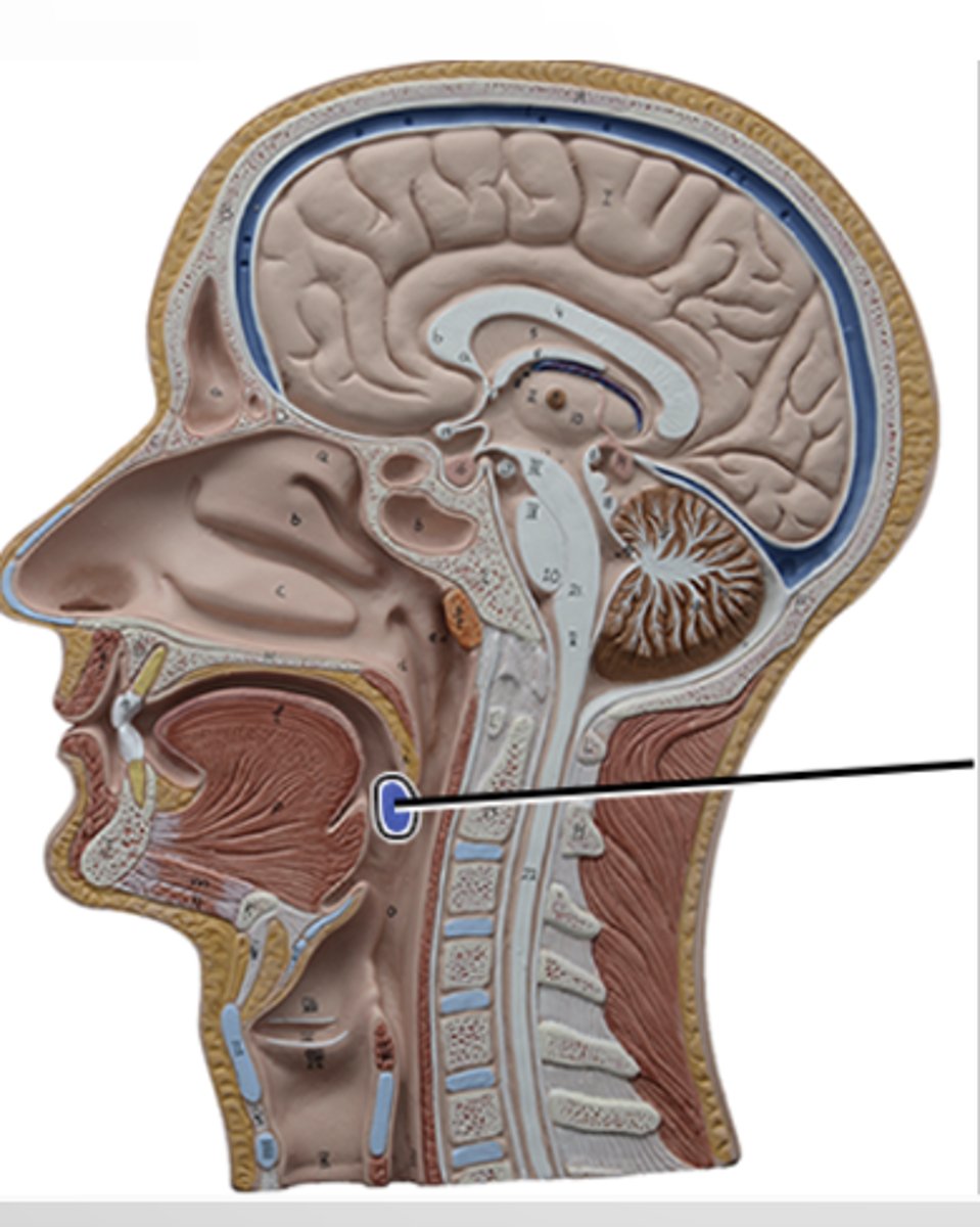
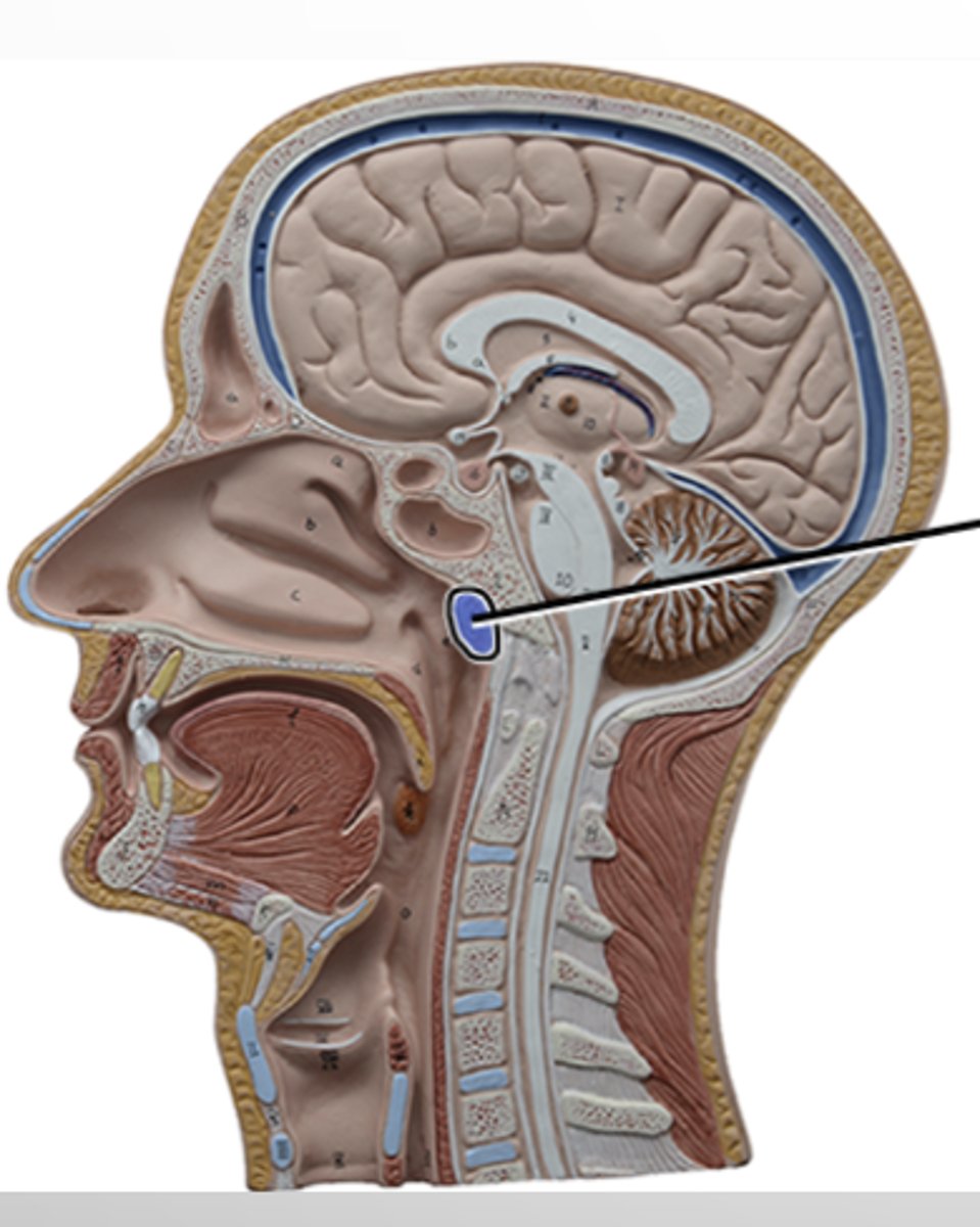
pharyngeal (adenoid) tonsil

pharyngeal tonsil
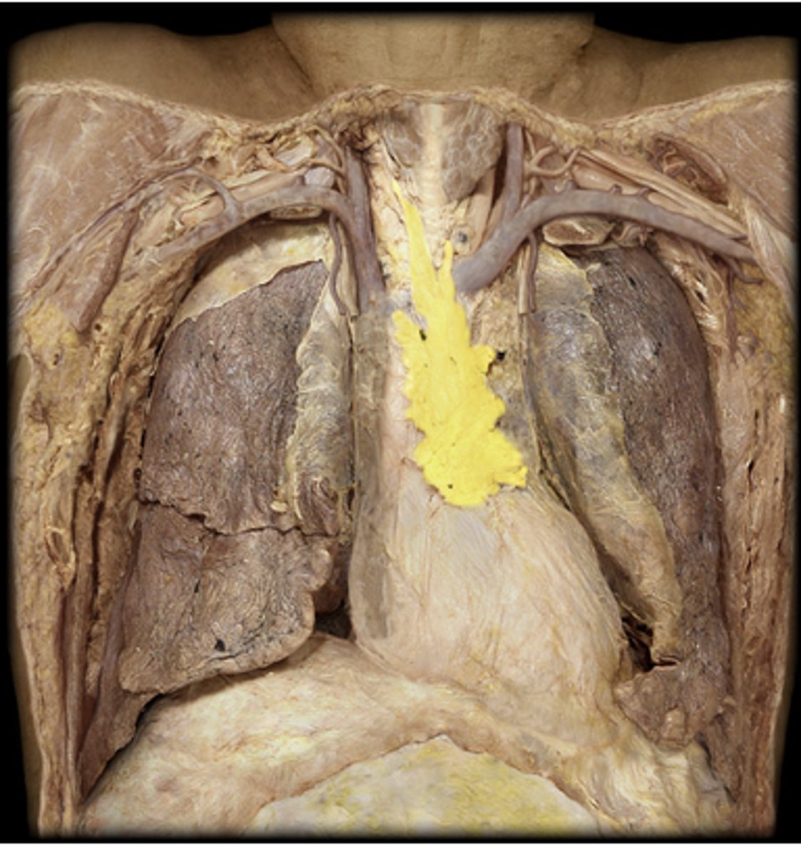
thymus
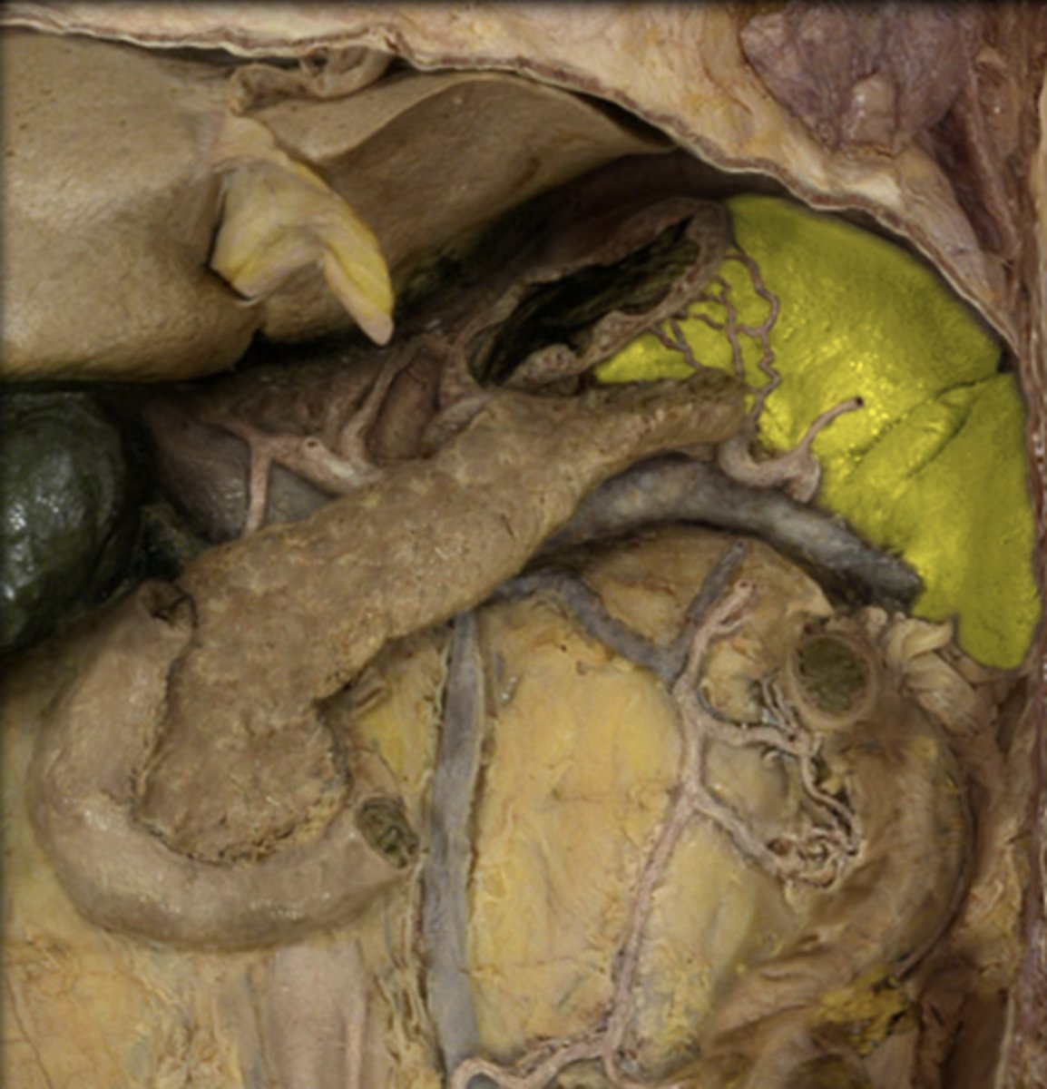
spleen
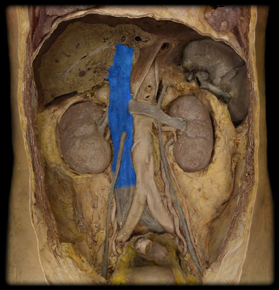
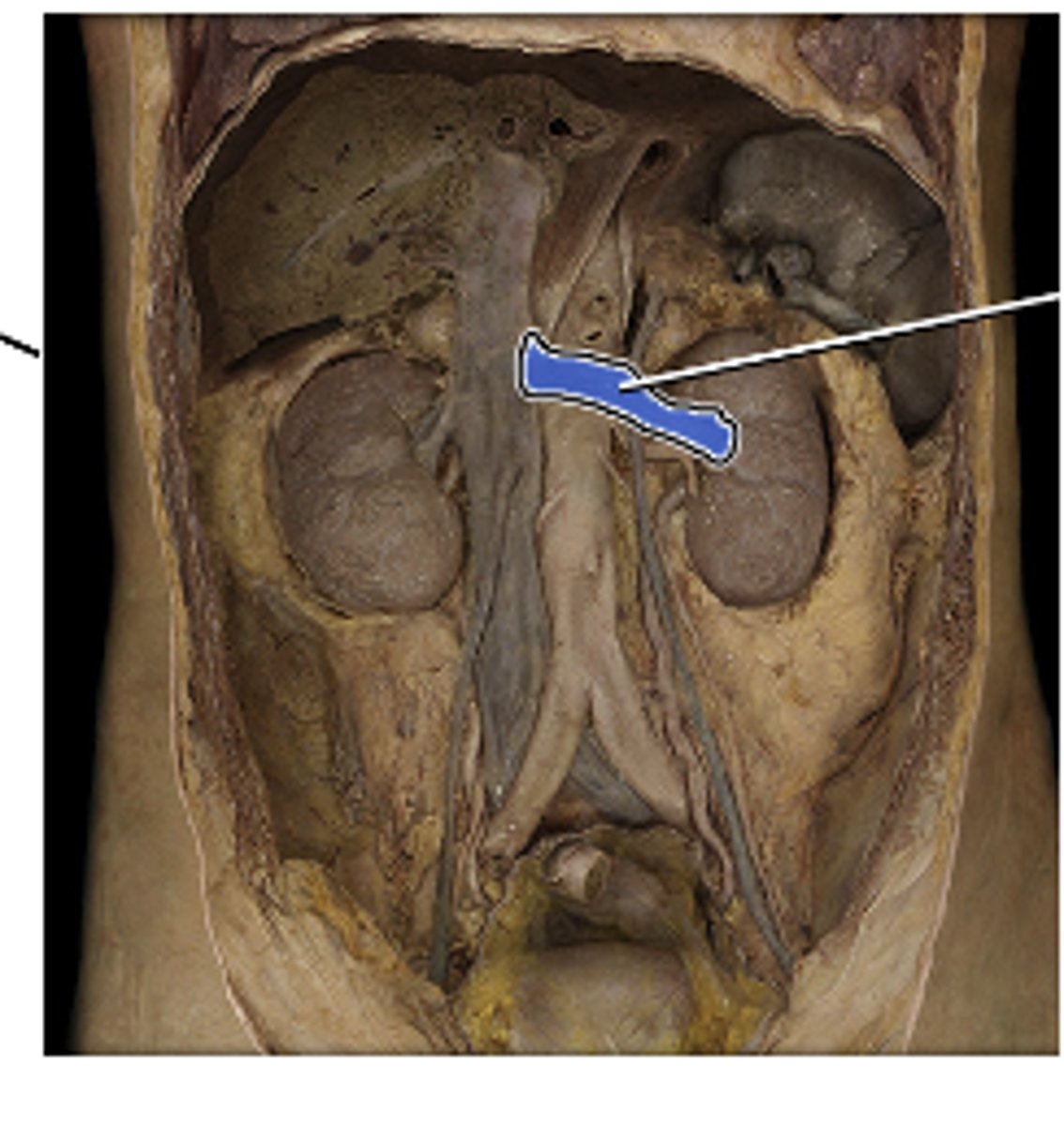
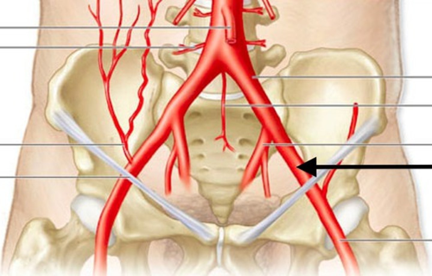
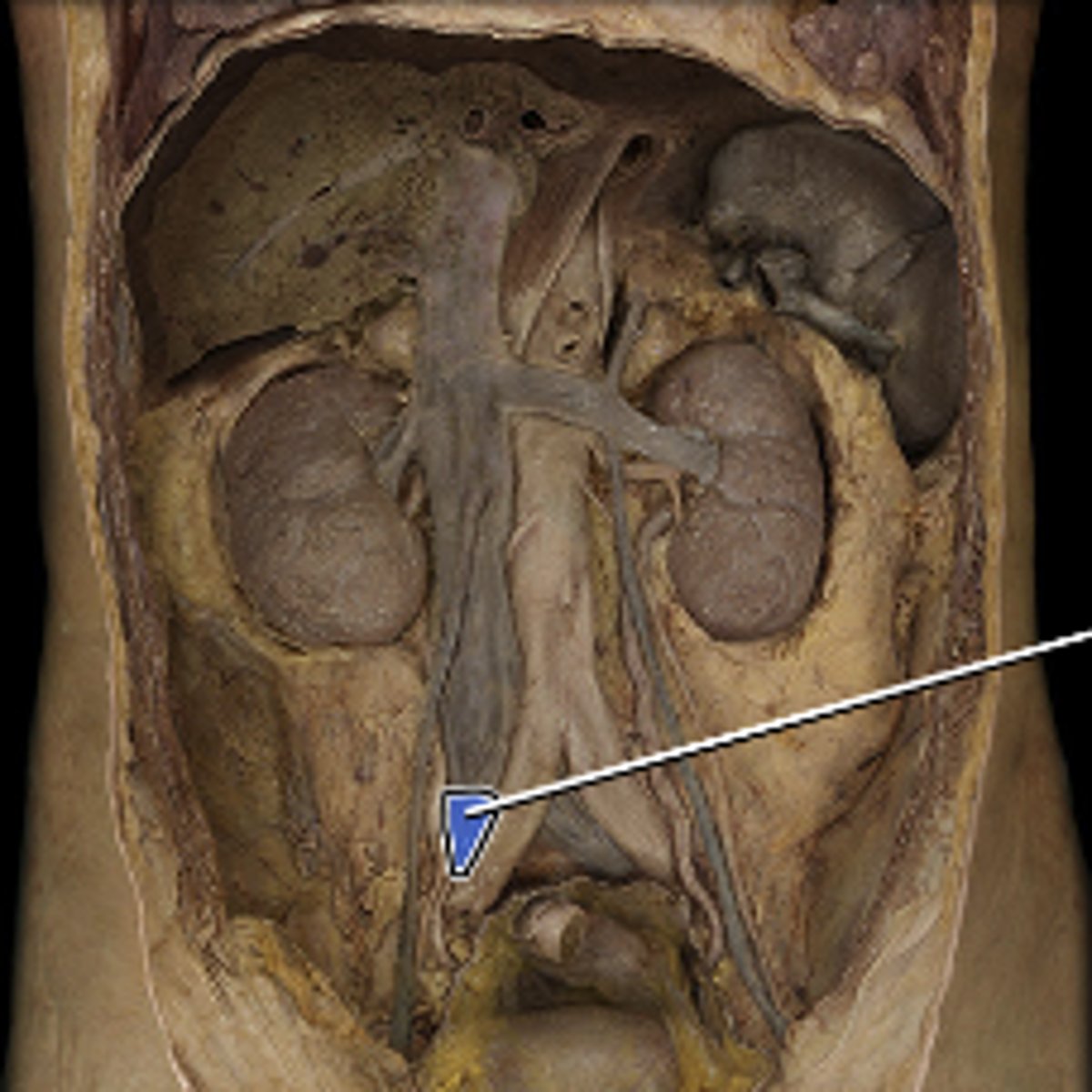
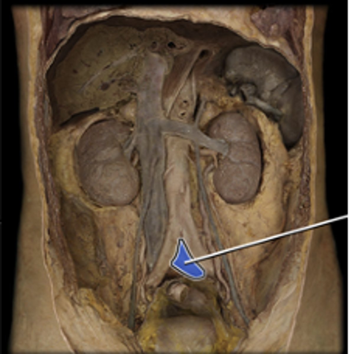
aorta
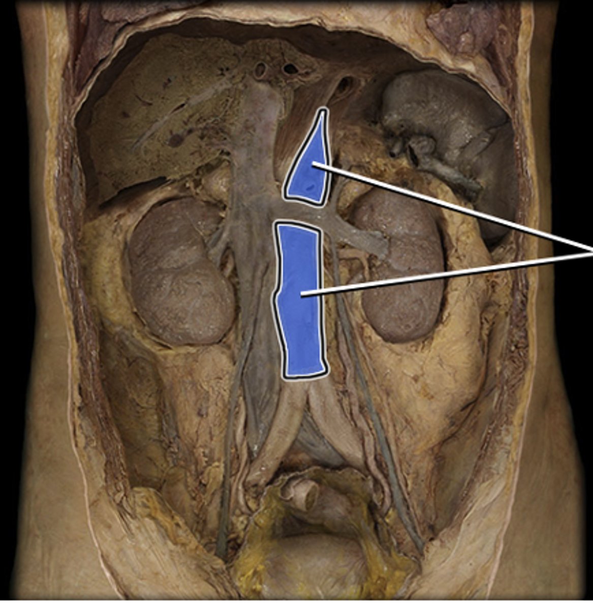
inferior vena cava
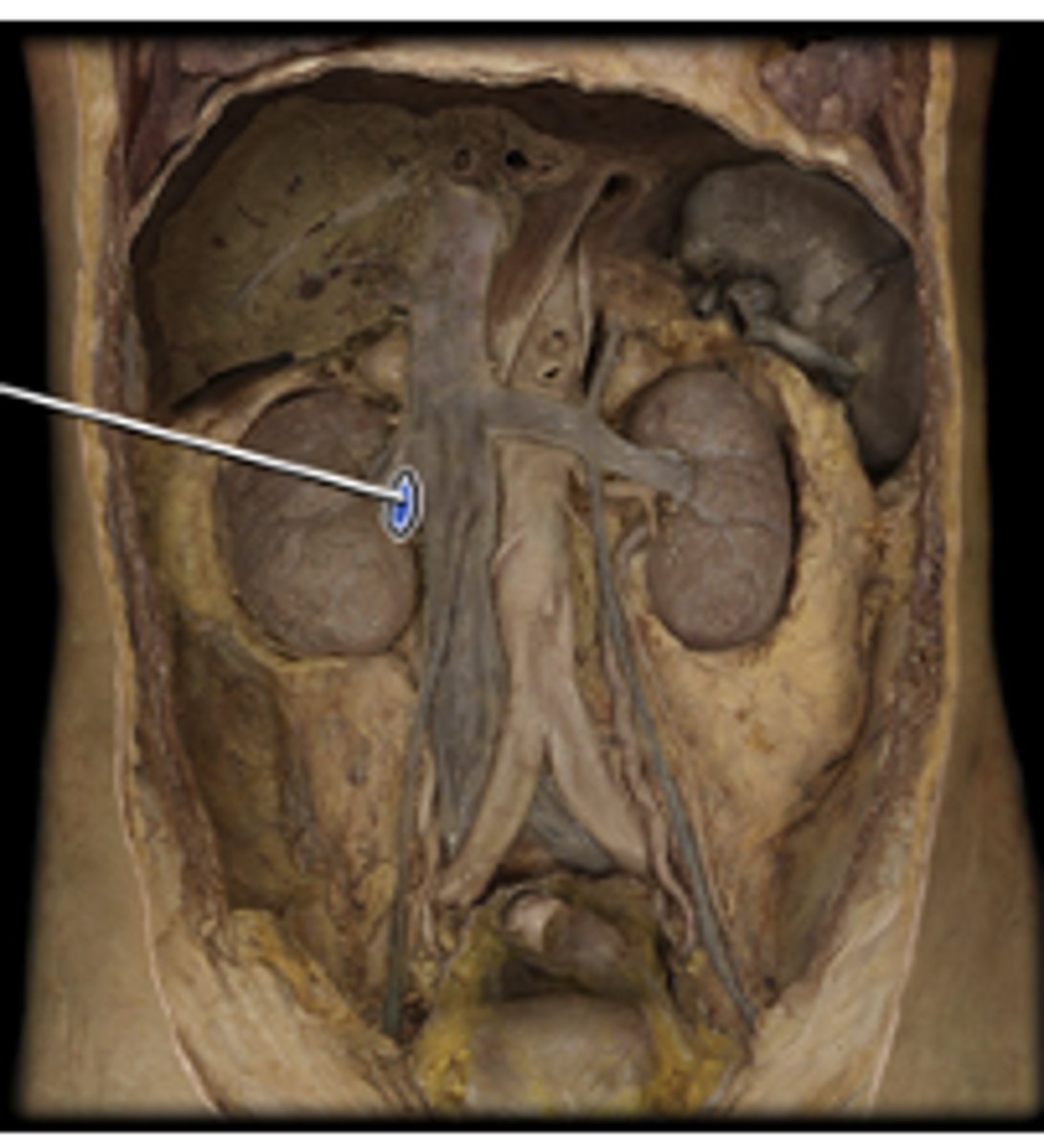
right renal artery
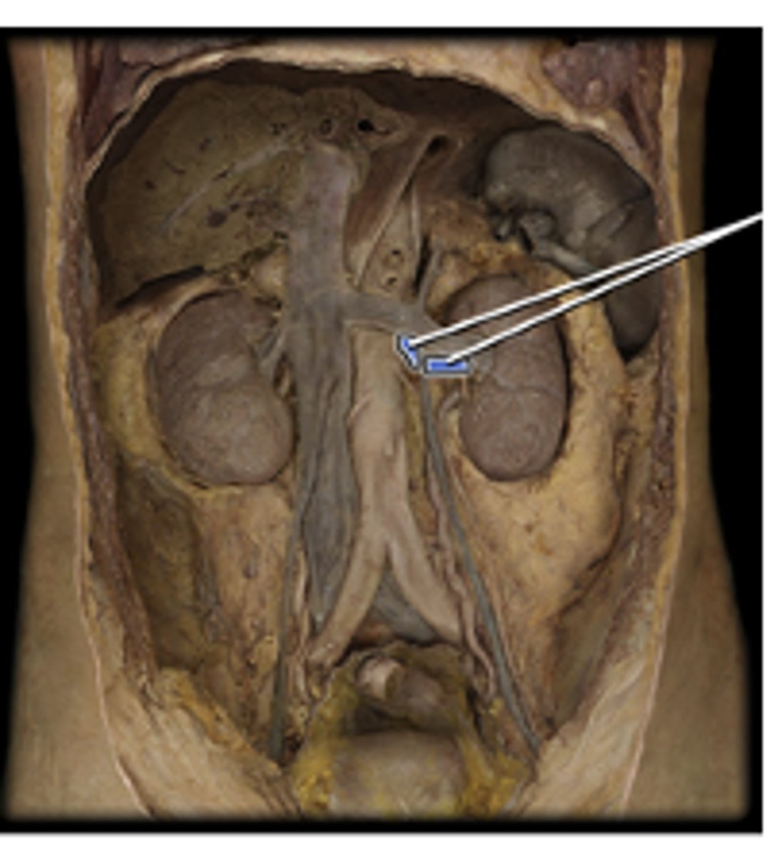
left renal artery
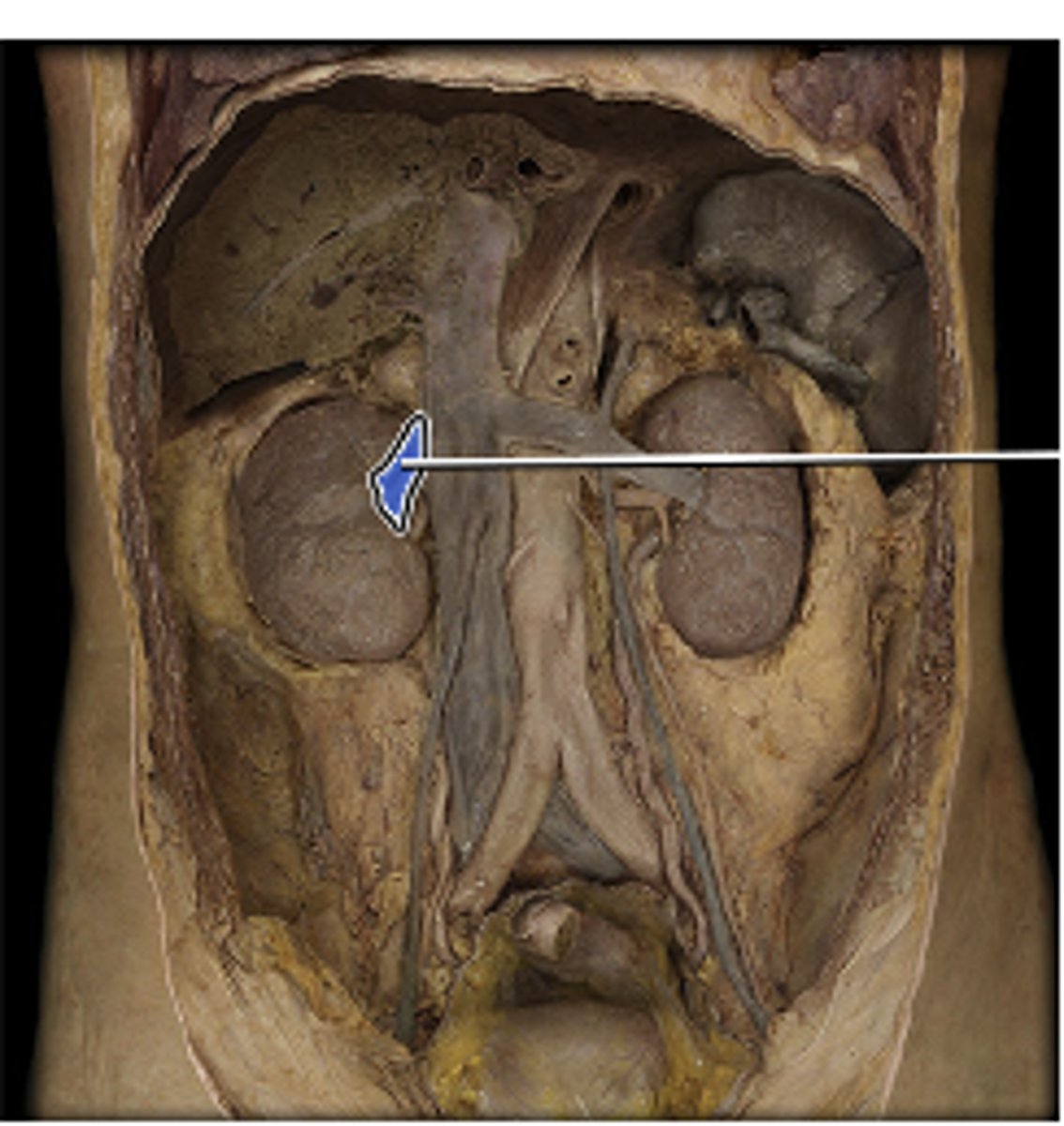
right renal vein
left renal vein
right common iliac artery
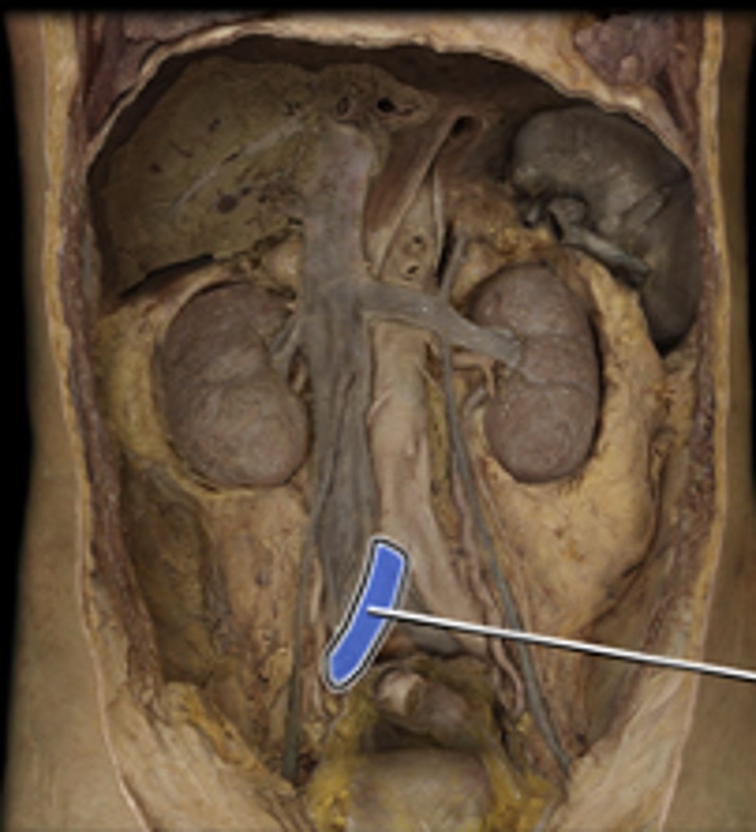
left common iliac artery
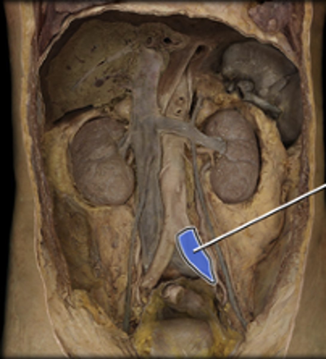
right external iliac artery
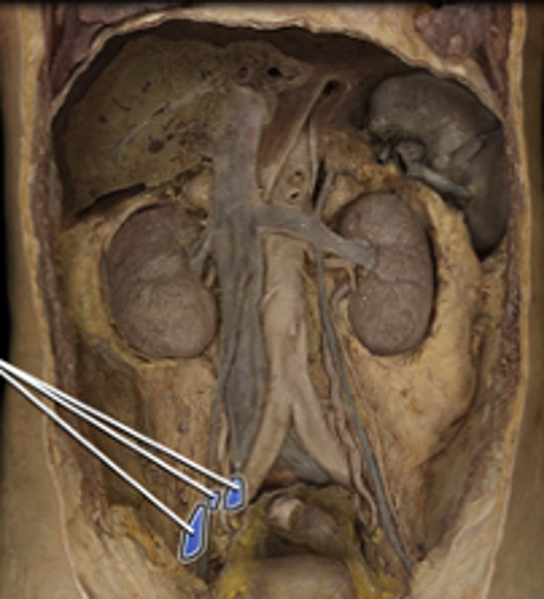
left external iliac artery
right common iliac vein
drains blood from pelvic walls, pelvic viscera and lower limb
left common iliac vein
drains blood from pelvic walls, pelvic viscera and lower limb
superior gluteal artery
supplies blood to the ilium, skin of gluteal region, piriformis, gluteus med-max-min
inferior gluteal artery
Supplies blood to the pelvic diaphragm, piriformis, quadratus femoris, biceps femoris, gluteus maximus, skin of buttocks and posterior thigh.
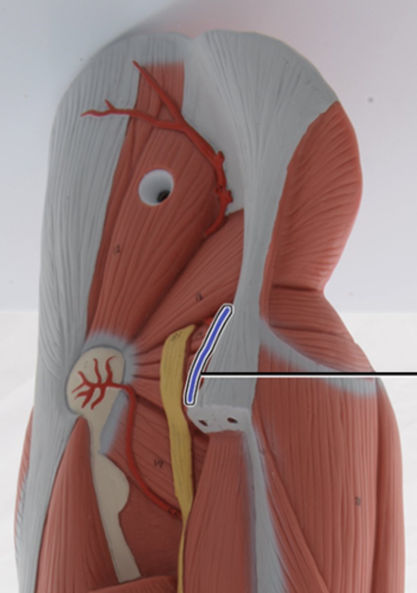
popliteal artery
Supplies blood to the knee joint, distal femur, proximal tibia, platella and proximal heads of the gastric, plantaris and the surrounding fascia and skin.
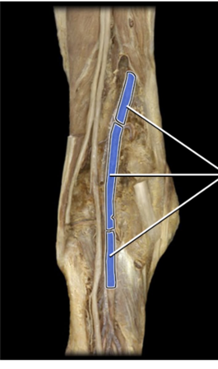
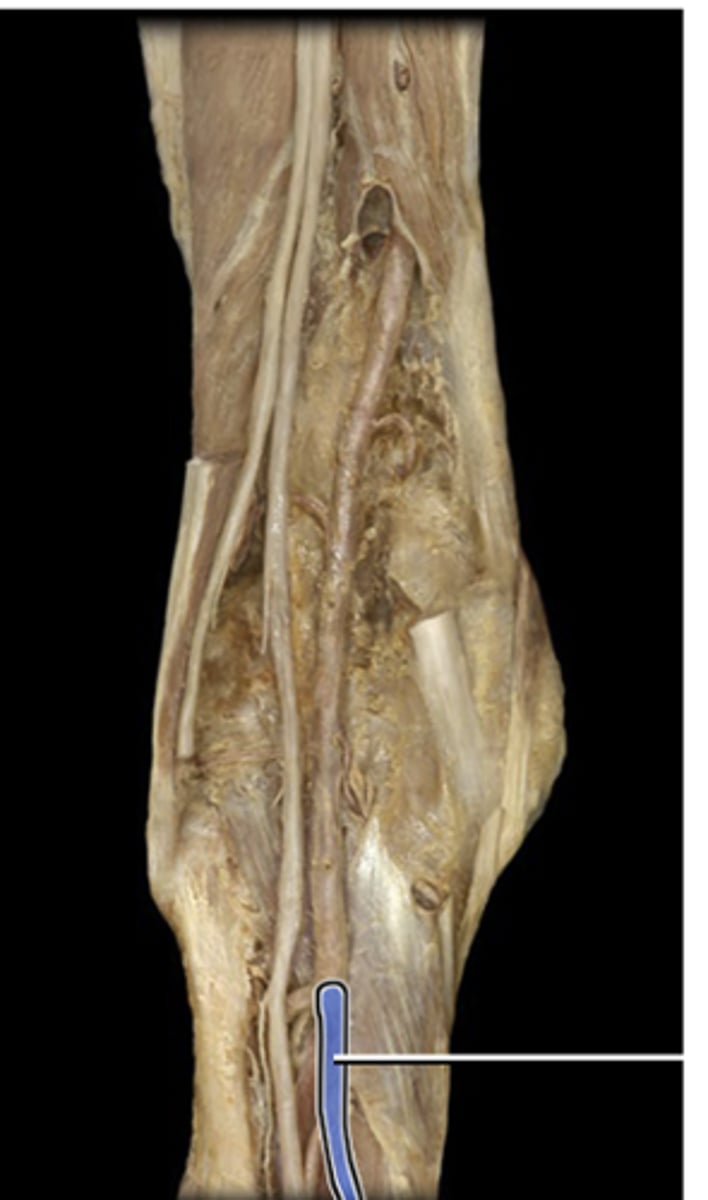
posterior tibial artery
Supplies blood to the muscles of the posterior and lateral lower leg, ankle and foot. Also supplies blood to the tibia and fibula.
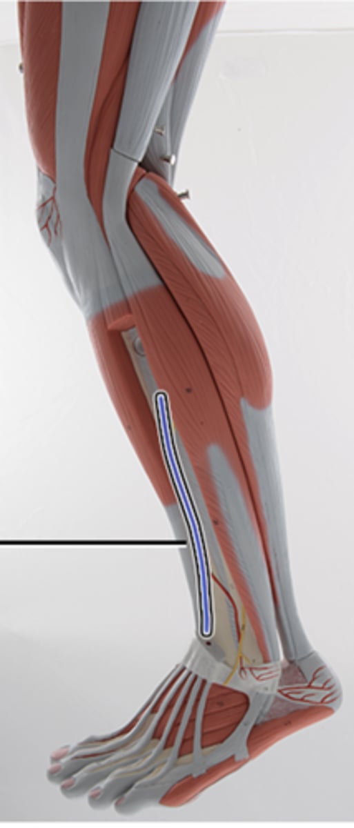
anterior tibial artery
Supplies blood to the muscles of the anterior compartment of the lower leg, ankle and foot
fibular artery
Supplies blood to the muscles of the anterior compartment of the lower leg, ankle and foot
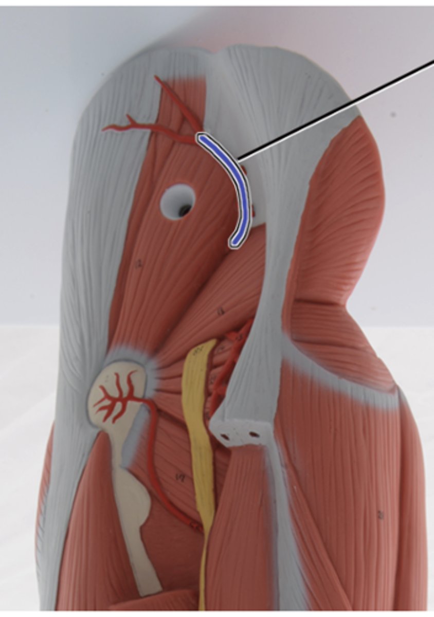
external iliac artery
Supplies blood to the lower abdominal wall, external genitalia, and lower limb
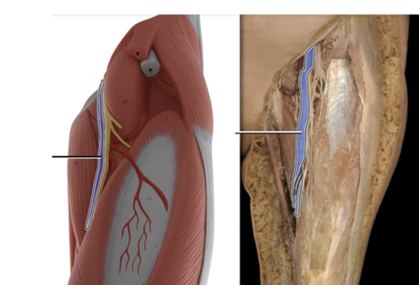
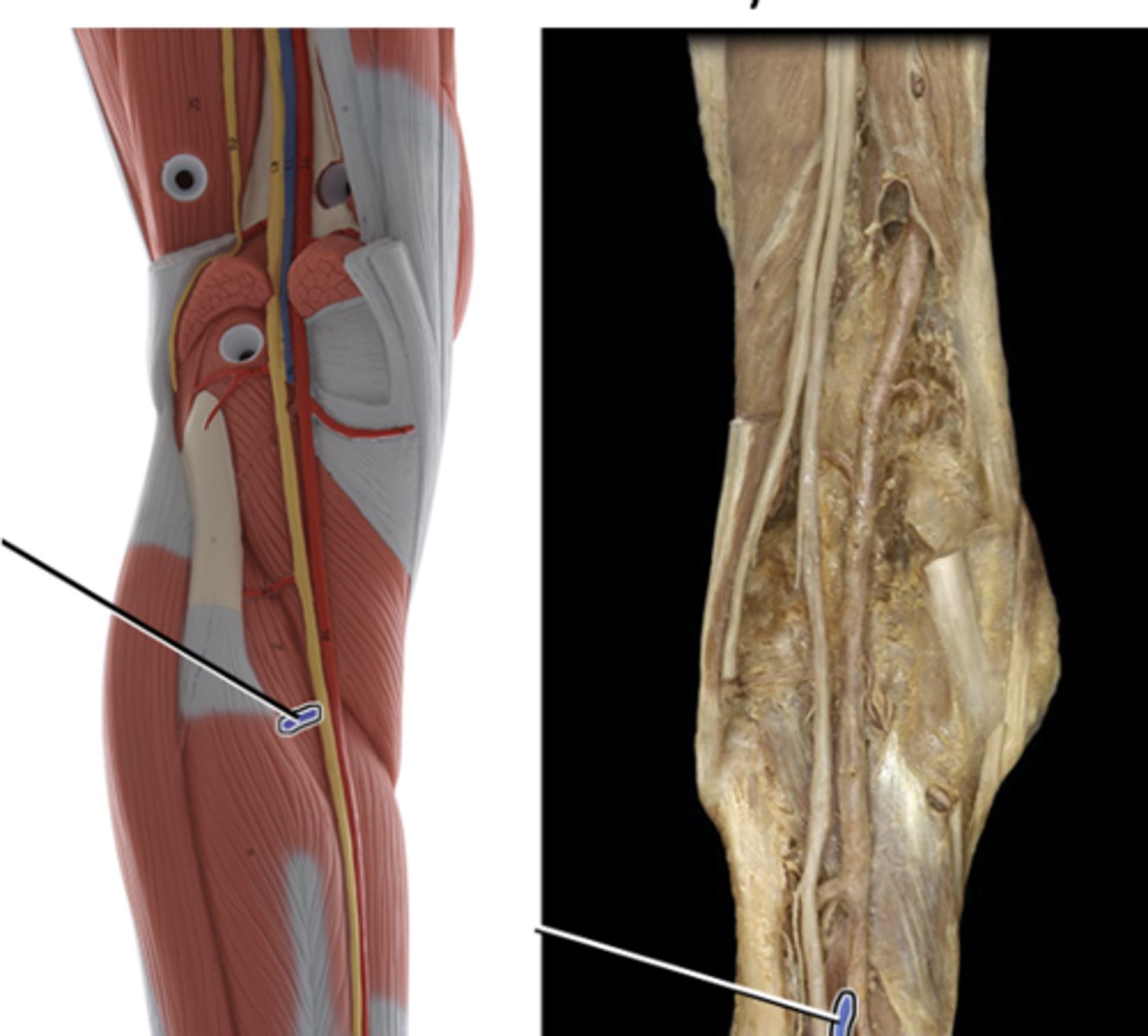
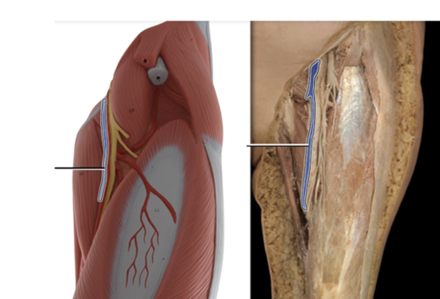
femoral vein
drains lower limb and delivers blood to the external iliac vein
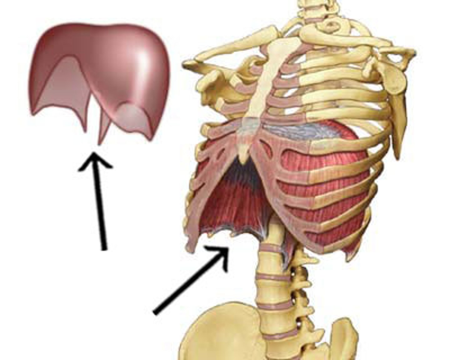
diaphragm
separates the thorax from abdomen. main muscle of respiration and is essential to the breathing process
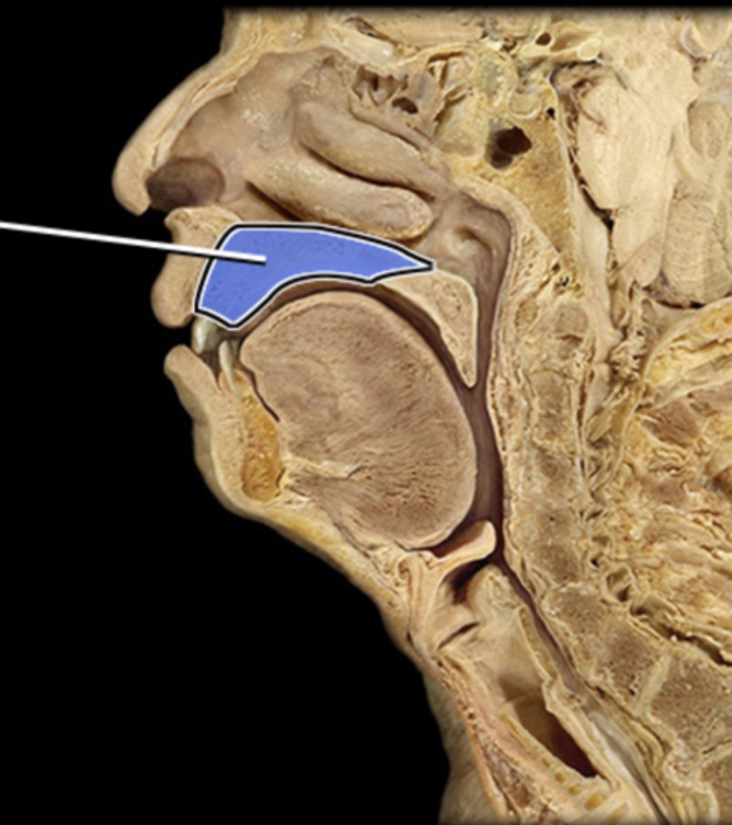
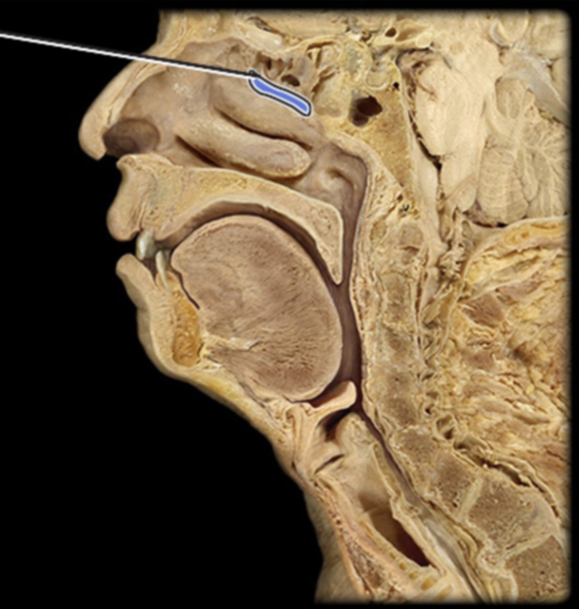
hard palate
rooms roof of the mouth and separates the oral cavity from the nasal cavity
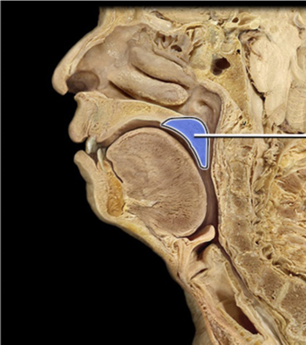
soft palate
comprised of muscle fibers, serves to elevate the nasopharynx
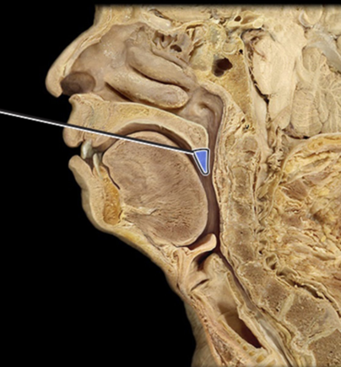
uvula
the end of the soft palate, touching it evokes gag reflex
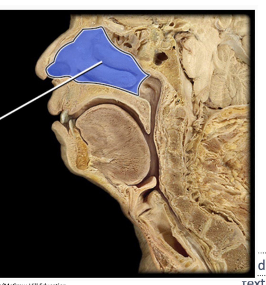
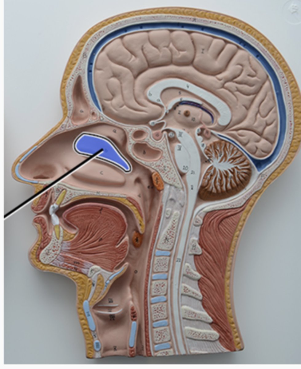
nasal cavity
humidifies and warms inspired air
superior nasal concha
middle nasal concha
inferior nasal concha
nasopharynx, oropharynx, laryngopharynx
what 3 regions make up the pharynx (throat)
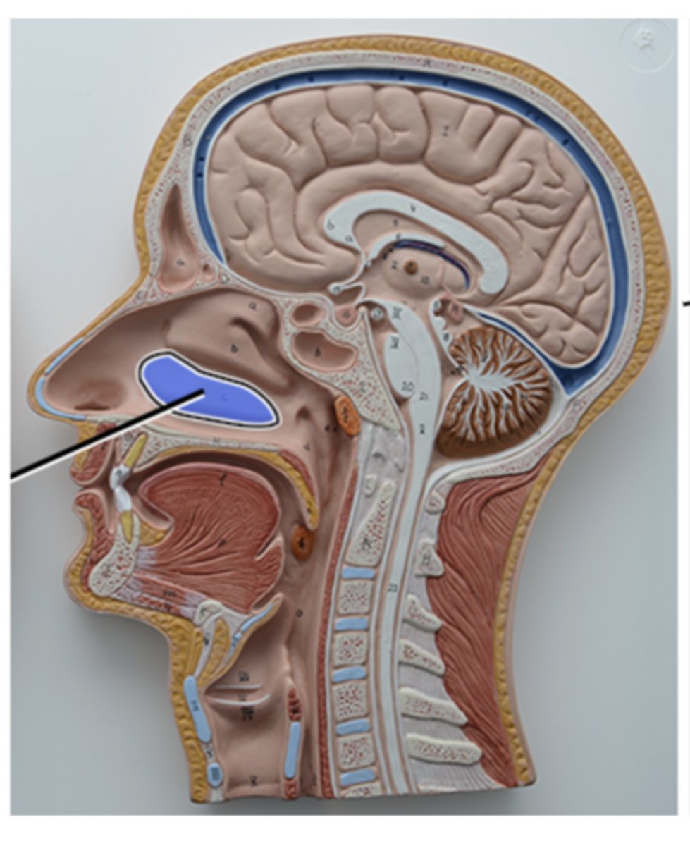
nasopharynx
receives auditory tubs and contains pharyngeal tonsil
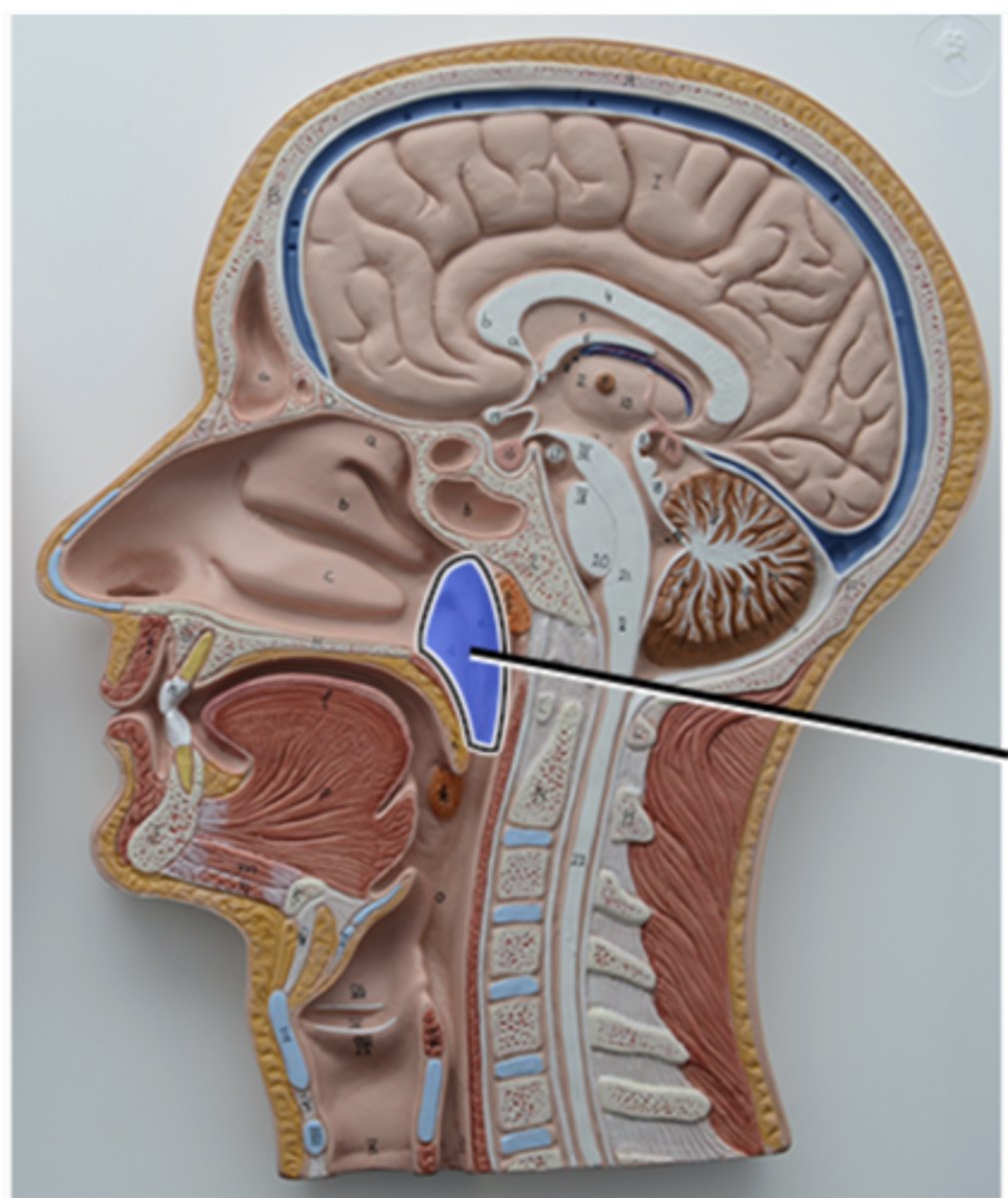
oropharynx
contains palatine tonsil
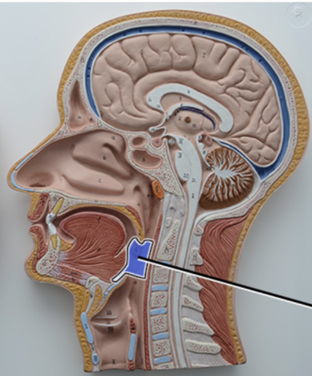
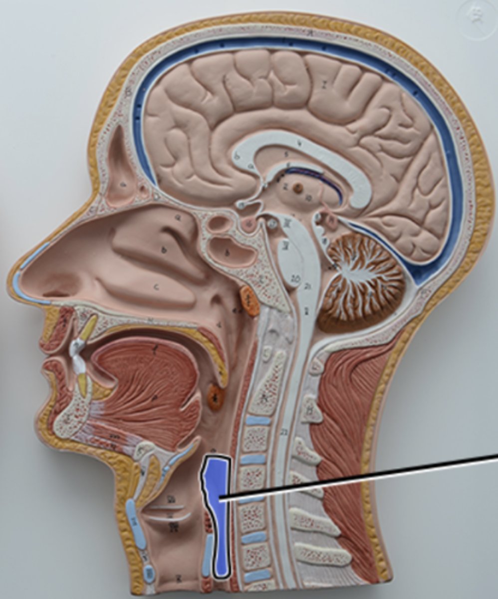
laryngopharynx
esophagus begins at this point
larynx (voice box)
houses muscles and ligaments that prevent aspiration and the production of voice
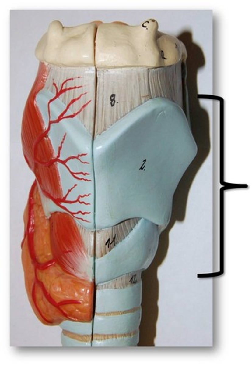
epiglottic cartilage, thyroid cartilage, and cricoid cartilage
what are the first three cartilages that make up the framework of the larynx
how many cartilages make up the framework of the larynx
9
epiglottis
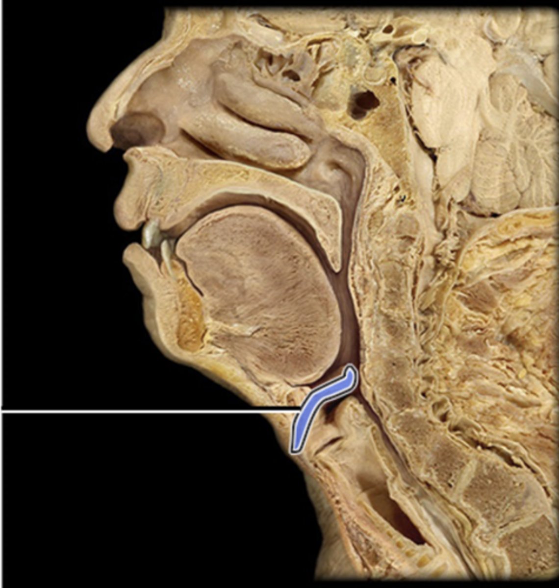
epiglottic cartilage
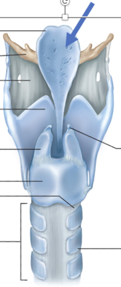
thyroid cartilage
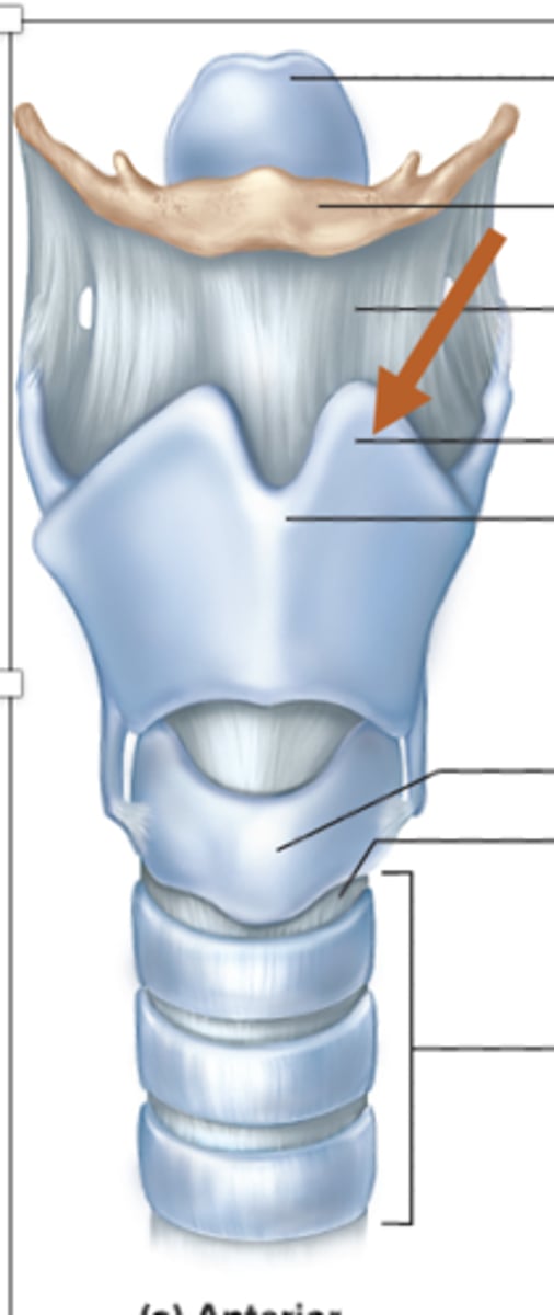
cricoid cartilage
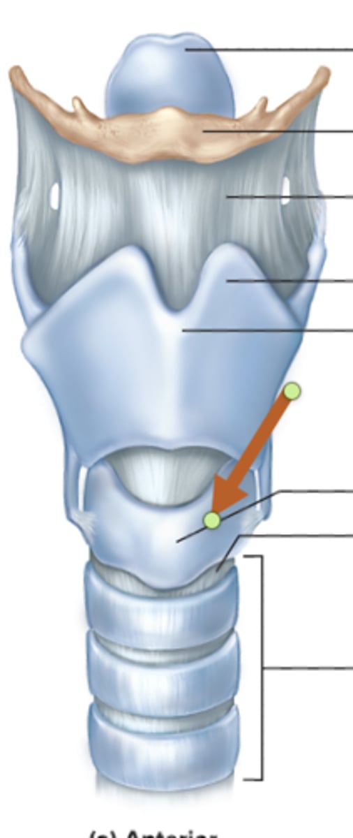
tracheal cartilage
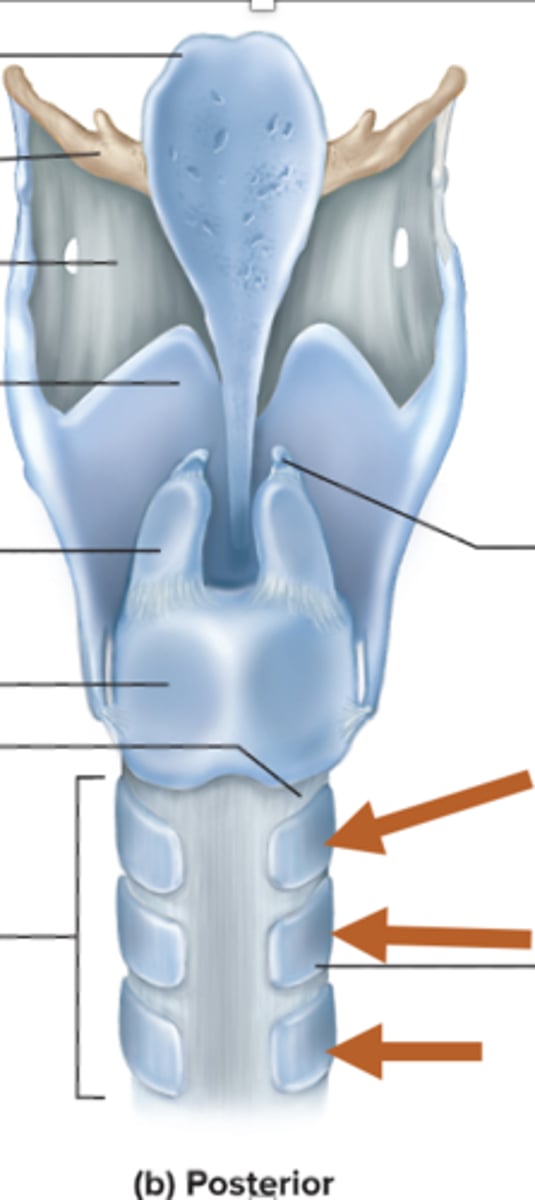
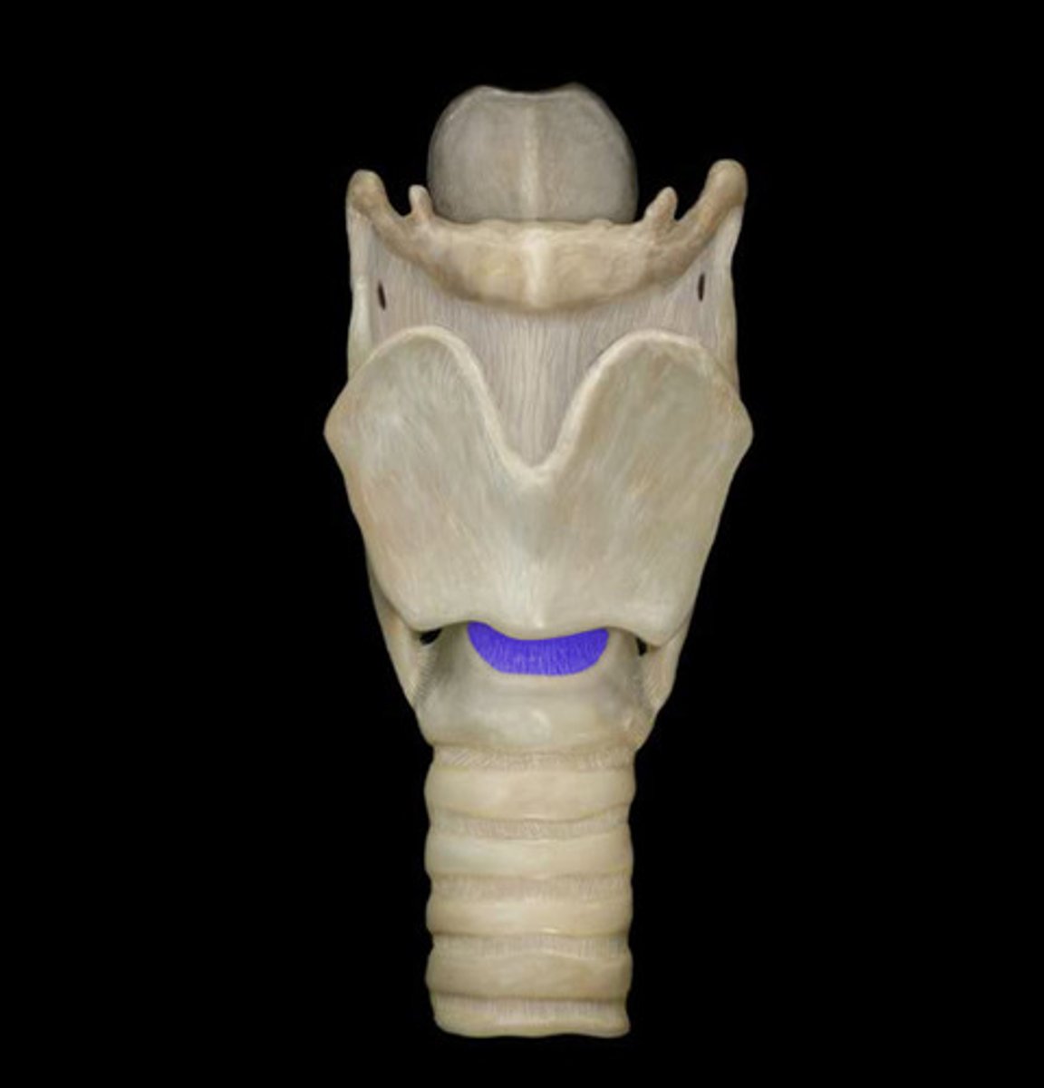
cricothyroid ligament
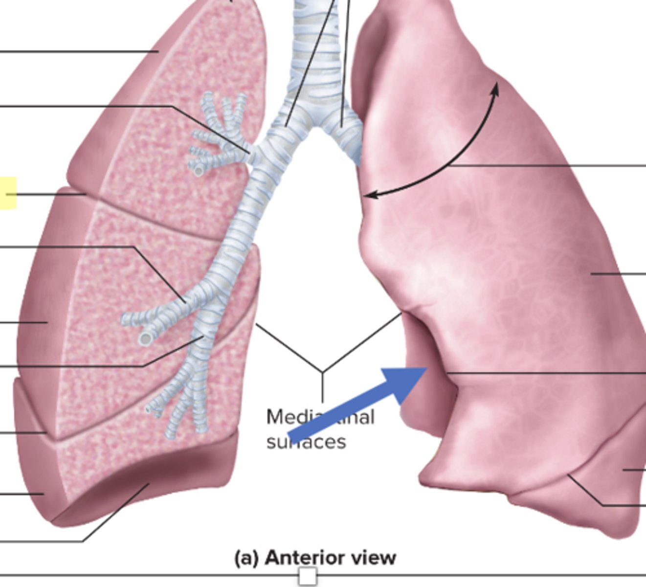
trachea
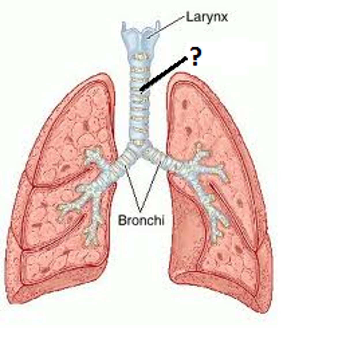
main bronchi
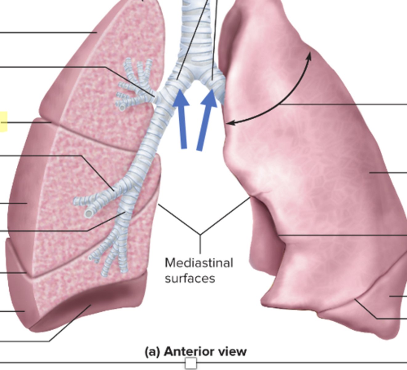
superior lobe
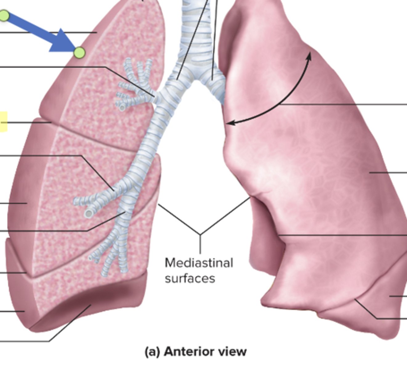
superior lobar bronchus
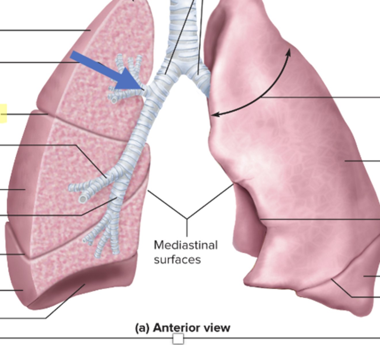
horizontal fissure
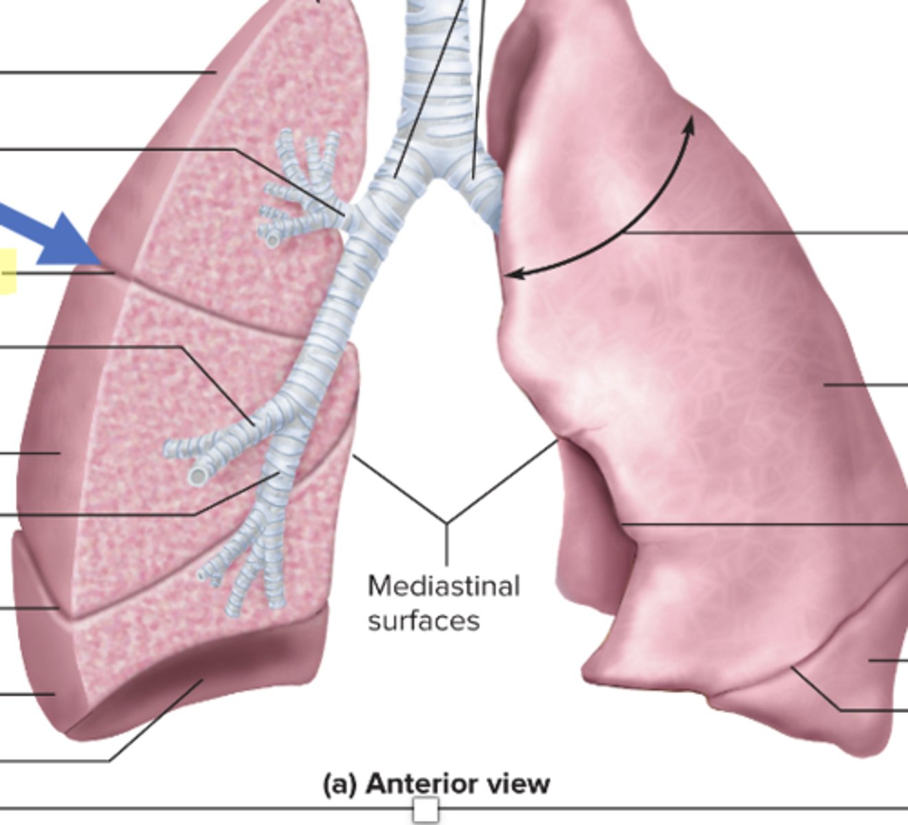
middle lobar bronchus
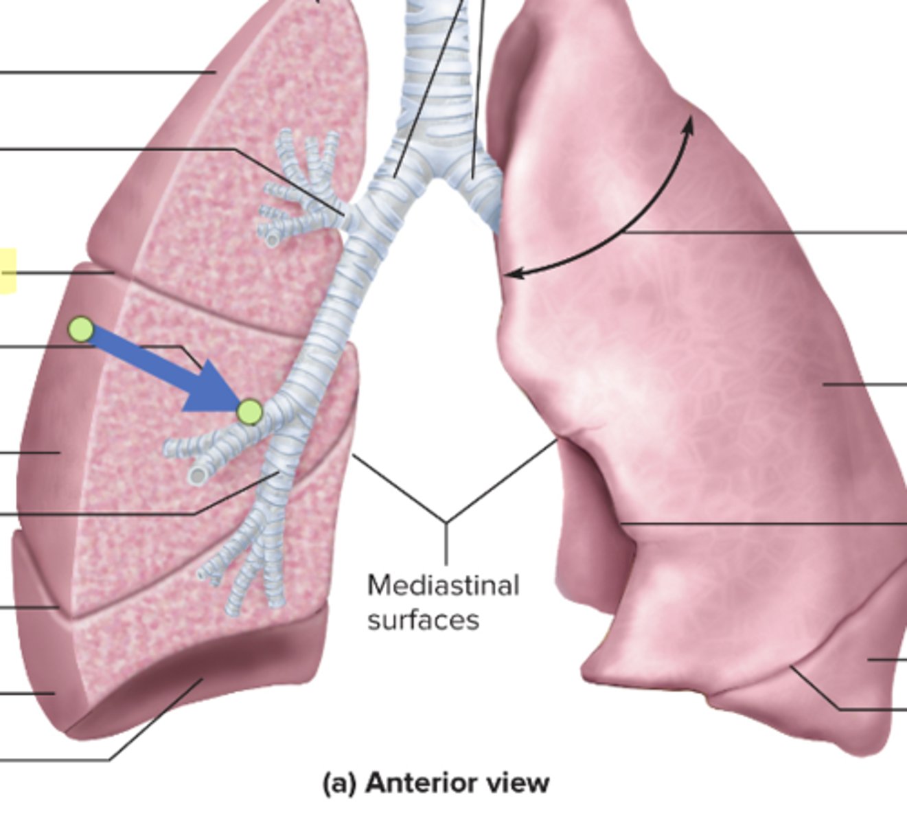
right middle lobe
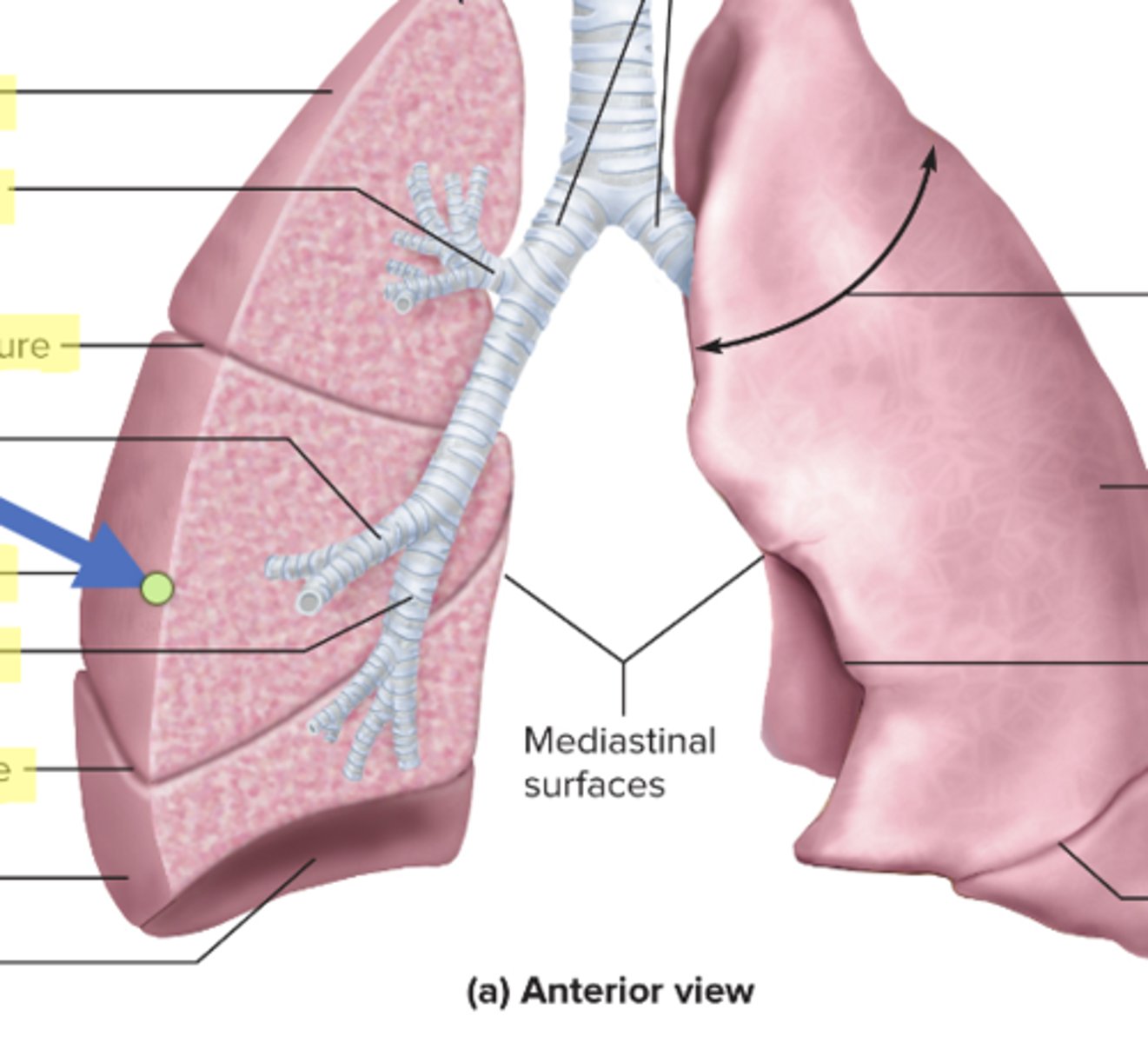
inferior lobar bronchus
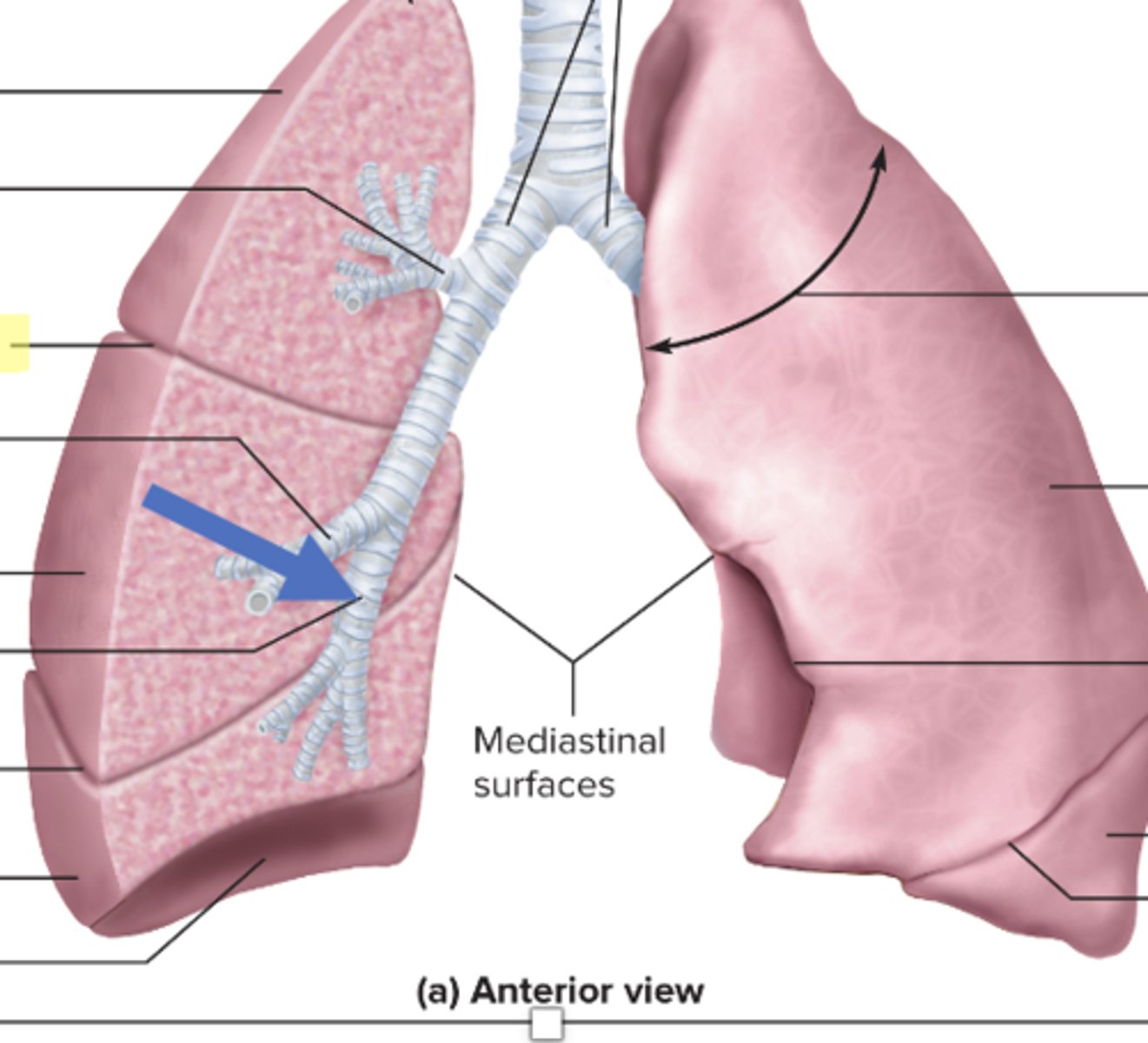
oblique fissure
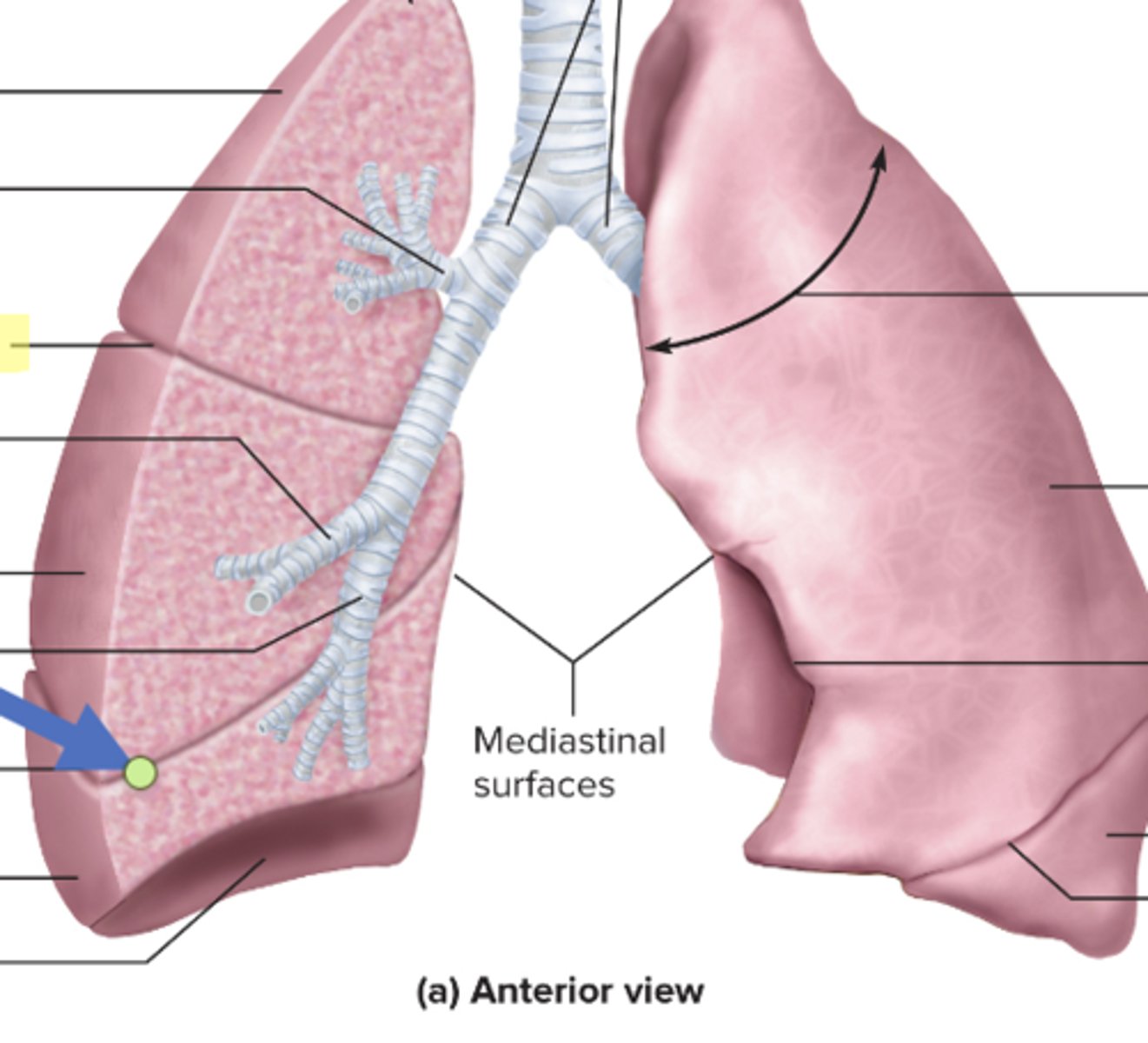
right inferior lobe
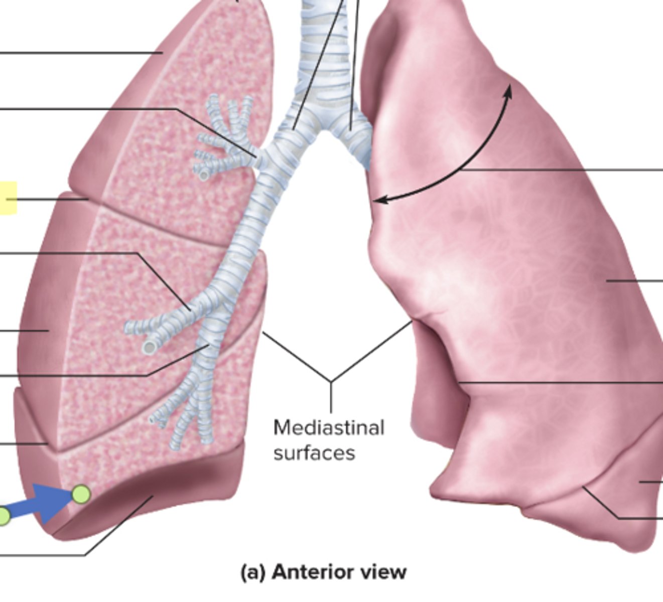
cardiac notch
tidal volume
amount of air inhaled during a normal breath
expiratory reserve volume
amount of air that can be exhaled after a normal exhalation
inspiratory reserve volume (IRV)
amount of air that can be further inhaled after a normal inhalation
residual volume
air left in the lungs after a forced exhalation
vital capacity
maximum amount of air that can be moved in or out of the lungs in a single respiratory cycle
inspiratory capactiy
volume of air that can be inhaled in addition to a normal exhalation
functional residual capacity
volume of air remaining after a normal exhalation
total lung capacity
total volume of air in the lungs after a maximal inspiration
forced expiratory volume
how much air can be forced out of the lungs over a specific time period, usually one second
Total Lung Capacity
In spirometry, TLC stands for
Inspiratory reserve, expiratory reserve, tidal volume, and residual volume
TLC is the sum of
Active process, inhalation
Diaphragm contracting
Passive process, exhalation
Diaphragm relaxing