Anatomy Final Exam
1/162
There's no tags or description
Looks like no tags are added yet.
Name | Mastery | Learn | Test | Matching | Spaced | Call with Kai |
|---|
No analytics yet
Send a link to your students to track their progress
163 Terms
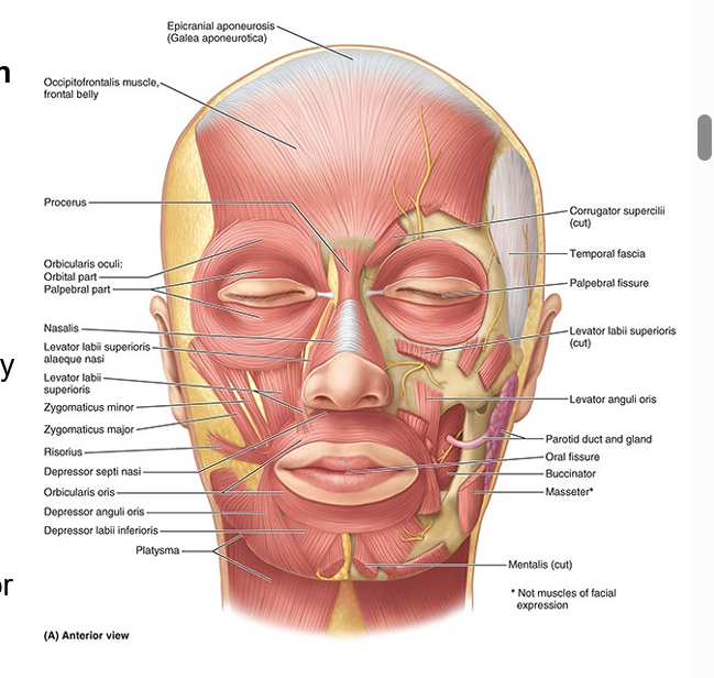
muscles of facial expression
in subcutaneous tissue of ant and post scalp, face, and neck
attach to bone or fascia and produce their effects by pulling the skin
innervated by CN VII & facial nerve
sensory innervation of facial skin
CN V
ophthalmic (V1) = upper face
superior orbital fissure
maxillary (V2) = mid face
foramen rotundum
mandibular (V3) = lower face
foramen ovale
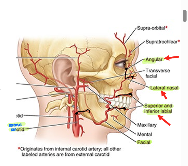
blood supply of face
facial artery = branch of external carotid
inferior labial
superior labial
lateral nasal
angular
superficial temporal = branch of external carotid
branches into transverse facial artery
feeds parotid gland & medial face
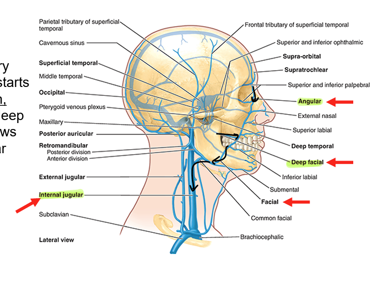
venous drainage of face
facial vein
primary drainage of face
starts at angular vein
collects from deep facial vein
flows into internal jugular vein
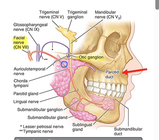
parotid gland
largest of 3 salivary glands
embedded structures
facial nerve
external carotid artery
parotid duct exits gland
parasympathetic innervation from CN IX via otic ganglion

parotid duct
exits parotid gland
pierces buccinator & enters oral cavity
delivers saliva
osteology of the neck
cervical vertebrae
manubrium of sternum
clavicles
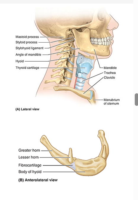
hyoid bone
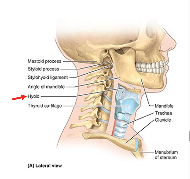
hyoid bone
lies in the ant part of the neck at the level of the C3-C4 vertebrae
does not directly articulate w/ other bone
only bone in body
attachment for ant neck muscles & and a prop to keep the airway open
superficial cervical fascia
layer of fatty connective tissue that lies between the dermis of the skin & the investing layer of deep cervical fascia
contains platysma anteriorly
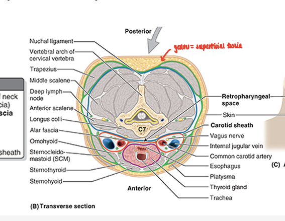
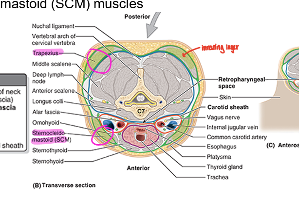
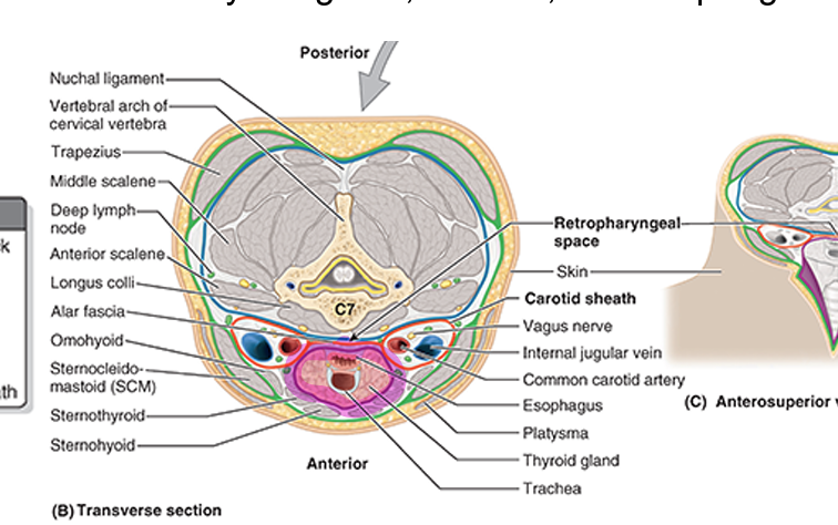
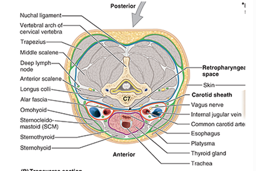
layers of deep cervical fascia
superficial → deep
investing
pretrachael
prevertebral
carotid sheath
investing layer of deep cervical fascia
most superfic deep fascia layer
surrounds entire neck deep to the skin & subcut tissue
splits into superfic & deeo layers to enclose trapezius & sternocleidomastoid (SCM) muscles
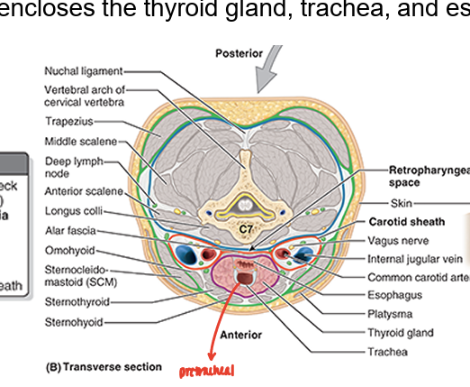
pretrachael layer of deep cervical fascia
ant part of neck
muscular part: encloses infrahyoid muscles
visceral part: encloses thyroid gland, trachea, & esophagus
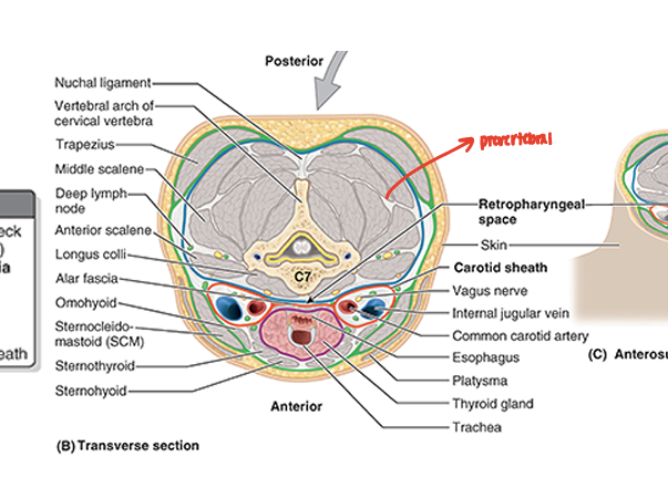
prevertebral layer of deep cervical fascia
tubular sheath for the vert column & the muscles associated w/ it
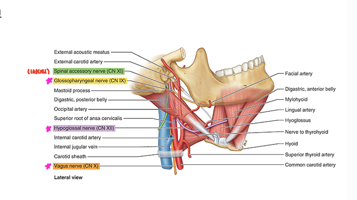
carotid sheath in deep cervical fascia
common & internal carotid arteries
internal jugular vein
vagus nerve (CN X)
carotid sinus nerve
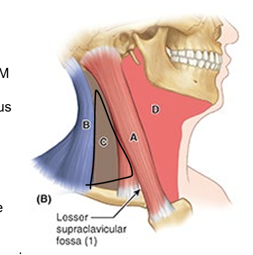
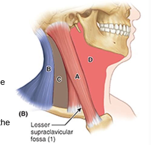
regions of superficial neck
sternocleidomastoid region (A)
post. cervical region (B)
ant. cervical region (D)
lateral cervical region (C)
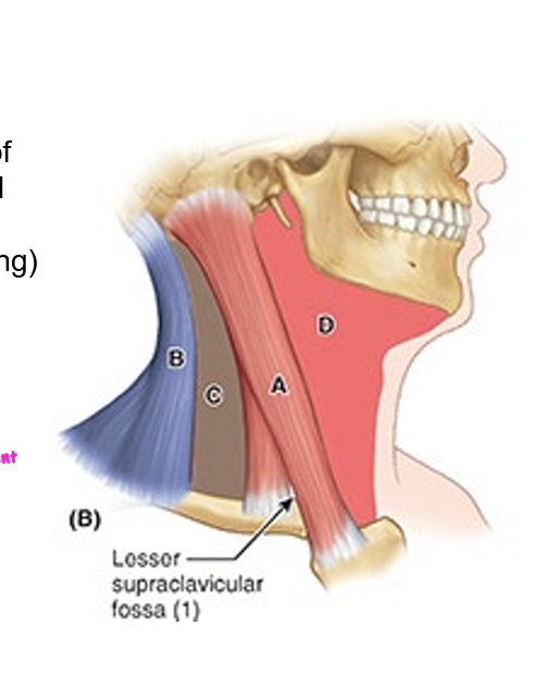
lateral cervical region boundaries
ant: post. border of SCM
post: ant. border of trapezius
inf: middle third of clavicle between trapezius & SCM
apex: where SCM and trapezius meet on superior nuchal line of occipital bone
roof: investing layer of deep cervical fascia
floor: muscles covered by prevert layer of deep cervical fascia
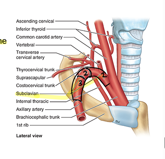
subclavian artery parts
named w/ respect to anterior scalene muscle
medial (1)
posterior (2)
lateral (3)
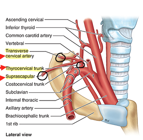
arteries of the lateral cervical region
two branches of thyrocervical trunk (1st part of subclavian)
transverse cervical trunk
suprascapular artery
3rd part of subclavian
occipital artery
branch of external carotid at apex of cervical region
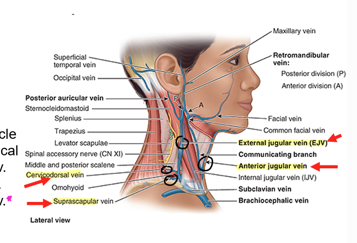
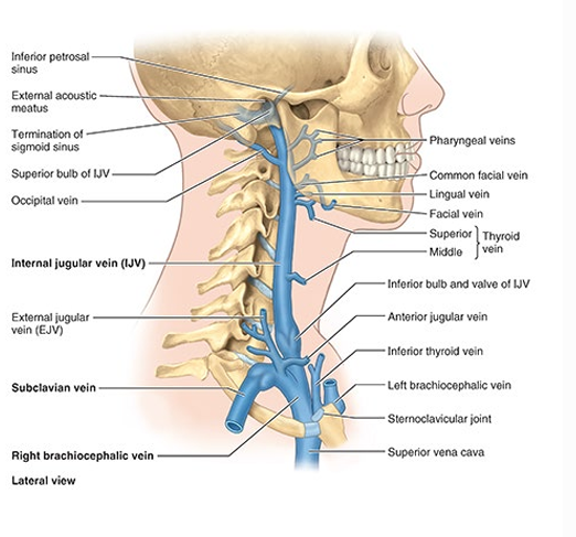
veins of the lateral cervical region
external jugular
transverse cervical (cervicodorsal) vein
suprascapular vein
anterior jugular vein (not in lateral region)
external jugular then drains into subclavian vein
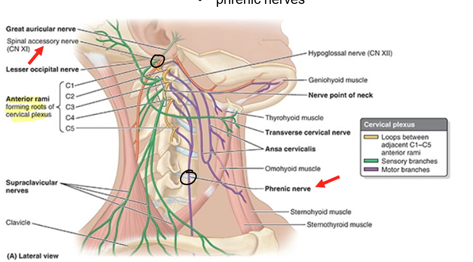
nerves of lateral cervical region
spinal accessory nerve (CN XI)
roots of the brachial plexus
suprascapular nerve
roots of cervical plexus
phrenic nerves
anterior cervical region boundaries
ant: median line of neck
post: ant border of SCM
sup: inf border of mandible
apex: jugular notch in manubrium
roof: subcut tissue containing the platysma
floor: pharynx, larynx, & thyroid gland
anterior cervical muscles
suprahyoid
infrahyoid
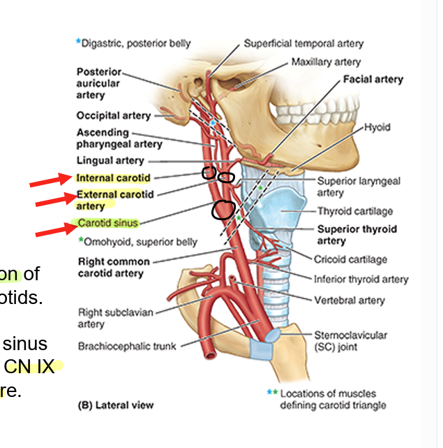
anterior cervical region arteries
common carotid
internal & external
carotid sinus at bifurcation of internal & external
baroreceptors in carotid sinus via carotid branch of CN IX to regulate bp
anterior cervical region veins
internal jugular vein & tributaries
anterior cervical region nerves
CNs IX, X, XII
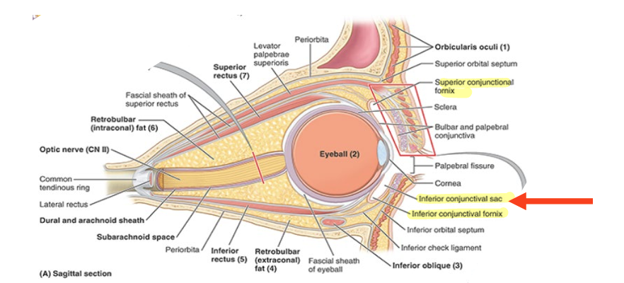
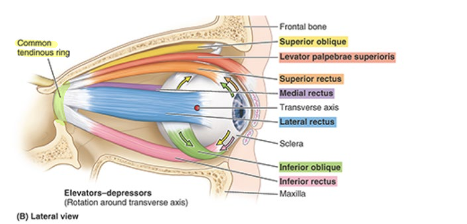
orbit
bony cavities shaped like quadrangular pyramids w/ their bases directed anterolaterally & their apices posteromedially
apex = optic canal
base = orbital margin (surrounds orbital opening)
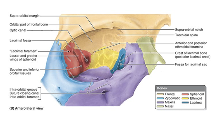
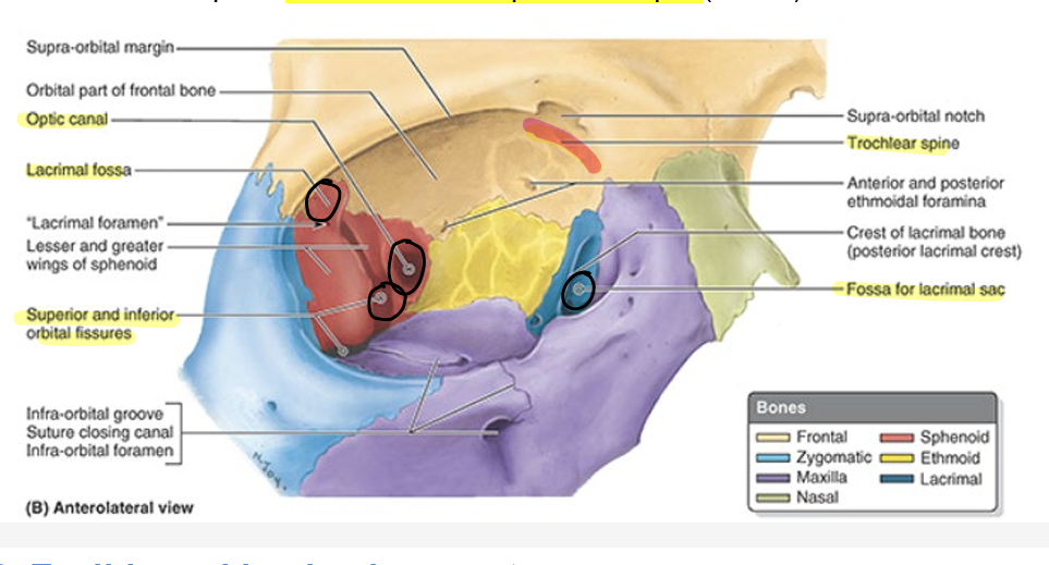
features of the orbit
optic canal: CN II, ophthalmic artery
superior orbital fissure: ophthalmic nerve (CN V1), CN III, IV, and VI
lacrimal fossa: lacrimal gland (frontal bone)
trochlear spine: for trochlear of sup. oblique (CN IV)
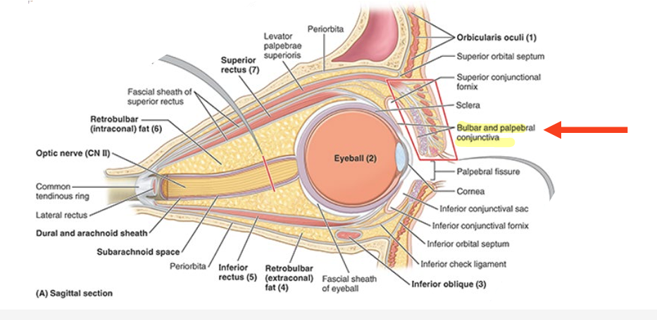
eyelids (conjunctiva)
eyelids are covered externally by thin skin
internally by transparent mucous memb = palpebral conjunctiva
palpebral conjunctiva is reflected onto the eyeball, where it is continuous w/ bulbar conjunctiva
attached to ant. surface (sclera) of the eyeball
eyelids part 2
lines of reflection of palpebral conjunctiva onto eyeball form recesses of pockets
= sup. & inf. conjunctival fornices
conjunctival sac (sup & inf) = space bound by palpebral & bulbar conjunctivae
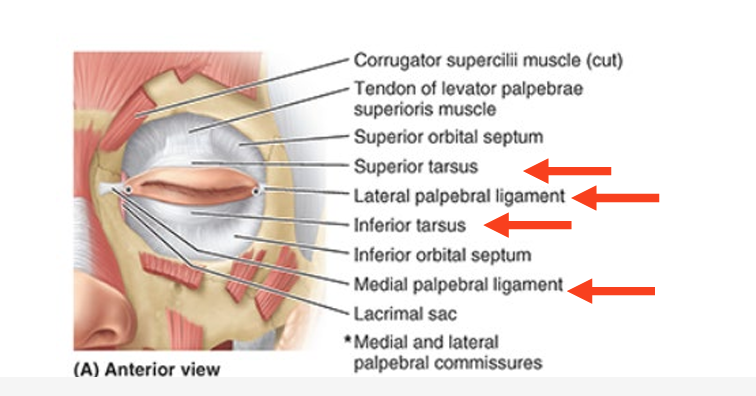
strengthened by dense bands of connective tissue = sup & inf tarsi
medial palpebral ligament
lateral palpebral ligament
medial & lateral palpebral ligaments
medial = connects tarsi to medial margin of orbit
between nose & medial angle of eye
lateral = attaches tarsi to lateral margin of orbit
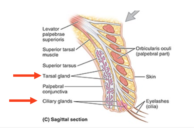
eyelid glands
tarsal glands
produce lipid secretion that lubricates edges of eyelids & prevent them from sticking tg when closed
ciliary glands
large sebaceous glands associated w/ eyelashes
lacrimal apparatus
includes:
lacrimal gland
excretory ducts of lacrimal gland
lacrimal canaliculi
nasolacrimal duct
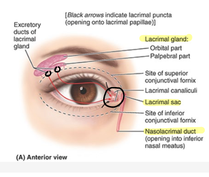
lacrimal gland
secretes lacrimal fluid (tears)
= watery physiological saline containing bactericidal enzyme lysozyme

excretory ducts of lacrimal gland
convey lacrimal fluid from lacrimal glands to conjunctival sac

lacrimal canaliculi
drain lacrimal fluid to lacrimal sac
dilated sup part of the nasolacrimal duct

nasolacrimal duct
conveys lacrimal fluid to inf. nasal meatus of nasal cavity
why nose drips when you cry

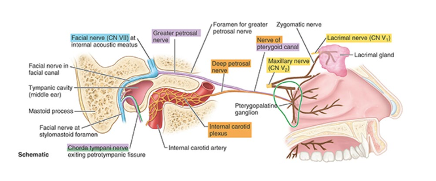
innervation of the lacrimal apparatus
parasymp secretomotor fibers are conveyed from the facial nerve
vasoconstrictive, postsynap symp fibers → brought from sup. cervical ganglion
branches of V1 and V2 = also autonomic innervation
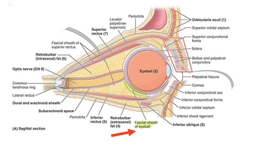
fascial sheath of eyeball
connective tissue layer surrounds the eyeball
post. forms actual socket for eyeball
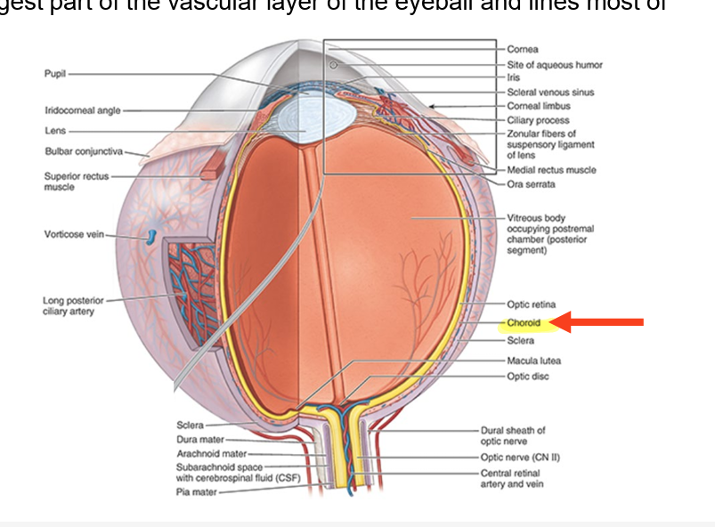
layers of eyeball
fibrous layer = outer coat
consists of sclera & cornea
vascular layer = middle coat
consists of choroid, ciliary body, & iris
inner layer = inner coat
consists of retina (optic & nonvisual part)
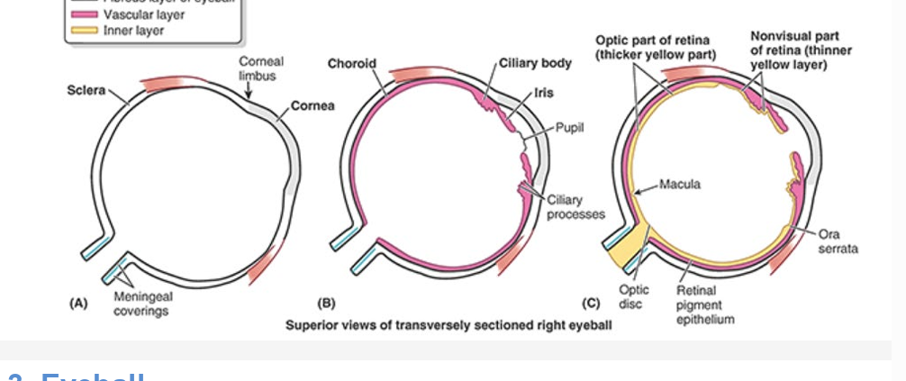
fibrous layer of eyeball
sclera
tough, opaque part
covers post. 5/6 of eyeball (white part)
cornea
transparent part
covers ant 1/6 of eyeball
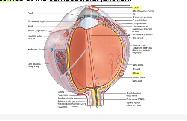
corneal limbus
angle formed by intersecting curvatures of sclera & cornea at corneoscleral jxn
sclera
tough, opaque part of fibrous layer
covers post. 5/6 of eyeball (white part)

cornea
transparent part of fibrous layer
covers ant 1/6 of eyeball

corneal limbus
angle formed by intersecting curvatures of sclera & cornea at corneoscleral jxn

vascular layer of eyeball
consists of choroid, ciliary body, and iris
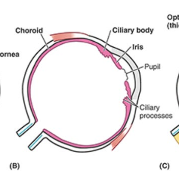
choroid
dark reddish brown layer between sclera & retina
forms largest part of vascular layer of eyeball
lines most of sclera
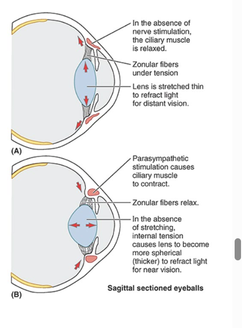
ciliary body
ring-like thickening of vascular layer post to corneoscleral jxn
contraction & relaxation of smooth muscle of ciliary body controls thickness of the lens

ciliary processes
in vascular layer of eyeball
secrete aqueous humor
clear watery fluid
fills ant. segment of eyeball
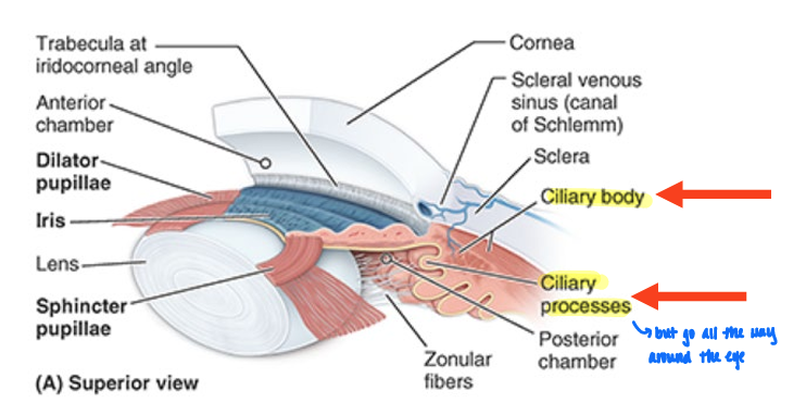
iris
lies on ant. surface of lens
thin contractile diaphragm w/ central aperture (pupil)
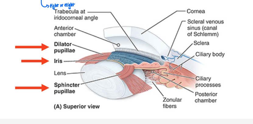
stimulation of iris
parasymp (feeding and breeding) stimulated
circularly arranged sphincter pupillae
decreases diameter (constricts or contracts pupil)
symp (fight or flight) stimulated
radially arranged dilator pupillae
increases its diameter (dilated pupil)
inner layer of the eyeball
retina
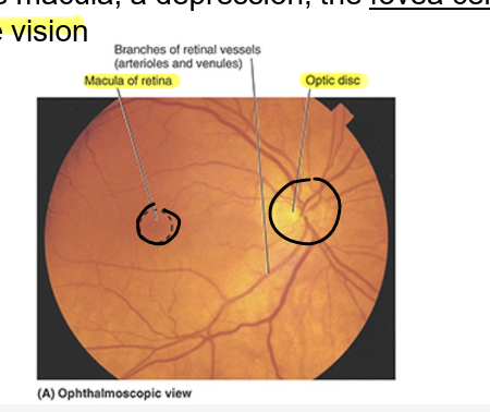
optic disc
macula

retina
sensory neural layer of eyeball
consists of two fxnal parts w/ distinct
optic part
nonvisual retina (retinal tissue not involved in vision)
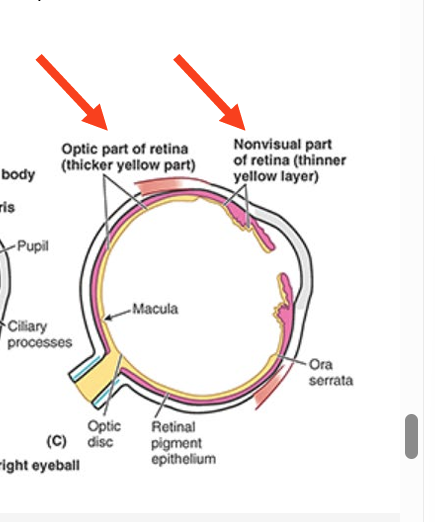
optic disc
where sensory fibers & vessels conveyed by optic nerve (CN II) enter eyeball
contains no photoreceps
optic disc insensitive to light (blind spot)

macula
small oval area of retina w/ special photoreceps cones that is specialized for acuity of vision
at center of macula = depression = fovea centralis
= area of most acute vision
chambers of the eye
anterior
space between cornea ant and iris/pupil post
aqueous humor
post
between iris/pupil ant and lens & ciliary body post
aqueous humor
vitreous chamber
post. 4/5of eyeball post to lens
vitreous humor
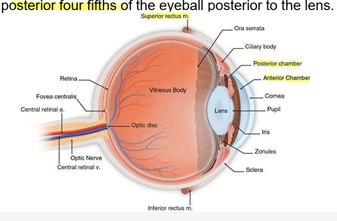
lens
post to iris and ant to vitreous humor
ciliary muscle changes shape of lens
para sympy stimulation via oculomotor nerve (CN III) → sphincter-like contraction of muscle
ring becomes smaller
tension on lens = reduced
thickness of lens increases w/ age
common tendinous ring
attachment for all four rectus muscle
arteries of the orbit
blood supply mainly from ophthalmic artery
branch of internal carotid artery
major branches
central retinal artery: branches from center of optic disc
lacrimal
short posterior ciliaries
long posterior ciliaries

central retinal artery supplies:
optic retina
lacrimal artery supplies
lacrimal gland
conjunctiva
eyelids

short posterior ciliaries supplies
choroid
in turn supplies cones & rods
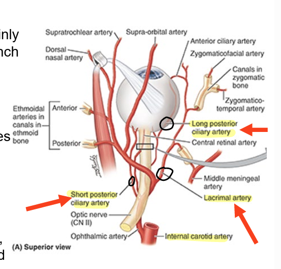
long posterior ciliaries supplies
ciliary body
ribs

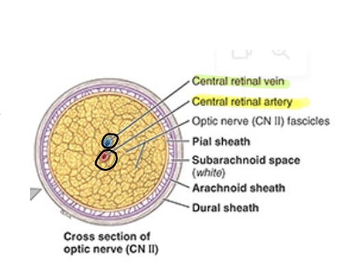
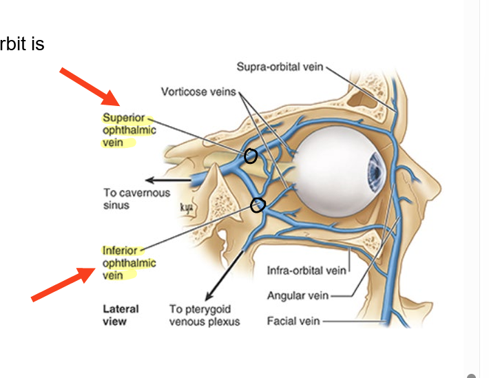
veins of the orbit
through superior & inferior ophthalmic veins
pass thru orbital fissure
also central retinal vein
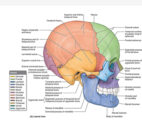
temporal region fossae
temporal fossa
infratemporal fossa
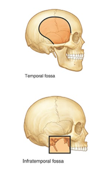
temporal fossa borders
posterior & superior: by temporal lines
ant: frontal & zygomatic bones
lateral: zygomatic arch
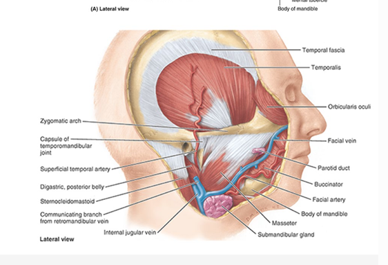
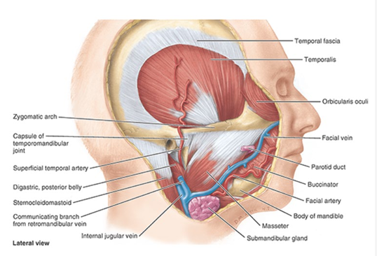
temporal fossa contents
upper part of temporalis muscle
infratemporal fossa contents
inferior to zygomatic arch
inf part of temporalis muscle
lat and med pterygoid muscles
maxillary artery
pterygoid venous plexus
nerves:
mandibular (V3)
inf alveolar
lingual
buccal
chorda tympani nerves (CN VII)
otic ganglion
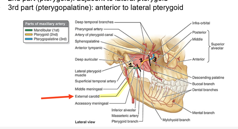
maxillary artery
branch of ext carotid
named according to relationship w/ lateral pterygoid
supplies lateral-deep face
1st part (mandibular): post to lat pterygoid
2nd part (pterygoid): adj to lat pterygoid
3rd part (pterygopalatine): ant to lat pterygoid
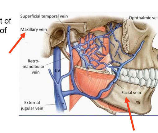
pterygoid plexus
drains most of region supplied by branches of maxillary artery
drains post into maxillary vein
drains ant into facial vein
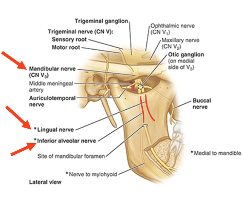
mandibular nerve (V3)
inf alveolar = innervation of mandibular teeth
lingual nerve = sensory to ant 2/3 of tongue
not taste
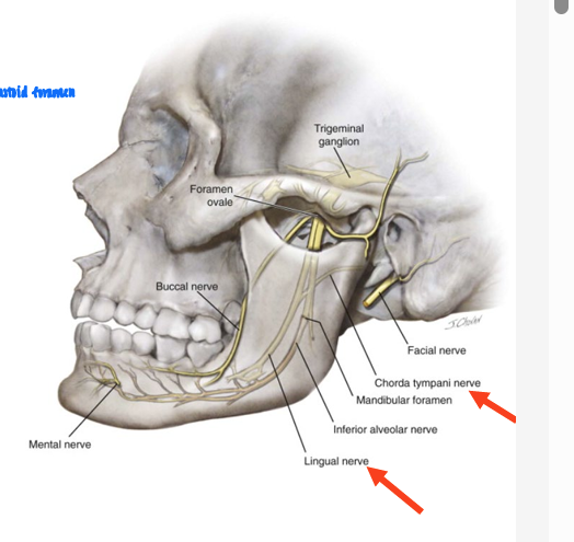
chorda tympani nerve
branch of CN VII
carries taste sensation from ant 2/3 of tongue
joins lingual nerve in infratemporal fossa
bony articular surfaces of tempromandibular joint
mandibular fossa of temp bone
articulates w/ head of mandible
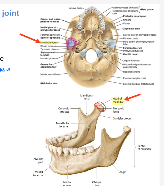
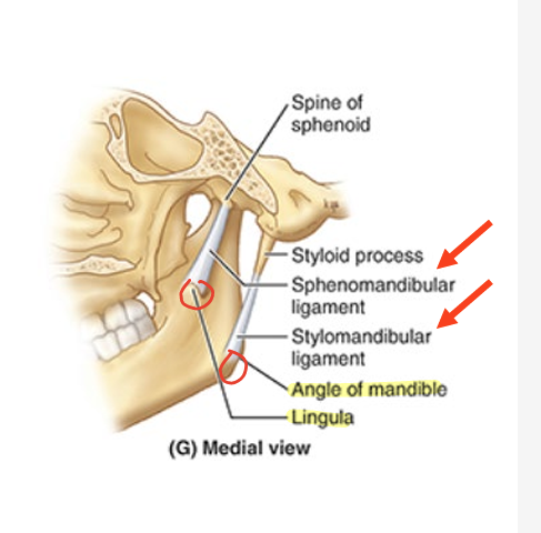
ligaments of tempromandibular joint
lat ligament = thickened part of joint capsule
2 extrinsic ligaments = connect mandible to cranium
stylomandibular lig = from styloid process to angle of mandible
sphenomandibular ligament = from spine of sphenoid to lingula of mandible
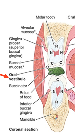
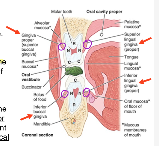
oral cavity
oral vestibule
oral cavity proper
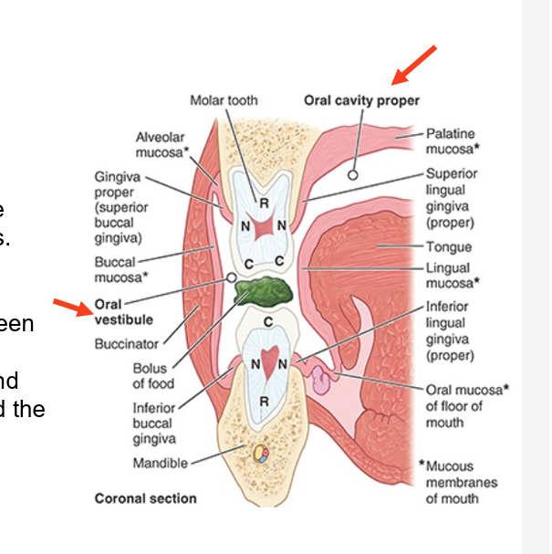
oral vestibule
slit-like space between teeth and gingivae (gums) and lips and cheeks
oral cavity proper
space between upper and lower dental arches of arcades
maxillary & mandibular alveolar arches and teeth they bare
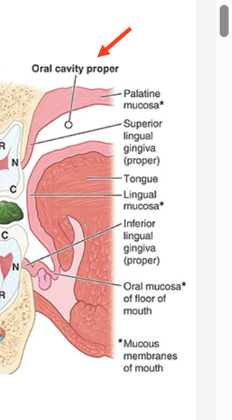
gingivae (gums)
composed of fibrous tissue covered w/ mucous memb
gingiva proper
firmly attached to alveolar part of mandible & alveolar process of maxilla & necks of teeth
ging proper adj to tongue = sup & inf lingual gingivae
ging proper adj to lips & cheeks = buccal gingiva
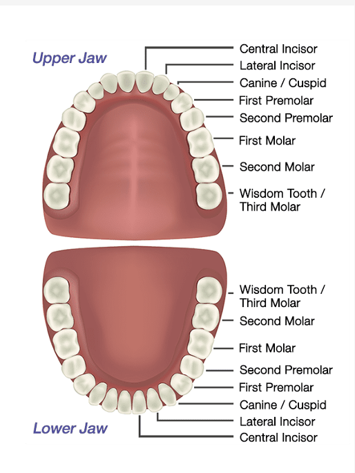
number of teeth
children = 20 deciduous
adults = 32 permanent
on each side of jaw (x4) (from ant → post)
2 incisors (central & lateral)
1 canine
2 premolars (1st and 2nd)
3 molars (1st-3rd)
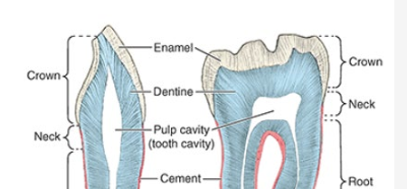
parts of teeth
crown = projects from gingiva (gums)
neck = between crown and root
root = fixed in tooth socket by periodontium
connective tissue surrounding roots
dental alveolus = tooth socket
most of tooth = composed of dentine
covered by enamel over crown & cement over the root
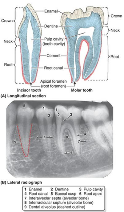
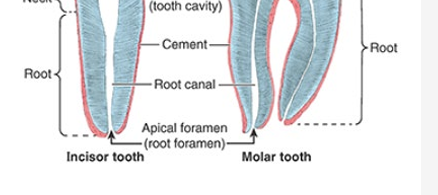
teeth pulp cavity
contains connective tissue, blood vessels, & nerves
teeth root canal
aka pulp canal
transmits nerves & vessels to and from pulp cavity thru apical foramen
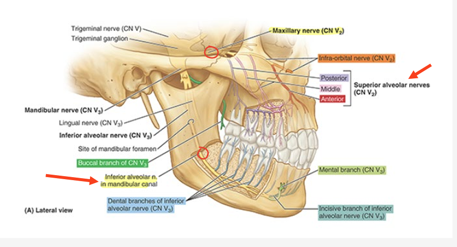
innervation of teeth
branches of sup (CN V2) and inf (CN V3) alveolar nerves
give rise to dental plexuses that supply maxillary & mandibular teeth
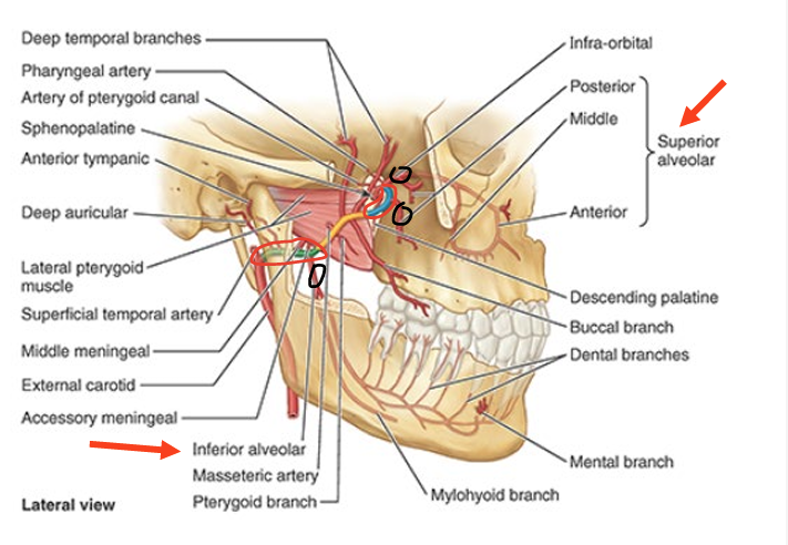
blood supply of teeth
sup alveolar artery = supplies maxillary teeth
inf alveolar artery = supplies mandibular teeth
branches of maxillary artery
palate
sep oral cavity from nasal cavities and nasopharnyx
hard palate ant
soft palate post
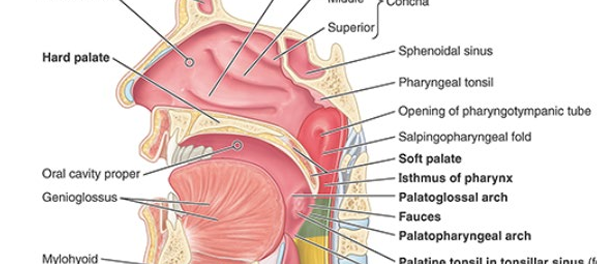
hard palate
ant 2/3 of palate
bony skeleton formed by
maxillae bones ant
palatine bones post
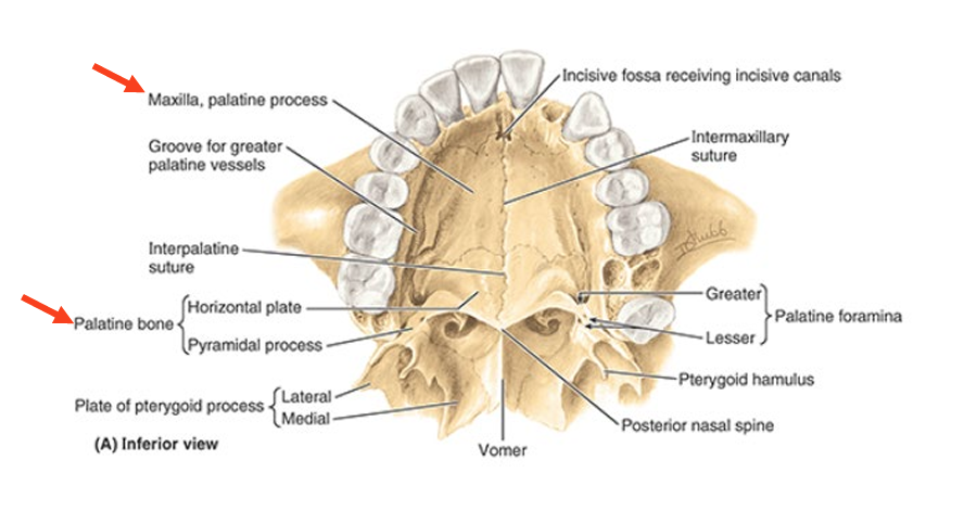
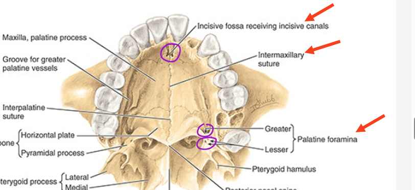
hard palate features
incisive fossa
intermaxillary suture
greater palatine foramina
lesser palatine foramina
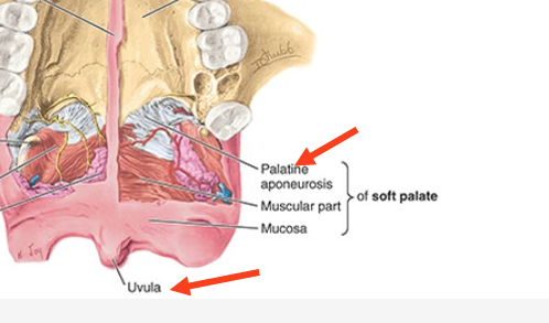
soft palate
movable post 1/3 of palate & is suspended from post border of hard palate
palatine aponeurosis
attaches to post edge of hard palate
blends w/ post muscular part of soft
structural support
uvula
conical process hanging from free margin of post soft palate
blood supply of oral cavity
from maxillary artery
greater and lesser palatine arteries
enter thru corresponding greater and lesser palatine foramina
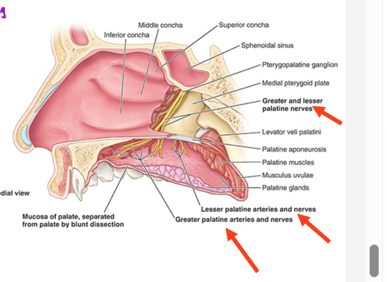
innervation of oral cavity
from maxillary nerve (V2)
greater and lesser palatine nerves
enter thru corresponding greater and lesser palatine foramina

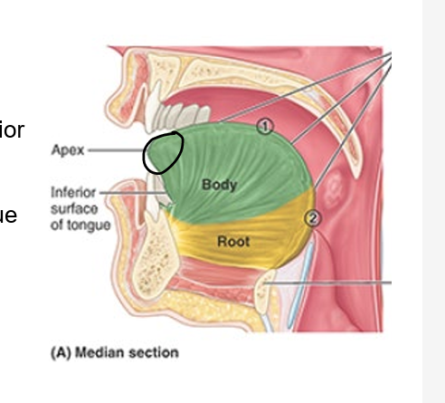
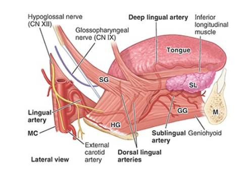
parts of the tongue
root = attached post portion
extends between mandible, hyoid, and nearly vertical post surface of tongue
body = ant 2/3 of tongue (between root and apex)
apex = ant end of body
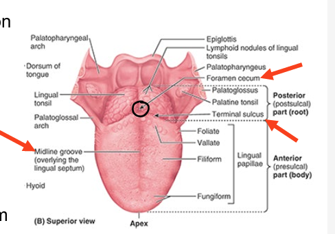
features of the tongue
terminal sulcus = V shaped groove on post dorsum of tongue
foramen cecum = depression at vertex of terminal sulcus
midline groove = divides ant part of tongue into L and R
lingual papillae = bumps on dorsum of tongue
some house taste buds
frenulum = connects underside of tongue from floor of mouth posteriorly
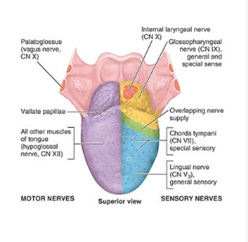
tongue innervation
motor
all intrinsic and extrinsic muscles of tongue (except 1) = supplied by CN XII
sensory
ant 2/3
general sensory = lingual nerve of CN V3
taste = CN VII (chorda tympani)
post 1/3
general sensory and taste = CN IX
blood supply of tongue
derived from lingual artery
arise from external carotid artery
pretrachael layer of deep cervical fascia
ant part of neck
muscular part: encloses the infrahyoid muscles
visceral part: encloses thyroid gland, trachea, & esophagus
visceral part of pretracheal fascia (layers)
endocrine layer = thyroid & parathyroid glands
respiratory layer = larynx & trachea
alimentary layer = pharynx & esophagus
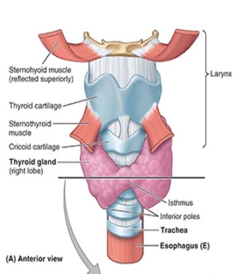
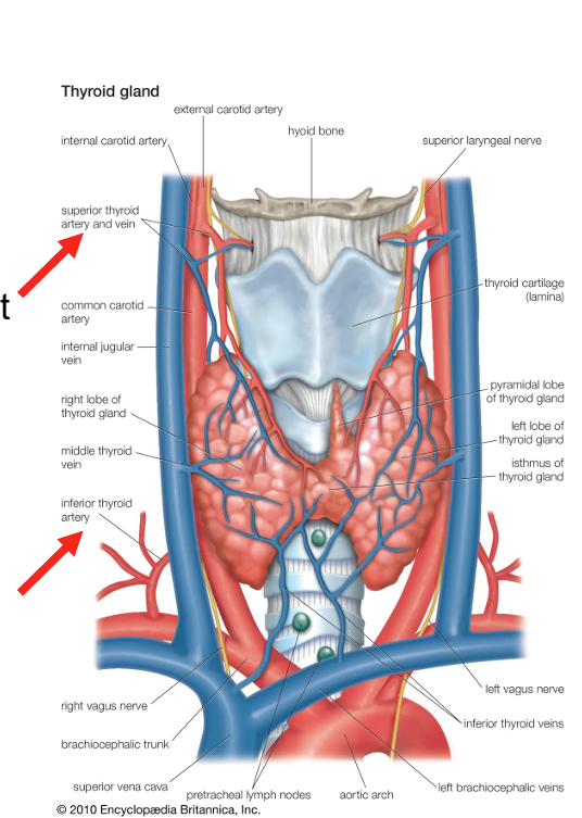
thyroid gland
part of endocrine layer of visceral pretrach fascia
produces
thyroid hormone = controls rate of metabolism
calcitonin = hormone that controls calcium metabolism
lies deep to sternothyroid & sternohyoid muscles
consists of R and L lobes
anterolateral to larynx and trachea
relatively thin isthmus unites lobes over trachea
blood supply of thyroid gland
sup thyroid arteries
1st branches of external carotid arteries
supply anterosuperior aspect of gland
inf thyroid arteries
largest branch of thyrocervical trunks
arise from subclavian arteries
supply posteroinf aspect of thyroid
parathyroid glands
part of endocrine layer of visceral pretrach fascia
produces parathyroid hormone = controls metabolism of phosphorous & calcium
usually 4 → lie on medial half of post surface of each lobe of thyroid gland
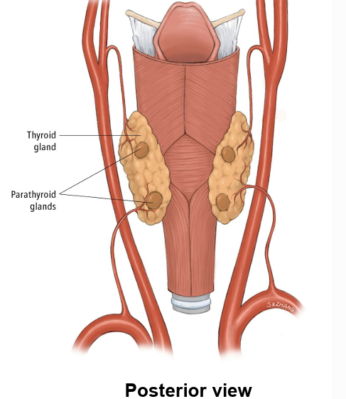
blood supply of parathyroid glands
inf thyroid arteries
provide primary blood supply to post aspect of thyroid land were parathyroid glands are located
branches of these arteries usually supply these glands
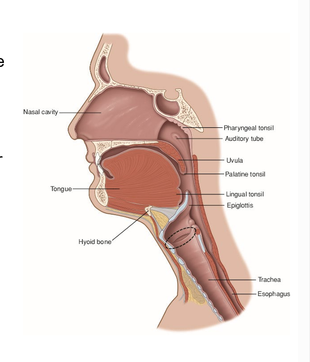
larynx
part of resp layer of visceral part of pretrach fascia
organ or voice production (the “voice box”)
guards the air passages → especially during swallowing when it serves as the “sphincter” or “valve” of the lower resp tract
connects inf part of pharynx (oropharynx) w trachea
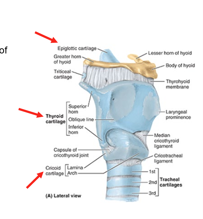
laryngeal skeleton
consists of 9 cartilages
3 single
thyroid
cricoid
epiglottic
3 paired
arytenoid
corniculate
cuneiform