Anatomy Lab Practical 2
1/105
Earn XP
Description and Tags
Skeletal II to Muscle III
Name | Mastery | Learn | Test | Matching | Spaced | Call with Kai |
|---|
No analytics yet
Send a link to your students to track their progress
106 Terms
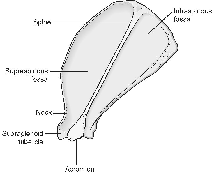
Spine of the Scapula
Thin narrow process on the middle of the scapula

Glenoid Fossa
Indented process where the humerus connects to the scapula
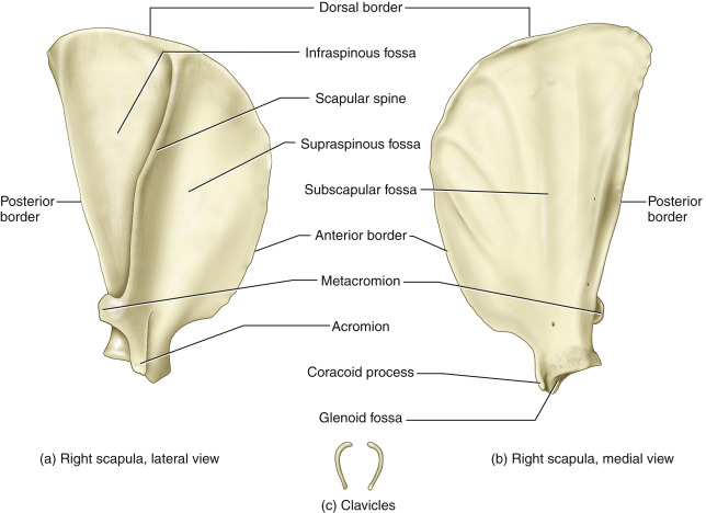
Acromion Process
Caudal bony extension of the spine of the scapula

Supraglenoid Tubercle
Tubercle that is superior (cranial) to the glenoid process
Wing of the Ilium
Most cranial part of the ilium; spans out to the sides

Body of the Ilium
Caudal part of the ilium; straight up and down

Tuber Coxae
Ventral thickening point of the ilium

Tuber Sacrale
Dorsal thickening point of the ilium; where the sacrum attaches to the ilium
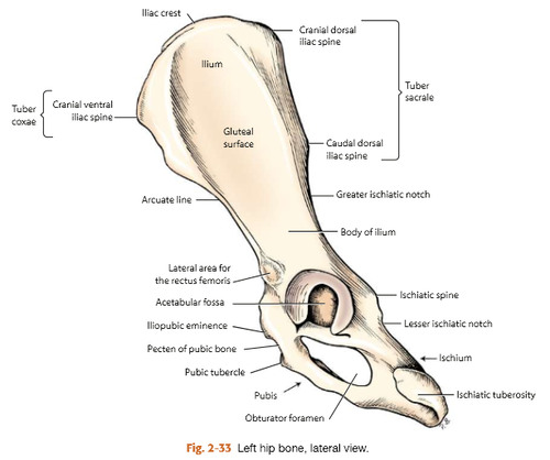
Ischiatic Tuberosity
Lateral thickening point of the ischium

Pelvic Symphysis
Where the two pubic bones join
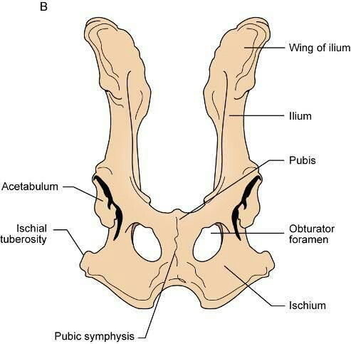
Obturator Foramen
Two holes on the caudal part of the os coxae

Acetabulum
Where the three pelvic bones join; where the femur attaches to the os coxae

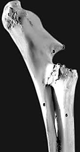
Head of the Humerus
Ball at the proximal end of the humerus
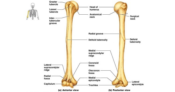
Greater Tubercle
Tubercle right next to the head of the humerus

Deltoid Tuberosity
Tuberosity on the side of the humerus

Olecranon Fossa
Divot on the posterior distal end of the humerus; where the olecranon attaches

Radial Fossa
Divot on the anterior distal end of the humerus; where the radius attaches

Head of the Femur
Ball at the proximal end of the femur

Greater Trochanter
Peak by the head of the femur
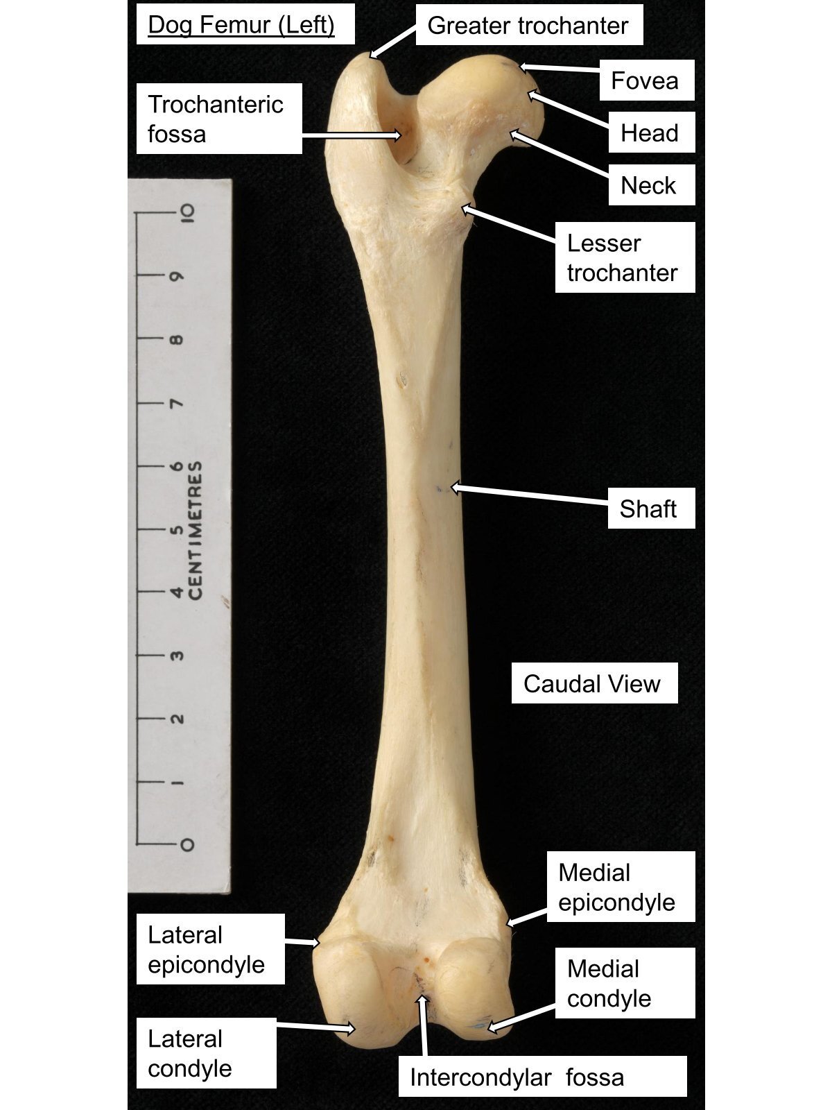
Medial and Lateral Condyles
The condyles on the distal end of the femur

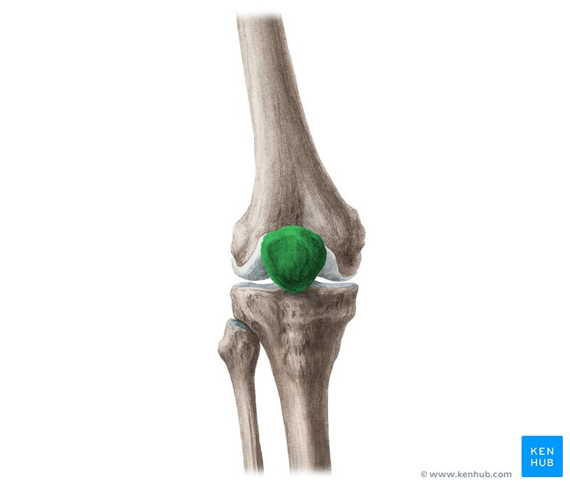
Patella
Kneecap
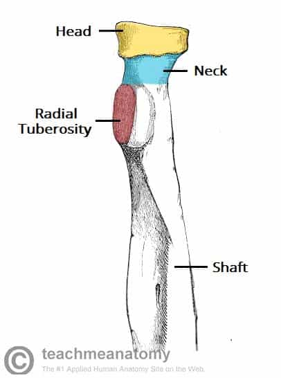
Head of the Radius
Proximal end of the radius; makes a circle shape
Tibia
T-shaped bone; larger of the two main distal leg bones
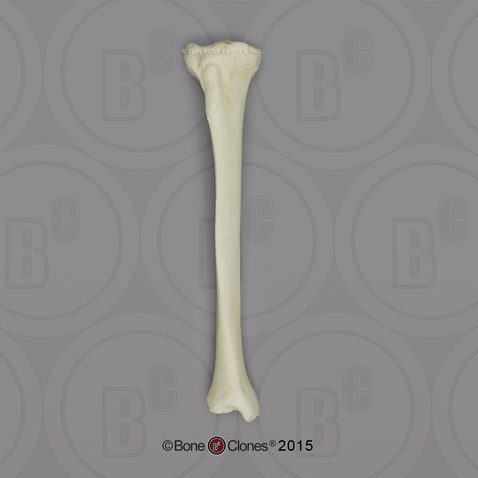
Olecranon
Elbow; process on the proximal end of the ulna
Fibula
Skinny bone in the distal hind leg

Carpus Bones
Bones in the wrist
Talus
Short bone in the proximal row of the tarsus

Calcaneus
Longer bone in the proximal row of the tarsus
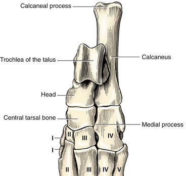
Metacarpal Bones
Bones distal to the carpals
Metatarsal Bones
Bones distal to the tarsus
Metacarpal/ Metatarsal/ Proximal Sesamoid Bones
Sesamoid bones between P1 and MC/MT
Distal Sesamoid Bones
Sesamoid bones between P2 & P3
Phalanges
Name for the digits
First Phalanx
Most proximal phalanx
Second Phalanx
Phalanx in the middle
Third Phalanx
Most distal phalanx
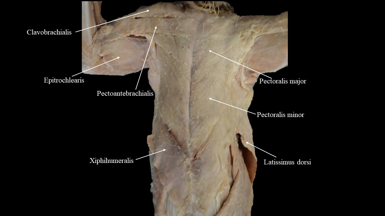
Pectoral Muscle Group
Ventral-cranial muscle group
Sternomastoid
V-shaped muscles in the neck
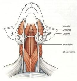
Sternohyoid
Vertical strap muscles in the neck

Latissimus Dorsi
Dorsal to the pectoral muscles

Atlanto-occipital Joint
Joint between the atlas and the occipital condyles
Atlanto-axial Joint
Joint between the atlas and axis
Vertebral Articulations
Joint between vertebrae
Glenohumeral Joint
Joint between scapula and humerus
Humeroradioulnar Joint
Elbow joint
Carpus
Wrist joint
Metacarpophalangeal/fetlock Joint
Joint between the MC and P1
Proximal Interphalangeal/pastern Joint
Joint between P1 and P2
Distal Interphalangeal Joint
Joint between P2 and P3
Coxofemoral Joint
Joint between os coxae and femur
Stifle Joint
Overall knee joint
Femoropatellar Joint
Joint between the femur and patella
Femorotibial Joint
Joint between the femur and tibia
Tarsus
Ankle joint
Metatarsophalangeal/fetlock Joint
Joint between MT and P1
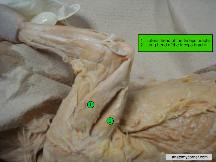
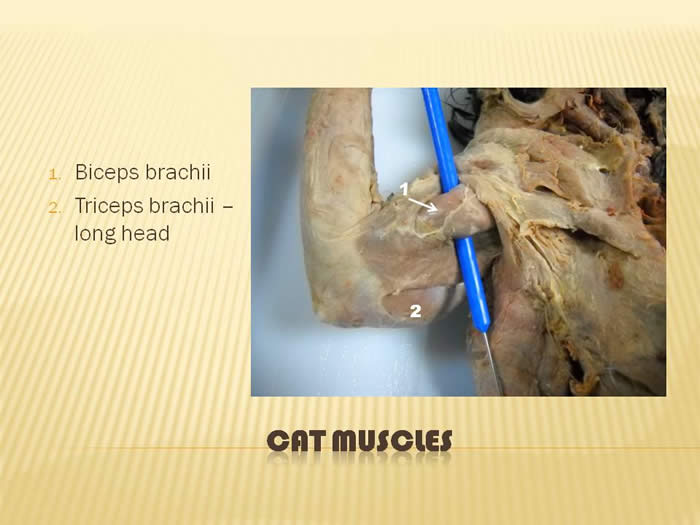
Triceps Brachii
3 headed muscle on the outside part of the proximal forelimb
Biceps Brachii
Thick muscle on the inner part of the humerus; only one head in animals
Serratus Ventralis
Large fan shaped muscle deep to the scapula
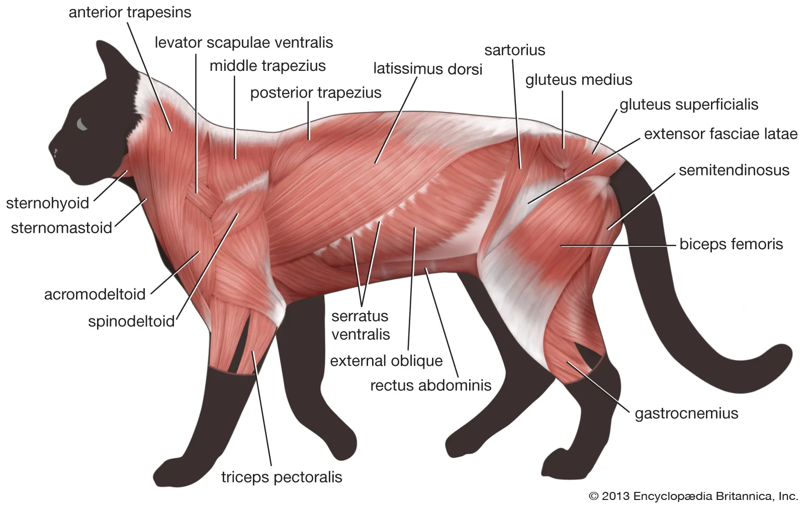
Gluteus Medius
Larger and more cranial glute muscle

Gluteus Maximus
Smaller and more caudal glute muscle

Caudofemoralis
Caudal to gluteus maximus; origin is the first coccygeal vert
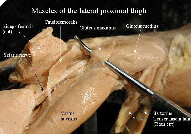
Biceps Femoris
Very large muscle on the outside of the hindleg
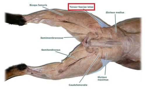
Semimembranosus
More medial of the muscles on the back of the hindleg

Semitendinosus
More lateral of the muscles on the back of the hindleg

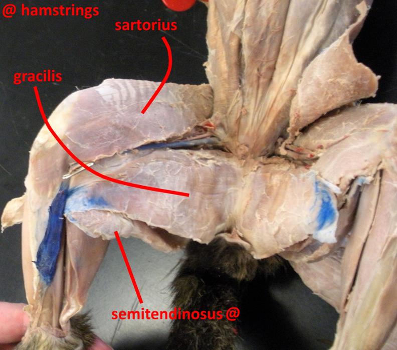
Gracilis
More caudal muscle on the inner thigh

Sartorius
More cranial muscle on the inner thigh
Vastus Lateralis
Most lateral muscle in the quadricep complex
Vastus Medialis
Most medial muscle in the quadricep complex
Vastus Intermedius
Deep to rectus femoris
Rectus Femoris
Most cranial of the quadricep complex
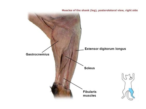
Gastrocnemius
Calf muscle; back of the lower hindleg
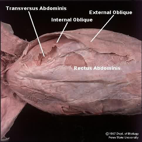
External Abdominal Oblique
Most superficial oblique muscle; runs caudoventrally
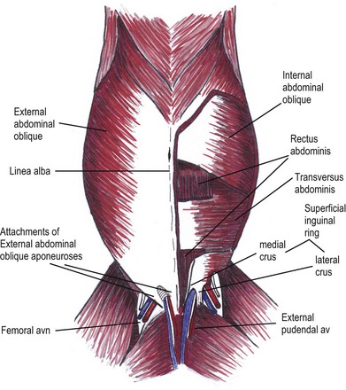
Internal Abdominal Oblique
Just deep to the external abdominal oblique muscles; runs cranioventrally

Transversus Abdominis
Most deep oblique muscle; fibers fun transversely (ventrally)

Rectus Abdominis
Most ventral abdominal muscle; long and narrow strap
External/internal Intercostals
Muscles between the ribs
Visceral Pleura
Lines the lungs directly

Parietal Pleura
Lines the ribs and diaphragm
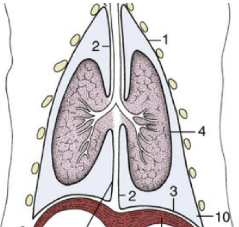
Pericardial Sac
Sac surrounding the heart

Heart
Superficial to the lungs; responsible for the circulation of blood

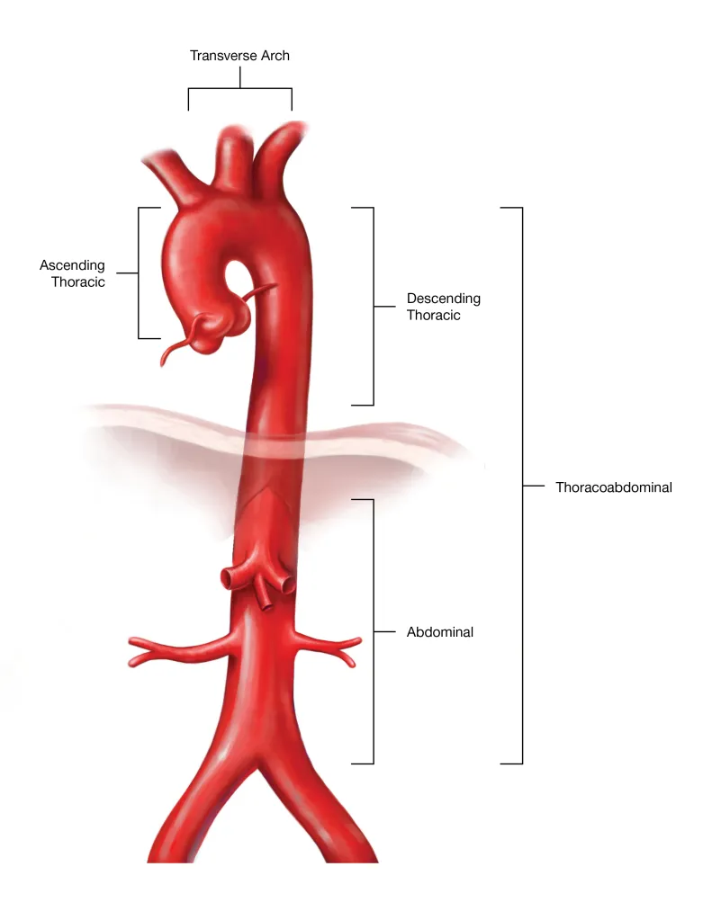
Aortic Arch
Part of the aorta that comes straight off of the heart; makes a bridge shape

Brachiocephalic Trunk
Branches into 3 branches; most medial branch of the aortic arch
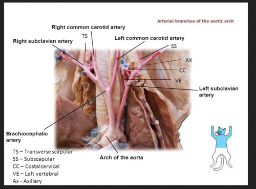
Common Carotid Arteries
Supply blood to the head; cross the trachea and esophagus to ascend the neck

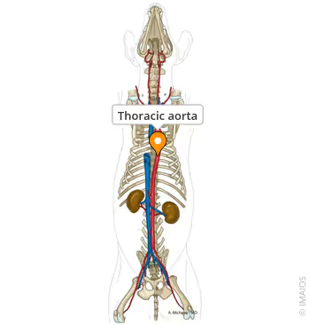
Thoracic Aorta
The cranial part of the aorta
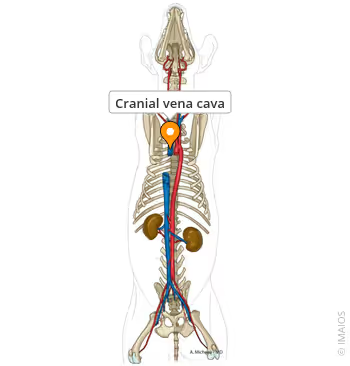
Cranial Vena Cava
Returns blood from the head, neck, and thoracic limbs; connects to the right atrium
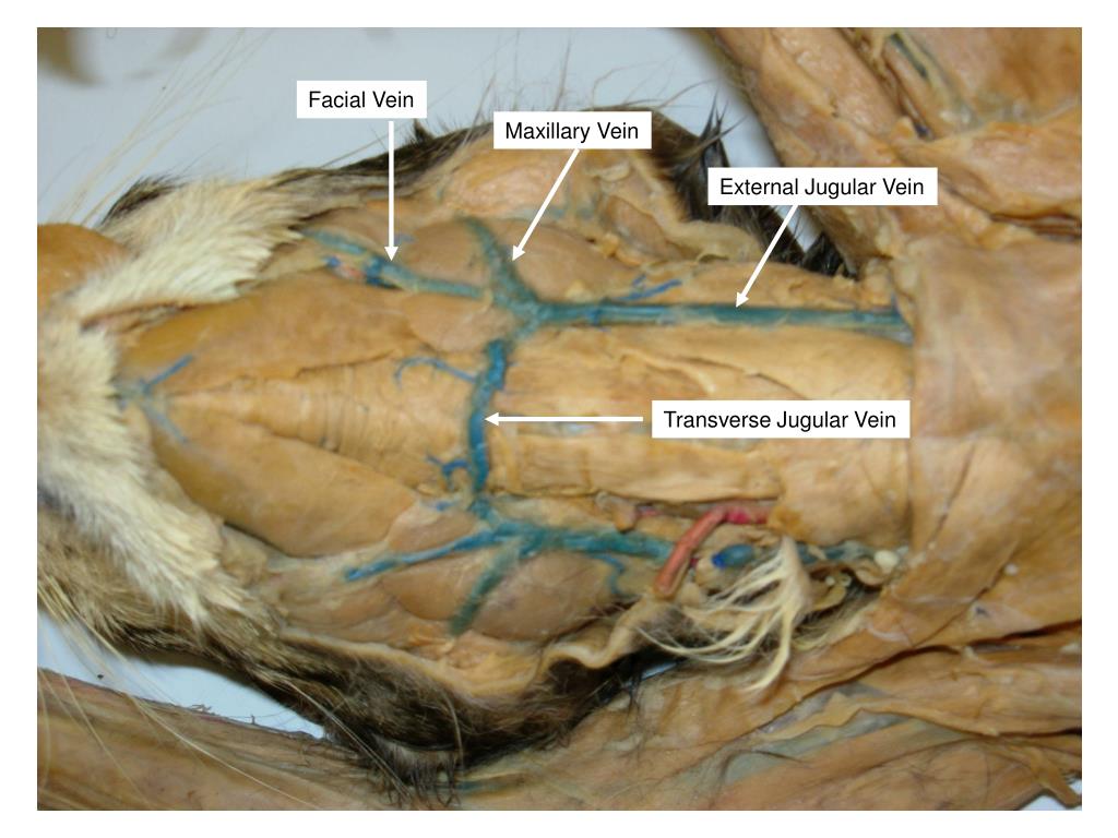
External Jugular Veins
Within the jugular groove of the neck
Abdominal Aorta
Part of the aorta in the abdominal cavity
Renal Arteries
Part of the abdominal aorta that connects to the kidneys

Renal Vein
Part of the vena cava that connects to the kidneys
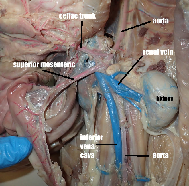
Cranial Mesenteric Artery
Connects to the small intestine; caudal to the celiac artery
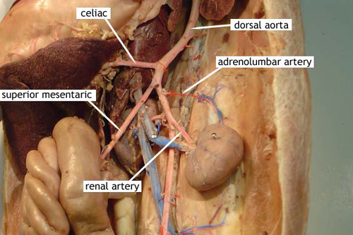
Celiac Artery
First branch off of the abdominal aorta; has 3 branches

Hepatic Portal Vein
Vein from liver to heart
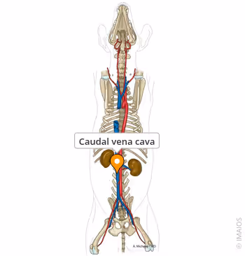
Caudal Vena Cava
Returns blood from the thorax, abdomen, and hindlimbs; connects to the right atrium
Right Atrium
Cranial chamber on the right side of the heart
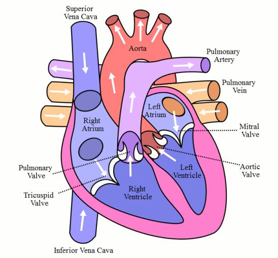
Right Ventricle
Caudal chamber on the right side of the heart

Left Atrium
Cranial chamber on the left side of the heart

Left Ventricle
Caudal chamber on the left side of the heart

Tricuspid Valve / Right Atrioventricular Valve
Valve between the right atrium and ventricle

Mitral Valve / Left Atrioventricular Valve
Valve between the left atrium and ventricle

Pulmonary Semilunar Valve
Between the right ventricle and pulmonary artery
