lab 19
1/75
There's no tags or description
Looks like no tags are added yet.
Name | Mastery | Learn | Test | Matching | Spaced | Call with Kai |
|---|
No analytics yet
Send a link to your students to track their progress
76 Terms
occurs when the sperm are transferred directly to the female’s body, and the secondary oocytes are fertilized inside of the female
internal fertilization
when the embryos are retained in the female’s body
viviparity
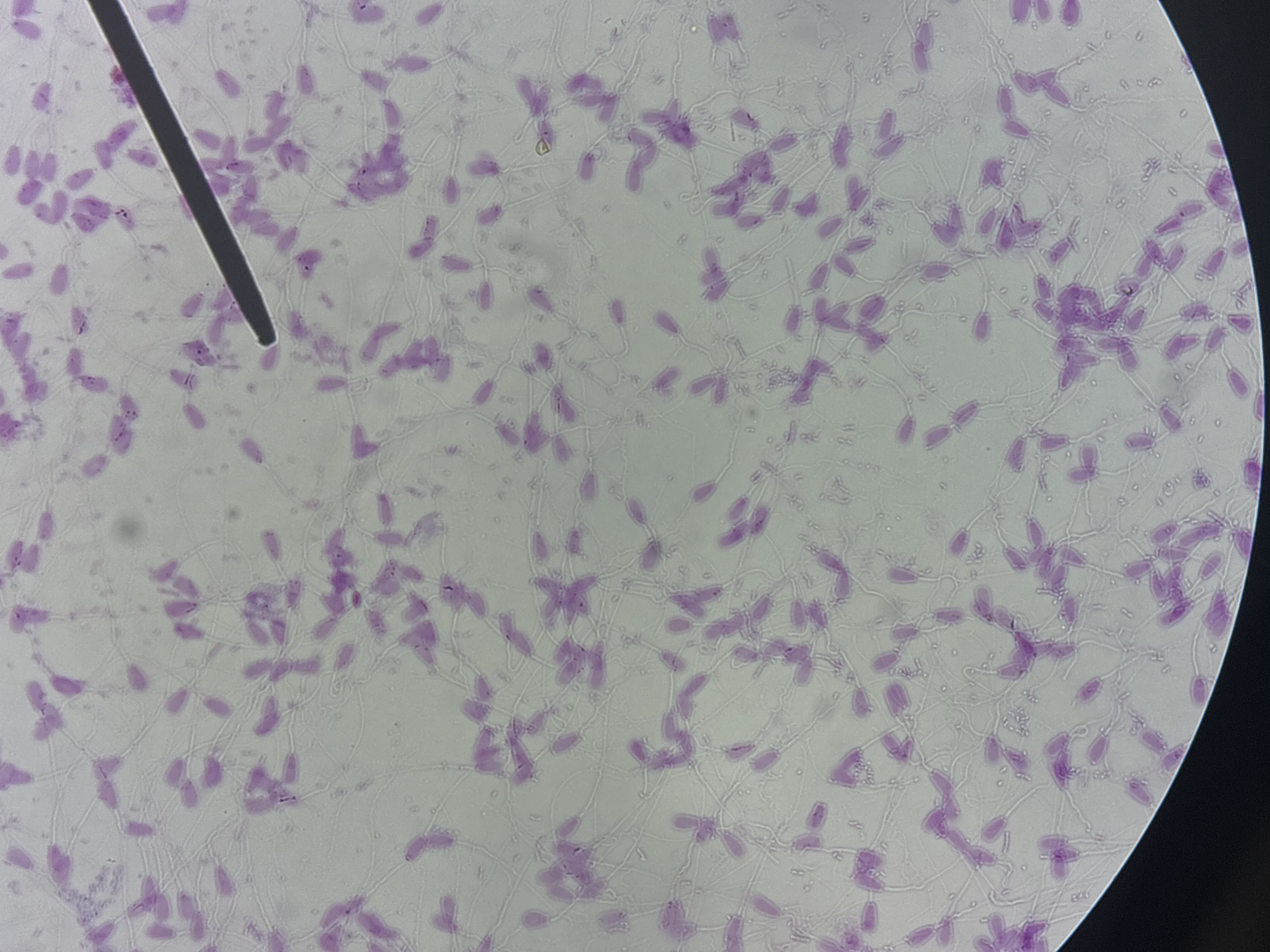
these cells contain three parts:
the head, mid-piece, and tail. The head is the body of the sperm cell. It contains the nucleus and is tipped with the acrosome. The acrosome is composed of granular enzymes to dissolve the zona pellucida, which surrounds the secondary oocyte.
Below the head is the midpiece. It contains mitochondria for energy production.
The tail is a flagellum below the midpiece. It propels the sperm cell.
sperm
the body of the sperm cell. It contains the nucleus and is tipped with the acrosome. The acrosome is composed of granular enzymes to dissolve the zona pellucida, which surrounds the secondary oocyte.
head
contains mitochondria for energy production.
location: below the head
midpiece
Flagellum below the midpiece, it propels the sperm cell.
tail
granular enzymes used to dissove the zona pellucida, which surrounds the secondary oocyte
acrosome
the male copulatory organ and contains erectile tissues.
These tissues engorge with blood during an erection. The two corpora cavernosa extends along the length of the penis and are located dorsally to the urethra. This tissue makes up the bulk of the penis.
The corpora spongiosum extends along the length of the penis and is located beneath the corpora cavernosa. It encircles the urethra.
penis
what two tissues engorge with blood during an erection?
corpora cavernosa
corpora spongiosum
the two ______ _____ extends along the length of the penis and are located dorsally to the urethra
this tissue makes up the bulk of the penis
corpora cavernosa
extends along the length of the penis and is located beneath the corpora cavernosa. It encircles the urethra.
corpora spongiosum
contain a series of lobules which contain the seminiferous tubules.
The lumen of the tubules is lined with seminiferous epithelium, which produces the spermatids.
Interstitial cells, which produce testosterone are also associated with the lobules.
testes
the lumen of the tubules is lined with, ____ ____, which produces the spermatids
seminiferous epithelium
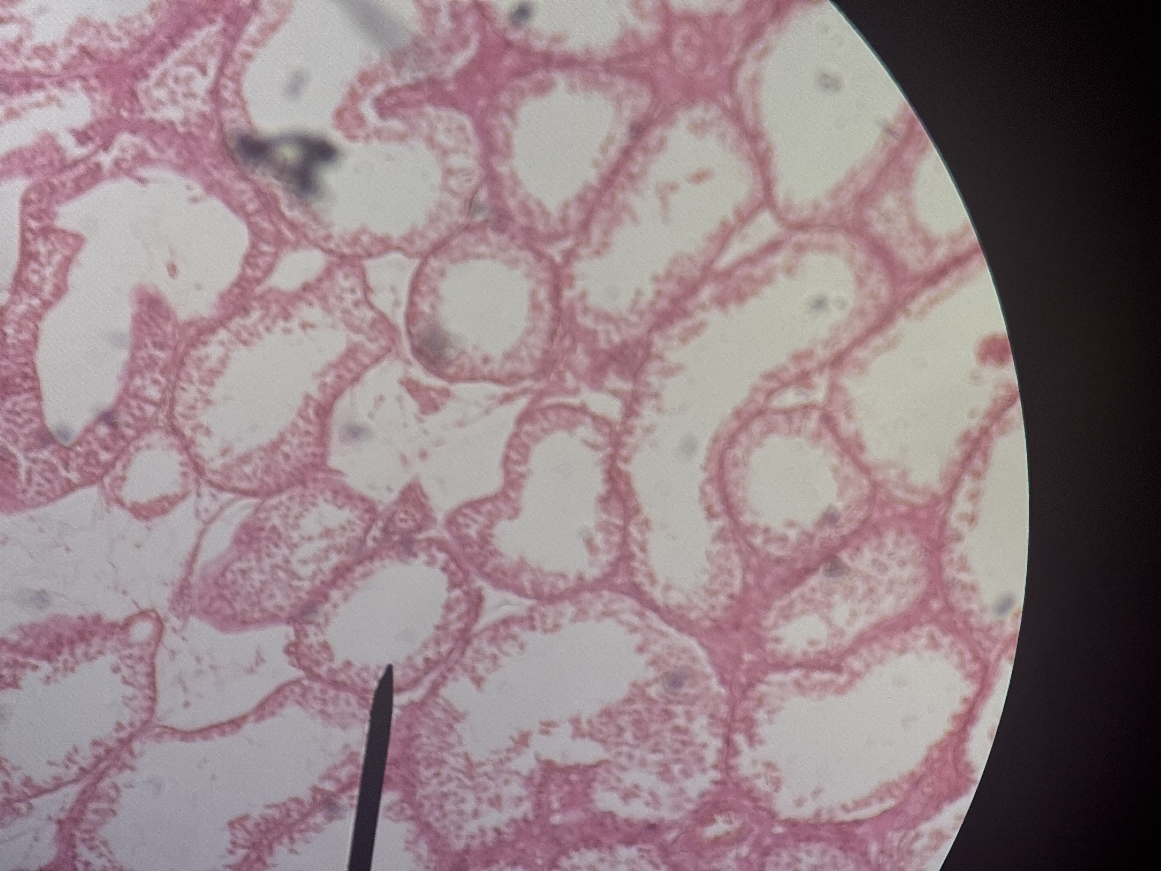
produce testostrone and are also associated with tubules
interstitial cells
the male gamete
sperm
This structure is an external sac of skin and smooth muscle containing the testes. Associated smooth muscles raise and lower the scrotum to control the temperature of sperm.
scrotum
These are the primary reproductive organs of males. They produce sperm and hormones like testosterone.
testes
These are a series of convoluted ducts combining into a comma-shaped organ located on the posterior surface of each testis. Spermatids differentiate into sperm and are stored in the epididymis.
epididymis
These ducts connect the epididymis with the ejaculatory ducts. They serve as sites for sperm storage and conduct sperm to the ejaculatory ducts during an ejaculation.
ductus deferens
These are short ducts formed by the merging of the ducts from the seminal vesicle and the vas deferens. They conduct sperm to the urethra
Ejaculatory ducts
This is a large tube connecting the urinary bladder with the distal end of the penis. It conducts both sperm and urine, although not at the same time.
Urethra
These sac-like glands are about 5 cm long. Their ducts empty into the ductus deferens. They produce about 60% of the semen’s volume.
seminal vesicles
This is a gland surrounding the urethra where it exits the bladder. It produces about 30% of the semen’s volume.
prostate gland
These are a pair of small glands located laterally on the urethra posterior to the prostate gland. They produce an alkaline mucus which lubricates the head of the penis.
Bulbourethral gland
The male copulatory organ that transfers the sperm directly to the female’s vagina.
penis
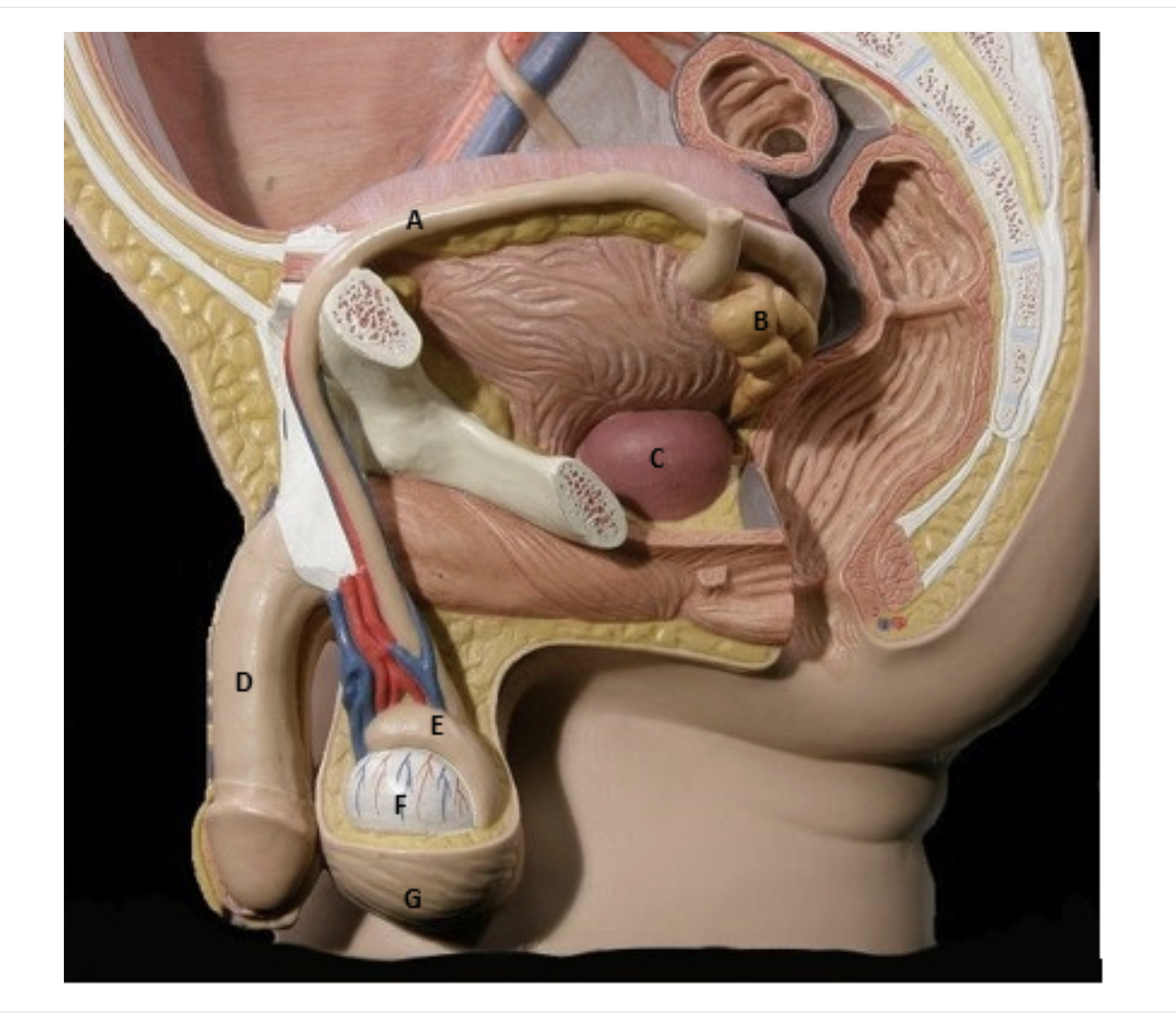
Label these structures of the male reproductive system:
A: ductus deferens
B: seminal vesicles
c: prostate
d: penis
e: epididymis
f: testes
g: scrotum
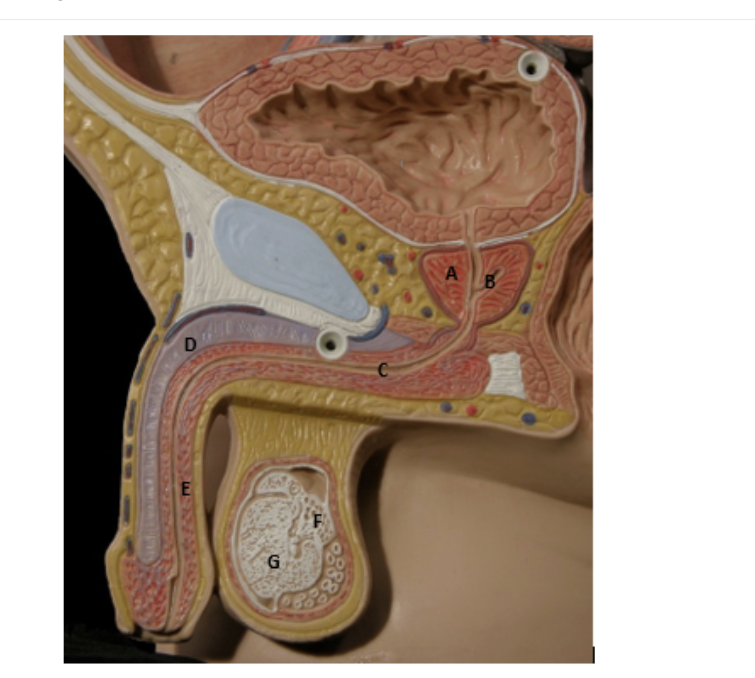
Label these structures of the male reproductive system:
A: prostate
b: ejaculatory duct
c: urethra
d: corpora cavernosa
e: corpora spongiosum
These structures are composed of a primary oocyte surrounded by a single layer of squamous cells.
Primordial follicles
These follicles are larger than the primordial follicles and the primary oocytes. They are surrounded by several layers of cuboidal cells.
primary follicles
These follicles develop a fluid-filled cavity called the antrum that surrounds the secondary oocyte.
Secondary follicles
fluid filled cavity
antrum
These large follicles have a well developed central chamber called the antrum. The granulosa cells surround the outside of the antrum. The secondary oocyte is pushed to one side and is surrounded by several layers of cells. The zona pellucida is a glycoprotein gelatinous covering of the oocyte. The corona radiata is a layer of cells that surround the zona pellucida on the inside of the antrum.
mature follicles
surround the outside of the antrum.
granulosa cells
a glycoprotein gelatinous covering of the oocyte.
zona pellucida
a layer of cells that surround the zona pellucida on the inside of the antrum.
corona radiata
This is the glandular tissue that develops from the mature follicle after ovulation of the secondary oocyte. It is important as a temporary endocrine gland in the secretion of hormones used to maintain pregnancy. If pregnancy does not occur, the corpus luteum degrades.
corpus luteum
These are the primary reproductive organs of females. They are located on the posterior wall of the female body cavity in the superior pelvic region. They contain the ovarian follicles that produce secondary oocytes and hormones like estrogen and progesterone.
ovaries
These are small ducts that open into the body cavity near each of the ovaries They extend from the ovaries to the uterus.
The secondary oocyte or zygote travels from the ovaries through these tubes to the uterus. Fertilization most often occurs in the uterine tubes.
The uterine tubes are also known as the fallopian tubes, or oviducts.
uterine tubes
The uterine tubes widen into a funnel-shaped structure that opens into the body cavity immediately next to the ovary.
The opening of the infundibulum has long, thin, feather-like projections called fimbriae. These structures facilitate the movement of the secondary oocyte into the uterine tube.
infundibulum
a thick muscular organ that lies between the uterine tubes and vagina. It is positioned posterior to the urinary bladder and anterior to the rectum. The innermost layer of the uterus is called the endometrium and serves as the location of fetal development.
uterus
This structure is the inferior part of the uterus, immediately superior to the vagina.
cervix
the tube that extends from the uterus to the outside of the female’s body. It is the female copulatory organ and is designed to receive the male’s penis. It also serves as the channel for childbirth and menstruation.
vagina
the female’s external genitalia. It is composed of the four structures listed below.
mons pubis
labia majora
labia minora
clitoris
vulva
a pad of fatty tissue that is superior to the pubic symphysis. It serves to cushion the pubic symphysis during intercourse.
mons pubis
a pair of thick folds of skin on either side of the vaginal opening. They partially protect the vaginal opening and are involved in sexual arousal.
labia majora
a pair of smaller folds of skin that lie beneath the labia majora and surround the urethral and vaginal openings.
labia minora
a small projection located between the labia majora just below the mons pubis. It is homologous to the glans penis in males and functions in sexual arousal.
clitoris
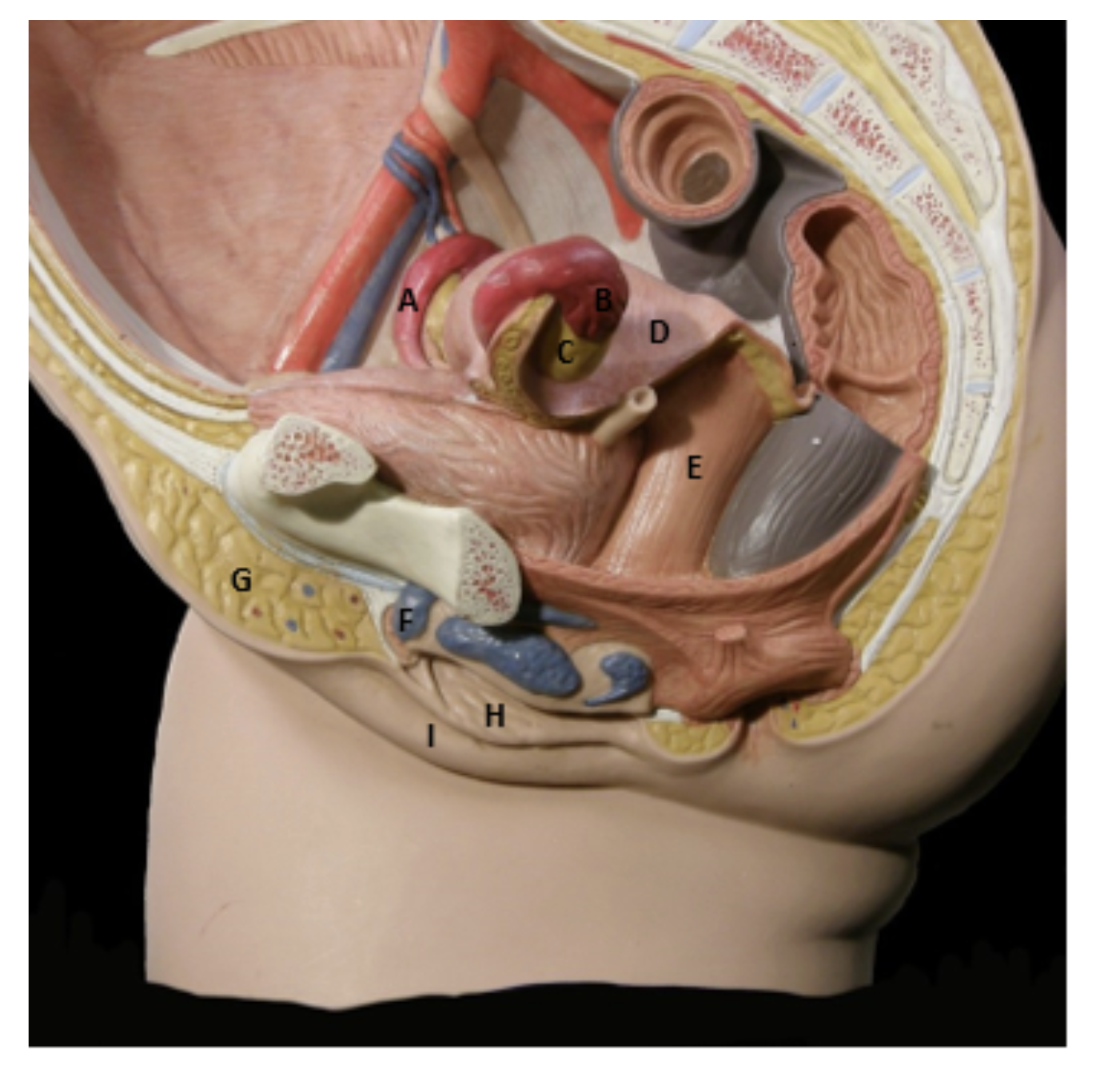
Identify the indicated structures of the female reproductive system
a: uterine tube
b: infundibulum
c: ovary
d: uterus
e: vagina
f: clitoris
g: mons pubis
h: labia minora
This trimester is the initial period of embryological and fetal development. By the end of this first three months, all of the organ systems have rudimentarily developed. Four major events occur during the first trimester: cleavage, implantation, placentation, and embryogenesis.
first trimester
clevage
When the secondary oocyte is fertilized it becomes a single cell that is called the zygote. The zygote then undergoes a series of subdivisions. These subdivisions reduce the amount of cytoplasm in the cells, which are called blastomeres. The blastomeres subdivide for about three days to form a solid ball of cells called the morula. The morula reaches the uterus around day four and during the next two days, it develops into a hollow ball of cells called the blastocyst. The blastocyst is composed of two cell layers. The outer layer develops into the placenta. The cells of the inner layer are clustered at one end of the blastocyst, and eventually develop into the embryo.
series of cellular subdivisions after the oocyte is fertilized
cleavage
When the secondary oocyte is fertilized it becomes a single cell that is called the ______ which undergoes a series of subdivisions
zygote
(zygote undergoes subdivisions)
These subdivisions reduce the amount of cytoplasm in the cells, which are called _______
these subdivide for about three days to form a solid ball of cells called the morula
blastomeres
this reaches the uterus around day four and during the next two days, it develops into a hollow ball of cells called the blastocyst.
morula
a hollow ball of cells
composed of two cell layers. The outer layer develops into the placenta. The cells of the inner layer are clustered at one end of the blastocyst, and eventually develop into the embryo.
blastocyst
implantation
occurs when the blastocyst becomes embedded in the endometrium of the mother’s uterus. The outer cell layer becomes intimately associated with the maternal circulatory system for nutrient and gas exchange. Meanwhile, the inner cell layer of the blastocyst also differentiates.
The inner cell layer becomes detached from the outer cell layer. The separation yields a fluid- filled cavity called the amniotic cavity. By the twelfth day, a third cell layer develops, and the blastocyst becomes a structure called the gastrula. During gastrulation, three germ layers form that give rise to the various organ systems: ectoderm, mesoderm, and endoderm. These three germ layers form a sheet of cells called the embryonic disk. The three germ layers then develop into the four extraembryonic membranes. These membranes support and protect the embryo.
when the blastocyte imbeds in the endometrium of the uterus
implantation
The inner cell layer becomes detached from the outer cell layer. The separation yields a fluid- filled cavity
amniotic cavity
By the twelfth day, a third cell layer develops, and the blastocyst becomes a structure called the
gastrula
During gastrulation, three germ layers form that give rise to the various organ systems:
ectoderm
mesoderm
endoderm
During gastrulation, three germ layers form that give rise to the various organ systems: ectoderm, mesoderm, and endoderm. These three germ layers form a sheet of cells called the
embryonic disk
This sac is a pouch formed from the endoderm and mesoderm. It is an initial site of blood cell formation.
yolk sac
This membrane is formed from the ectoderm and mesoderm. It lines the inner surface of the amniotic cavity. The fluid contained within the cavity is called the amniotic fluid.
The amnion and the amniotic fluid surround and protect the growing embryo.
amnion
This membrane is formed from the endoderm and mesoderm. It eventually gives rise to the urinary bladder.
Allantois
This membrane is formed from the mesoderm and the outer cell layer of the blastocyst. The chorion differentiates into the fetal portion of the placenta as the embryo enlarges.
Chorion
placentation
the development and differentiation of the fetal and maternal portions of the placenta. The chorion develops finger like projections called chorionic villi which make contact with the endometrium.
As the placenta enlarges it creates a large bulge on the surface of the endometrium. Blood from ruptured maternal blood vessels slowly leaks through small chambers in the maternal placenta called lacunae. Gas and nutrient exchange between the maternal blood and the fetal blood by diffusion. However, the blood streams of mother and baby never mix. The blood from the lacunae then reenters the maternal circulatory system through ruptured veins.
As the embryo grows, it moves further away from the endometrium, but it remains connected to the placenta by the umbilical cord. The umbilical cord contains two umbilical arteries and one umbilical vein.
the developmental and differentiation of the fetal and materna portions of the placenta
placentation
The chorion develops finger like projections called _____ _____, which make contact with the endometrium
chorionic villi
The blood from the _____ hen reenters the maternal circulatory system through ruptured veins.
lacunae
As the embryo grows, it moves further away from the endometrium, but it remains connected to the placenta by the ____ _____. It contains two umbilical arteries and one umbilical vein.
umbilical cord
the differentiation of the body of the embryo and the rudimentary development of the organ systems.
The body of the embryo begins to emerge from the embryonic disk and develops into a bulge that has head and tail folds. The dorsal, ventral, and lateral surfaces of the embryo can clearly be observed.
Embryogenesis
During the these semesters: the fetus gains weight and the organ systems continue to develop. By the end of the pregnancy, the uterus and the fetus occupy a large portion of the maternal abdominal cavity.
second and third trimesters
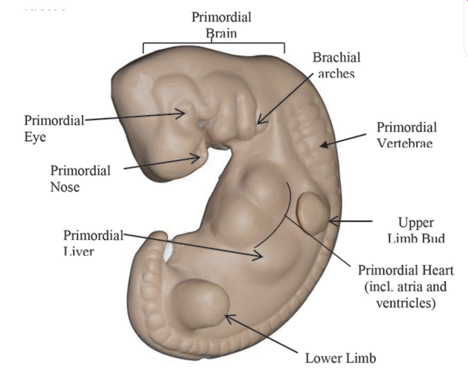
be able to identify the structure of the 4 week old embryo
i will be able to identify the structure of the 4 week old embryo
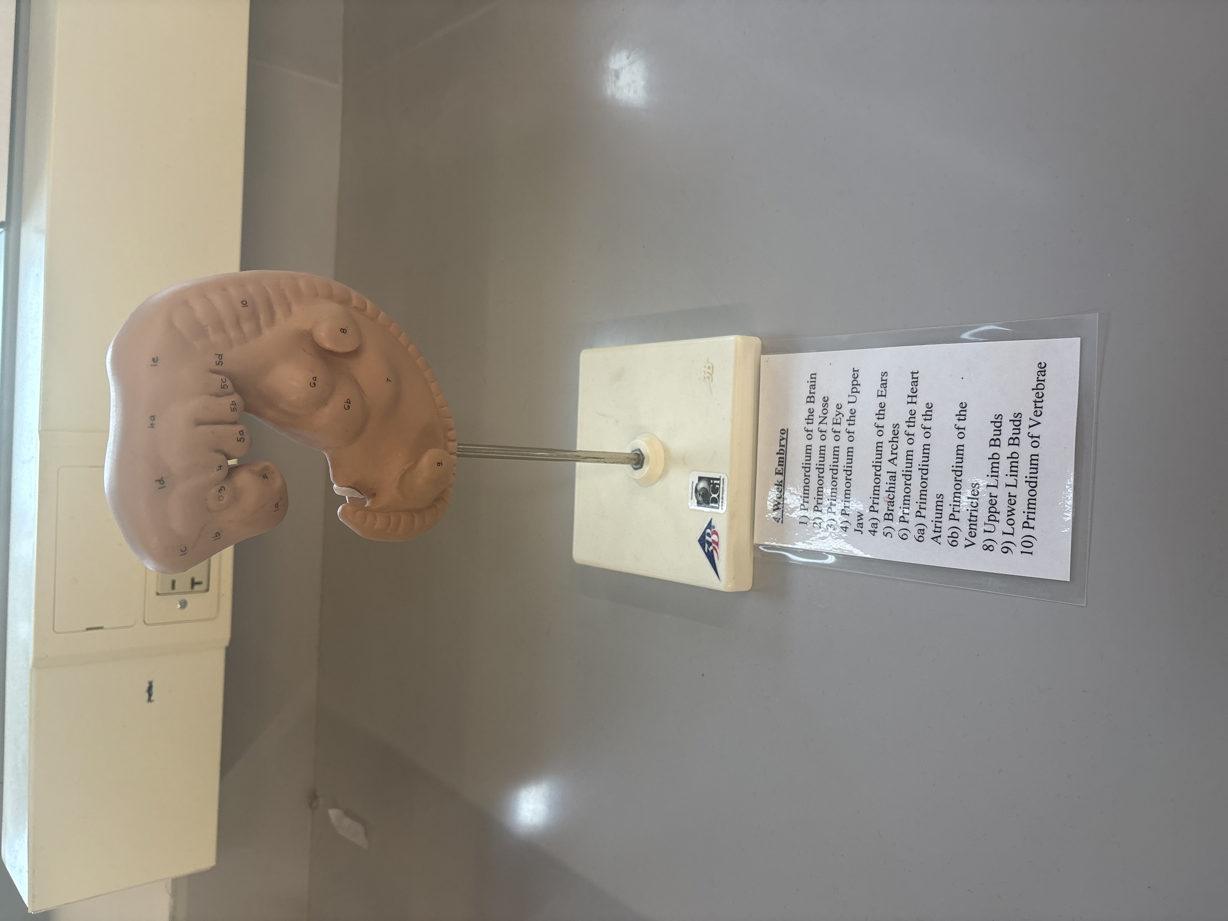
4 week embryo, identify structures
primordium of the brain
primordium of nose
primordium of eye
primordium of the upper jaw
4a. primordium of the eyes
brachial arches
primordium of the heart
6a. primordium of the atriums
6b. primordium of the ventricles
upper limb buds
lower limb buds
primordium of vertebrae
be able to identify the rest of the months of pregnancy
i will be able to identify the rest of the months of pregnancy
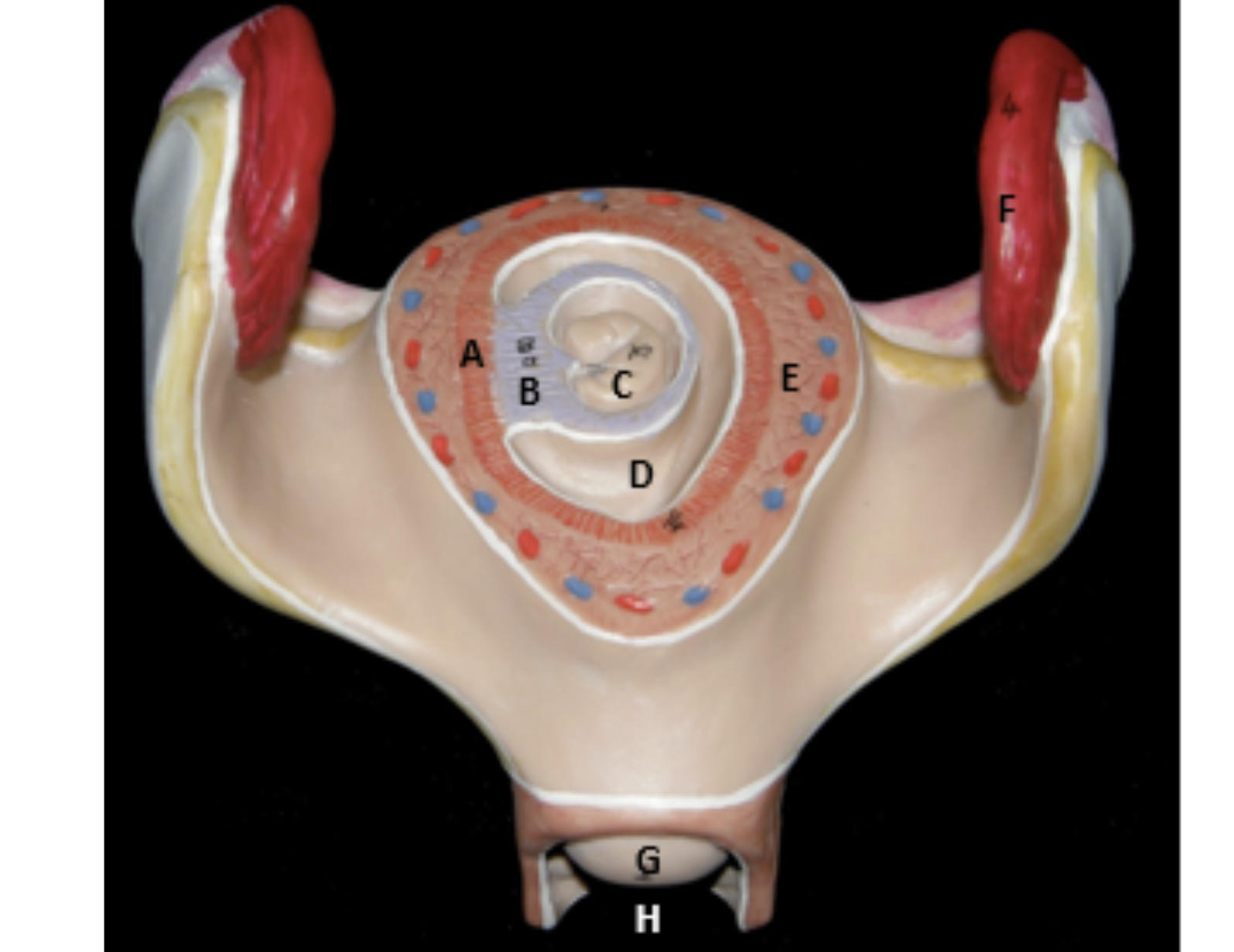
identify the indicated structures.
a: maternal placenta
b: fetal placenta
d: amniotic cavity
e: uterus
f: infundibulum
g: cervix
h: vagina