Basic LM, Optics & Resolution
1/13
There's no tags or description
Looks like no tags are added yet.
Name | Mastery | Learn | Test | Matching | Spaced | Call with Kai |
|---|
No analytics yet
Send a link to your students to track their progress
14 Terms
What is empty magnification?
Magnification beyond 500-1000x the numerical aperture of the objective
Increases image size but adds no detail → can lead to image degradation
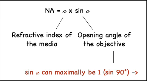
What is numerical aperture (NA)?
It is the light gathering ability of an objective
The higher the NA, the more powerful the system, and the better the resolution
NA = n x sin(θ)
n = refractive index of the immersion media,
θ = the half-angle of the maximum cone of light the objective can collect
What is contrast?
The visibility of structures against a background
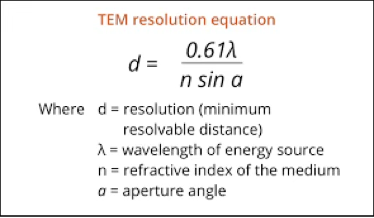
What is resolution? What two parameters determines reolsution?
The shortest distance between two point sources where they’re observed as two individual parts.
The smaller the resolution, the more powerful the system.
Determined by NA and wavelength
d = 0.61λ/NA
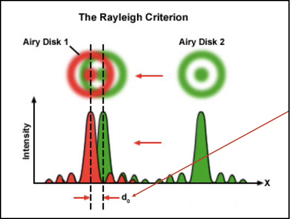
What is an Airy disk and how does it impact resolution?
It is the diffraction-limited distribution of intensities
Features a bright central maximum surrounded by diffraction rings
The resolution is limited by this diffraction, as it determines how close 2 Airy disks can get before they merge, and information is lost
What is maximum theoretical resolution?
Defined by when the first maximum of an airy disk coincides with the first minimum of another airy disk.
Dotted lines are the maximum theoretical resolution
What is a point spread function (PSF)?
Describes how the optical system blurs a point source
It characterises resolution in 3D (in Z direction too), and the PSF determines the minimum size you can resolve
Objects smaller than the PSF appear the same size as the PSF
Therefore, the smaller/narrower the PSF, the better the resolution, but it depends on what the experiment requires
What effect does a confocal pinhole has on the PSF?
The pinhole blocks out-of-focus emission, thus resulting in a narrower PSF, and a better resolution.
The wider the pinhole, the more light reaches the detector, and the opposite occurs
Why can you image a single fluorescent protein but NOT measure its size?
Because the protein is much smaller than the PSF (~250–300 nm).
The image shows only the Airy disk of the optical system, not the actual size of the molecule.
The PSF sets the measurement floor.
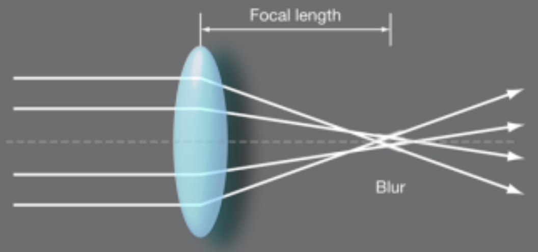
Spherical aberration?
Occurs when the focus at the centre of the lends differ from the periphery
Can be caused by a mismatch in refractive index between immersion medium and sample
Results in: blurry image, loss of resolution, loss of brightness, Z-position inaccuracy
Chromatic aberration?
Occurs when different wavelengths of light focus at different focal lengths
Happens as the lens has a wavelength-dependent refractive index
Results in: color around objects and multi-color images that are out of focus relative to each other (shifted)
No matter how advanced the microscope system is, there are 3 parameters that we add as users, what are they?
Sample
Immersion media
Coverslip
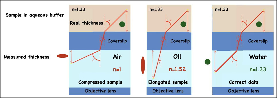
What is the refractive index mismatch, and what aberration does it cause?
Happens when the refractive index of the sample medium differs from the objective immersion medium
(e.g., aqueous sample n=1.33 with oil objective n=1.5).
Light rays bend differently at the interface, causing mainly spherical aberration
Describe 3 contrasting enhancing techniques in Brightfield microscopy?
Phase contrast:
Due to cells having different thicknesses, and thus refractive indices, the light gets delayed, creating a phase shift
Phase contrast converts the invisible phase shift into visible brightness differences
Parts of the cell appear darker/brighter than the background
Optimal for unstained living cells (like bacteria) or thin samples
Not optimal for thicker samples
Differential interference contrast (DIC):
Pseudo 3D method, with extremely thin focal depth
Based on polarisation microscopy, where polarisers are positioned perpendicularly to block out background light
Uses Normanski prism to split light into 2 beams that passes through the sample
If one beam passes through a thicker/denser area, and the other beam through a thinner, they are delayed differently, which is converted to intensity contrast by DIC
Optimal for unstained cells, edges, organelles, fine
Darkfield:
Provides an image formed entirely from diffracted light with a black/dark background
Blocks out direct transmitted light from entering the objective, so only light scattered from the sample enters
Optimal for viewing samples without labelling, like pollen, and for small objects