Cardiology (Chapter 13)
1/18
There's no tags or description
Looks like no tags are added yet.
Name | Mastery | Learn | Test | Matching | Spaced | Call with Kai |
|---|
No analytics yet
Send a link to your students to track their progress
19 Terms

Describe the structure of the heart in terms of its coverings and layers.
Coverings: Pericardium (Fibrous outer layer; Serous inner layers—parietal and visceral/epicardium). Layers:Epicardium (outer), Myocardium (middle muscle), Endocardium (inner lining).

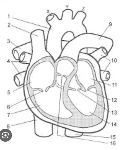
Describe the structure of the heart in terms of chambers, valves, and blood vessels.
Chambers: R/L Atria (receiving) and R/L Ventricles (pumping).
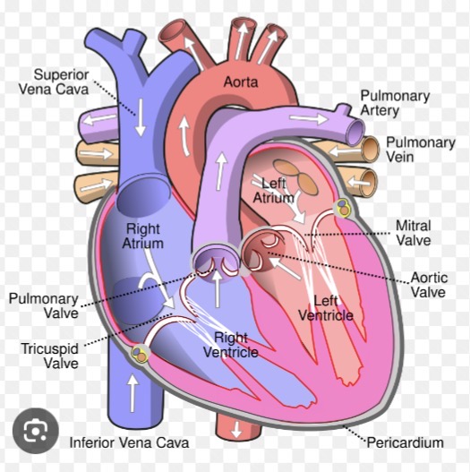
Valves: AV (Tricuspid/Mitral) and SL (Pulmonary/Aortic). Vessels: Vena cavae, Pulmonary trunk/arteries, Pulmonary veins, Aorta.

Name the function of heart valves.
To prevent the backflow of blood, ensuring a one-way flow through the heart.

Distinguish between AV and SL valves in terms of location, structure, and when they close.
AV (Atrioventricular): Between atria and ventricles; have cusps/flaps anchored by chordae tendineae; close during ventricular systole.
SL (Semilunar): Between ventricles and large arteries; have 3 pocket-like cusps; close during ventricular diastole.

Define/describe the terms chordae tendineae and papillary muscles.
Chordae tendineae: Collagen cords ("heart strings") that anchor AV valve cusps to the heart wall.
Papillary muscles:Muscles in the ventricles that contract to pull chordae tendineae taut, preventing valves from eversion (prolapse) into the atria.
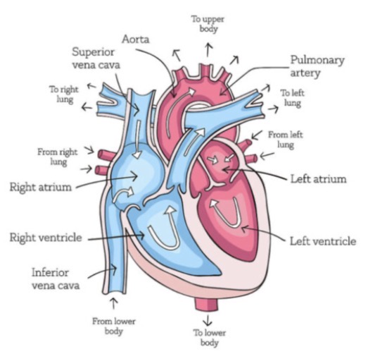
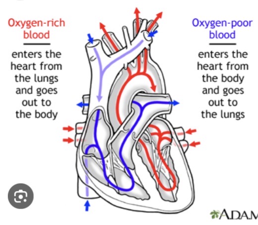
Name (and locate) the veins that deposit their blood into the atria of the heart (which atria? deox- or oxygenated?).
Right Atrium (Deoxygenated): Superior Vena Cava, Inferior Vena Cava, Coronary Sinus.
Left Atrium (Oxygenated): 4 Pulmonary Veins.

Name (and locate) the arteries that take blood away from the heart (from which ventricle? deox- or oxygenated?).
Right Ventricle (Deoxygenated): Pulmonary Trunk (leads to Pulmonary Arteries).
Left Ventricle (Oxygenated): Aorta.
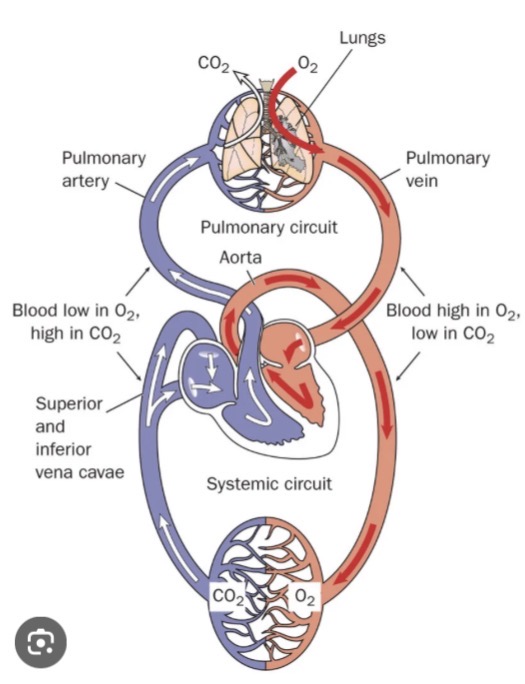
Distinguish between pulmonary, coronary and systemic circulation
Pulmonary: Right heart to lungs to Left heart (gas exchange).
Systemic: Left heart to body tissues to Right heart (nutrient delivery).
Coronary: Functional blood supply to the heart muscle itself.
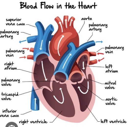
Track a drop of blood starting from the Vena Cava through the entire heart.
Vena Cava -> Right Atrium -> Tricuspid Valve -> Right Ventricle -> Pulmonary SL Valve -> Pulmonary Trunk/Arteries -> Lungs -> Pulmonary Veins -> Left Atrium -> Mitral (Bicuspid) Valve -> Left Ventricle -> Aortic SL Valve -> Aorta
Explain the significance of each component of the cardiac conduction system and trace how the cardiac impulse travels through the myocardium.
SA Node: Pacemaker, starts impulse.
AV Node: Delays impulse for atrial contraction.
AV Bundle (His): Connects atria/ventricles.
Bundle Branches: Carries impulse through septum. Purkinje Fibers: Depolarizes contractile cells of ventricles.
Name the common term for the sinoatrial (SA) node.
The Pacemaker.
Name the term referring to all of the events associated with one heartbeat.
The Cardiac Cycle.
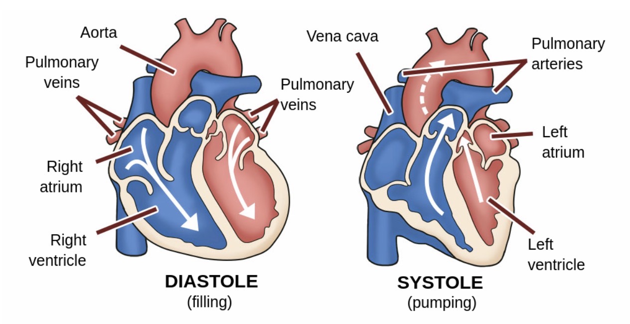
Define the terms systole and diastole.
Systole: Period of contraction (pumping). Diastole: Period of relaxation (filling).
Discuss heart sounds in terms of what they represent, how they sound, how they are detected and their significance.
Represent: Closing of valves.
Sound: "Lub" (S1, AV valves closing) and "Dup" (S2, SL valves closing).
Detected: Via stethoscope (Auscultation). Significance: Abnormal sounds (murmurs) can indicate valve problems.
Outline the phases of the cardiac cycle in terms of what is happening in the ECG trace.
P wave: Atrial depolarization.
QRS complex: Ventricular depolarization (atrial repolarization is hidden).
T wave:Ventricular repolarization.
Define the terms tachycardia and bradycardia.
Tachycardia: Abnormally fast heart rate (>100 bpm). Bradycardia: Abnormally slow heart rate (<60 bpm).
Name the longest vein in the body and the venipuncture site.
Longest vein: Great Saphenous Vein (leg). Venipuncture site: Median Cubital Vein (elbow).
Study Root Words: -hemo, -erythro, -leuko, -thrombo, -myo, -card.
Hemo: Blood.
Erythro: Red.
Leuko: White.
Thrombo: Clot.
Myo: Muscle.
Card: Heart.
Blood flow pathway
Tricuspid Valve (Right side)
Pulmonic Valve (Right side)
Mitral Valve (Left side)
Aortic Valve (Left side)
OX: Right side = OXygen-poor (Deoxygenated).
LOX: Left side = OXygen-rich (Oxygenated).
Full sequence:
Body to Vena Cava
Right Atrium
Tricuspid Valve
Right Ventricle
Pulmonary Valve
Pulmonary Artery to Lungs
Pulmonary Vein to Left Atrium
Mitral (Bicuspid) Valve
Left Ventricle
Aortic Valve
Aorta to Body
