APHY 101 Ch 6: Bone Tissue and the Skeletal System Ivy Tech Diagram | Quizlet
1/95
There's no tags or description
Looks like no tags are added yet.
Name | Mastery | Learn | Test | Matching | Spaced | Call with Kai |
|---|
No analytics yet
Send a link to your students to track their progress
96 Terms
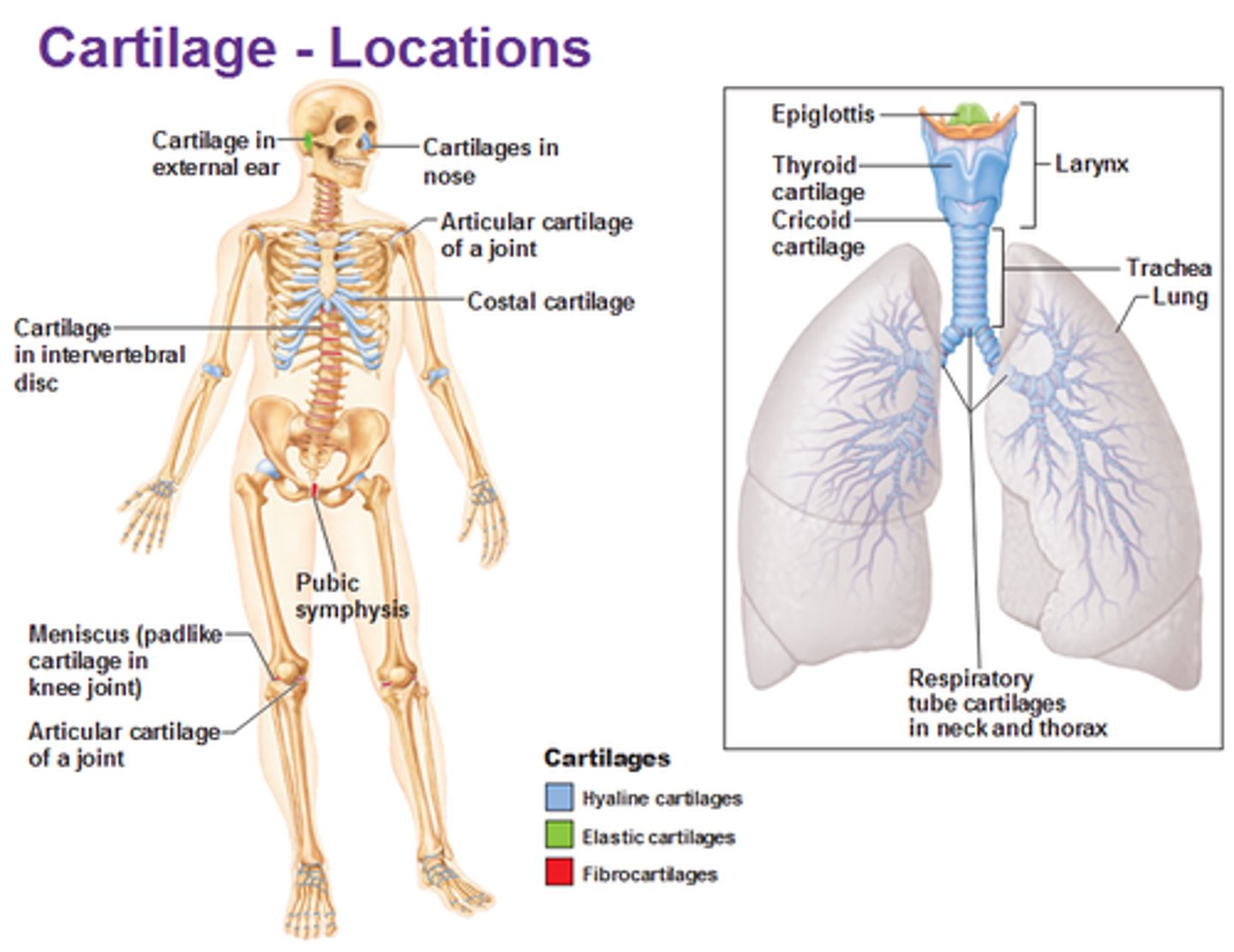
Skeletal cartilage
provides smooth surfaces, flexibility, and support;
water content of cartilage lends resiliency; contains no blood vessels or nerves; surrounded by a perichondrium
Perichondrium
surrounds the cartilage; dense connective tissue girdle; contains blood vessels for nutrient delivery; resists outward expansion
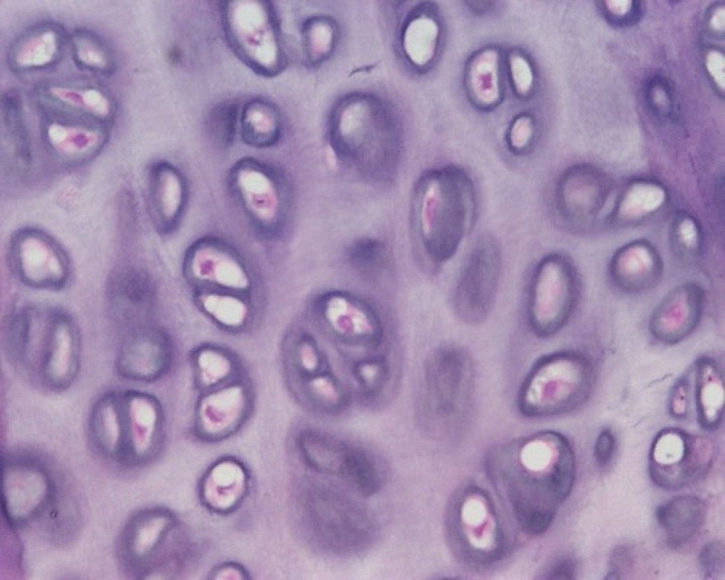
cartilage
all skeletal ____________ contains chondrocytes in lacunae and extracellular matrix
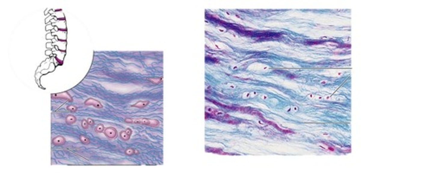
Types of Skeletal Cartilage
Hyaline, Elastic, and Fibrocartilage
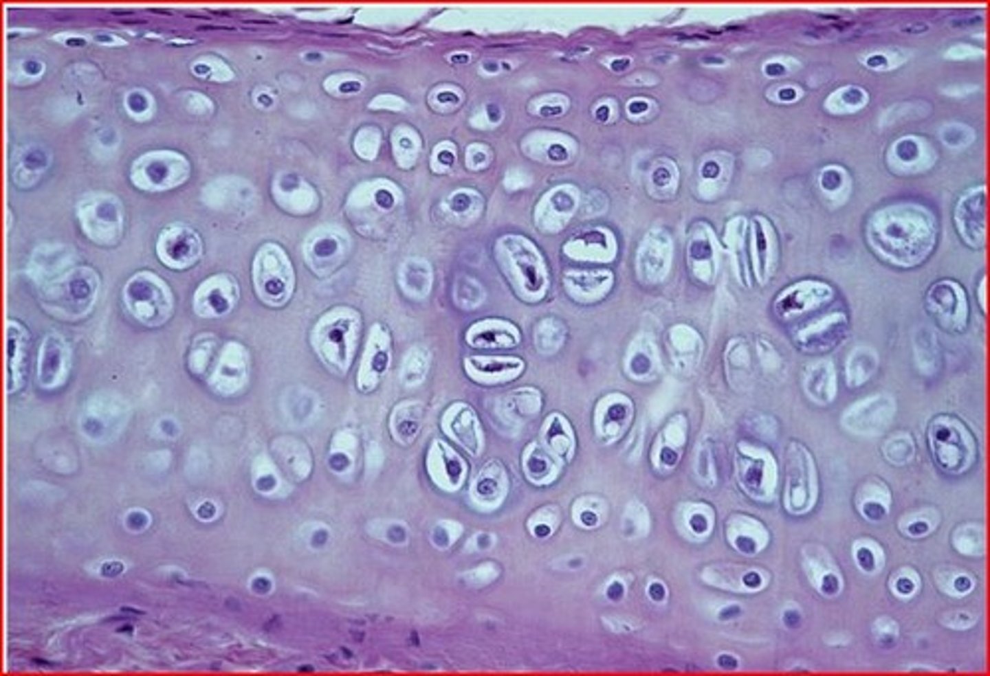
Hyaline Cartilage
provides support, flexibility, and resilience; collagen fibers only; most abundant type; articular, costal, respiratory, nasal cartilage
Elastic cartilage
similar to hyaline cartilage, but alsocontains elastic fibers (which stain dark); external ear and epiglottis
Fibrocartilage
thick collagen fibersgive great tensile strength; menisci of knee; vertebral discs
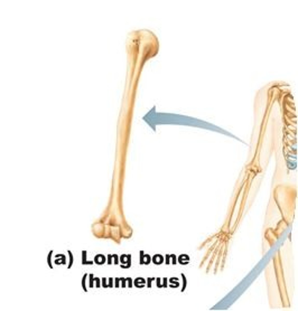
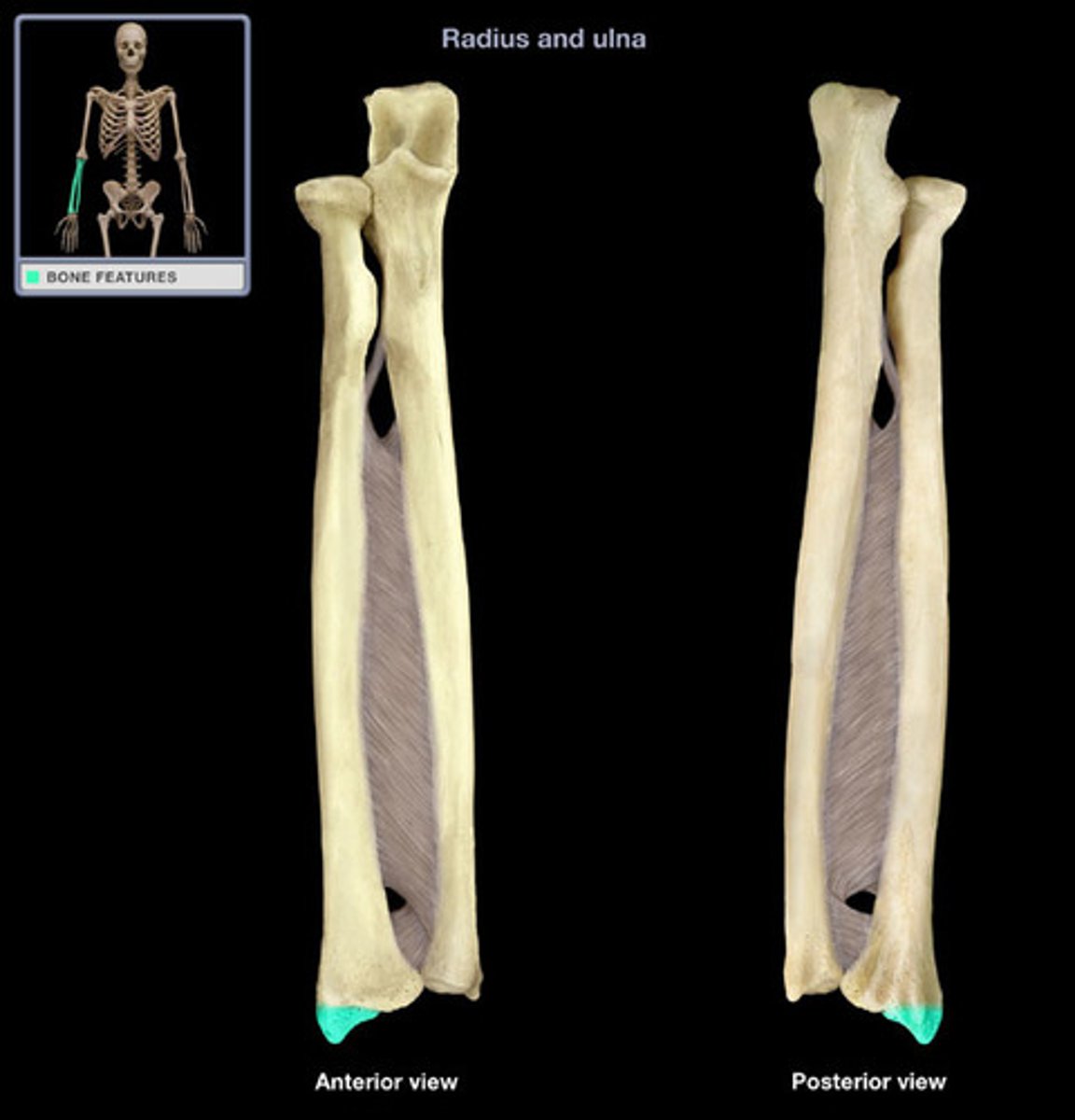
Long Bones
longer than they are wide; limb, wrist, ankle bones
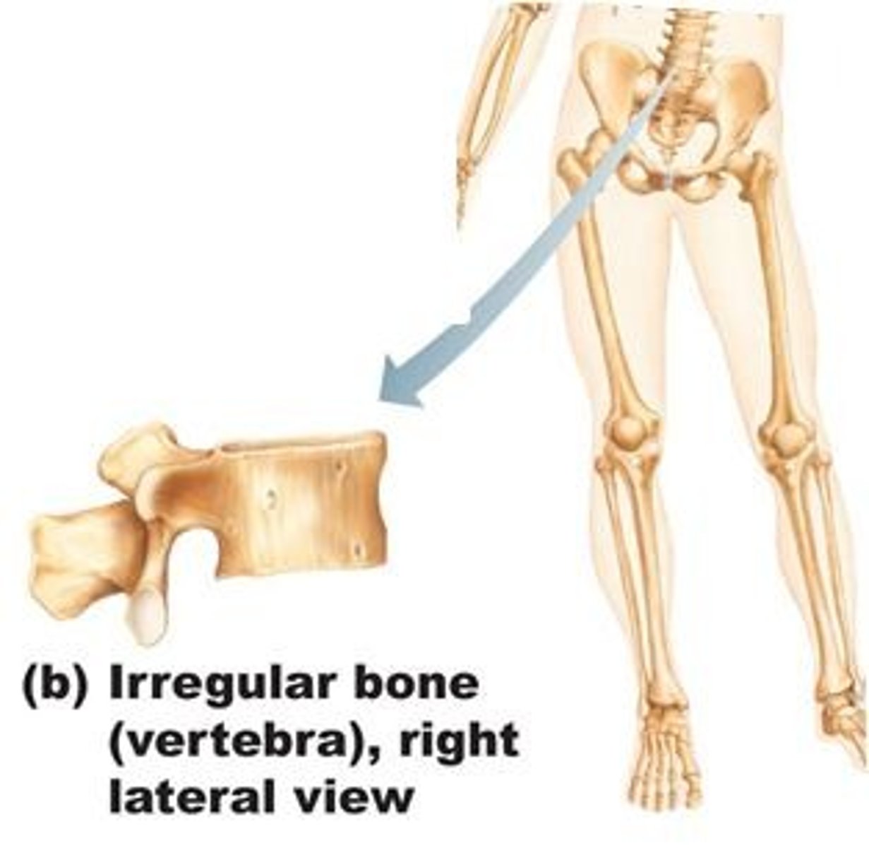
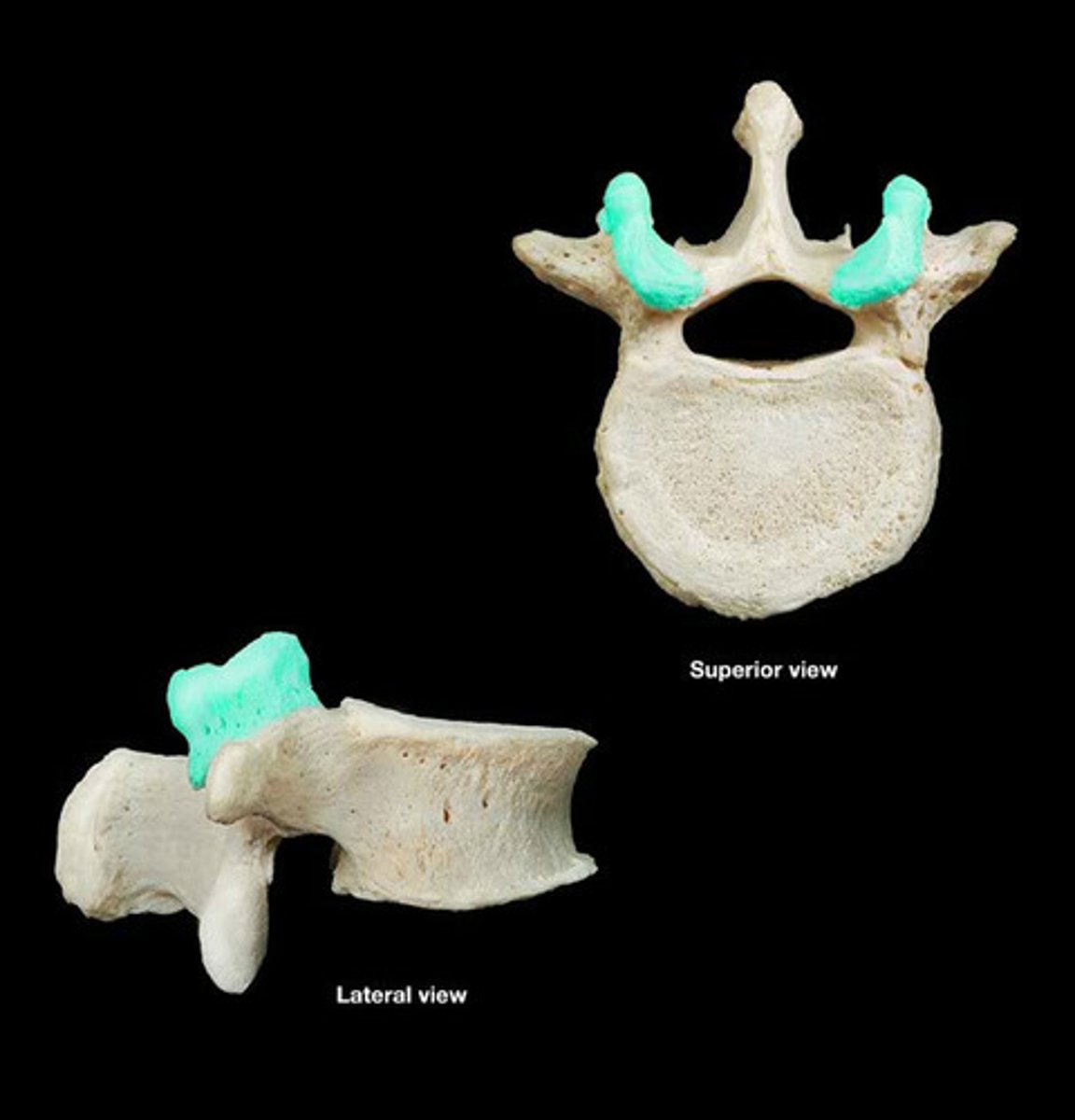
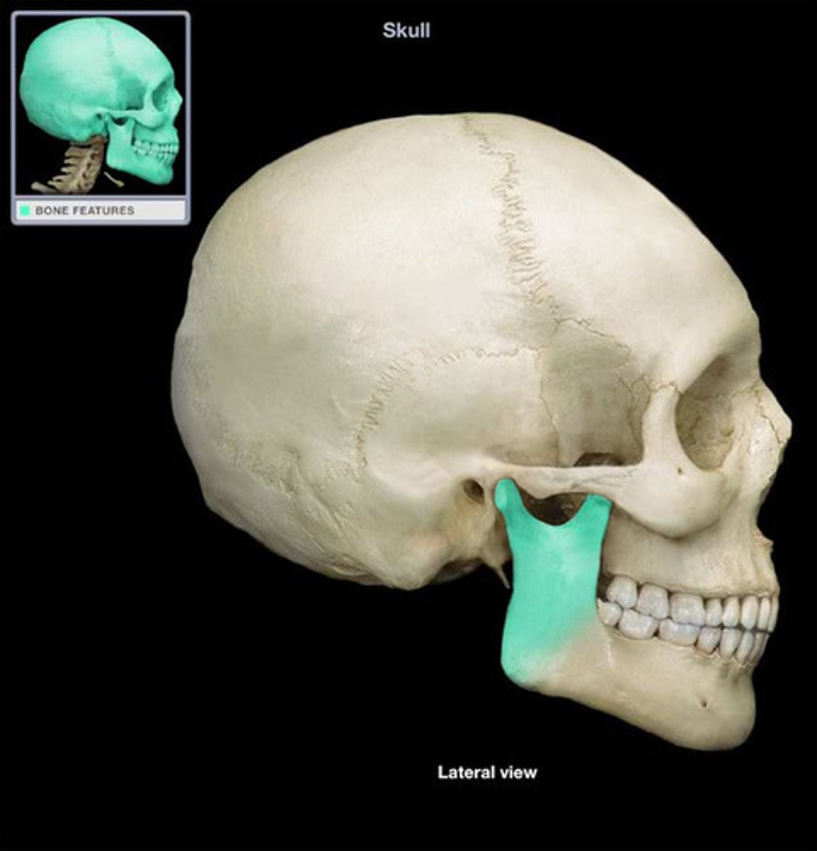
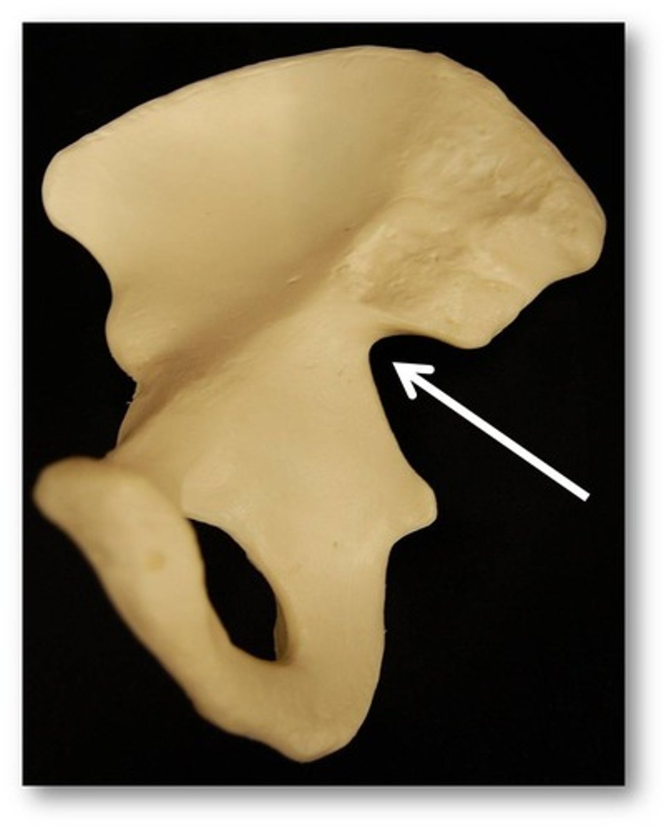
Irregular bone
complicated shapes; vertebrae, coxal bones
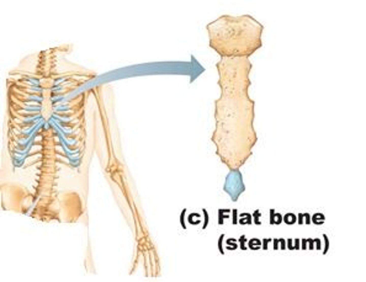
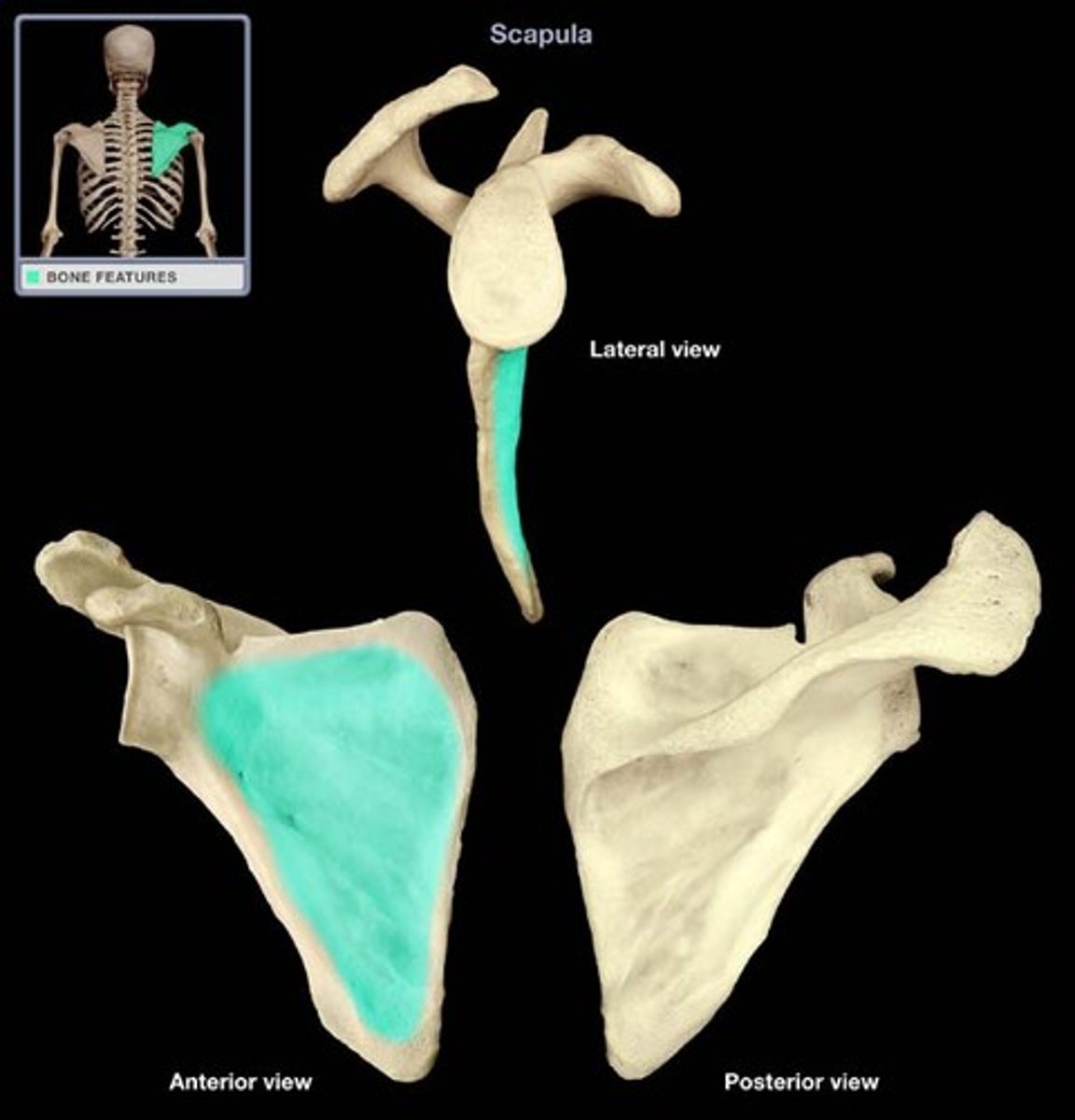
Flat bone
thin, flat, slightly curved; sternum, scapulae, ribs, most skull bones; bone that consists of a layer of spongy bone sandwiched between two thin layers of compact bone
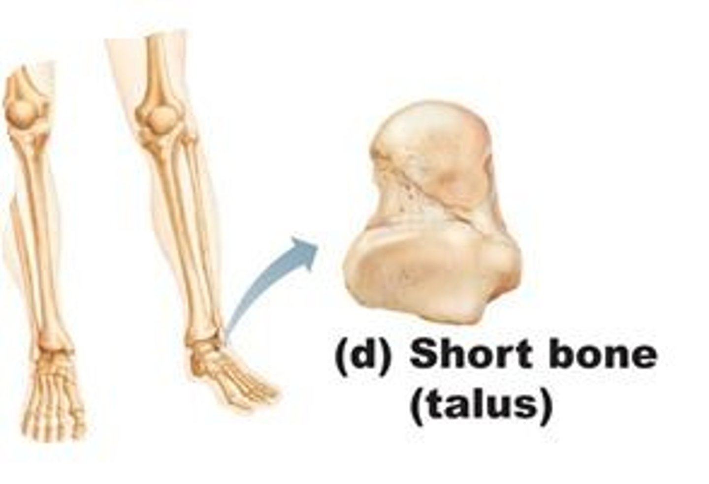
Short bone
cube-shaped bones (in wrist and ankle); sesamoid bones (within tendons, e.g., Patella); vary in size and number in different individuals
Functions of the bones
Support - maintain body shape
Protection - brain, spinal cord, heart, lungs
Movement - levers for muscle action
Mineral and growth factor storage - store calcium and phosphorous
Blood cell formation - hematopoiesis
Triglyceride (fat) storage
Bones are organs because?
contain different types of tissues: bone (osseous) tissue, nervous tissue, cartilage, fibrous connective tissue, muscle and epithelial cells in its blood vessels
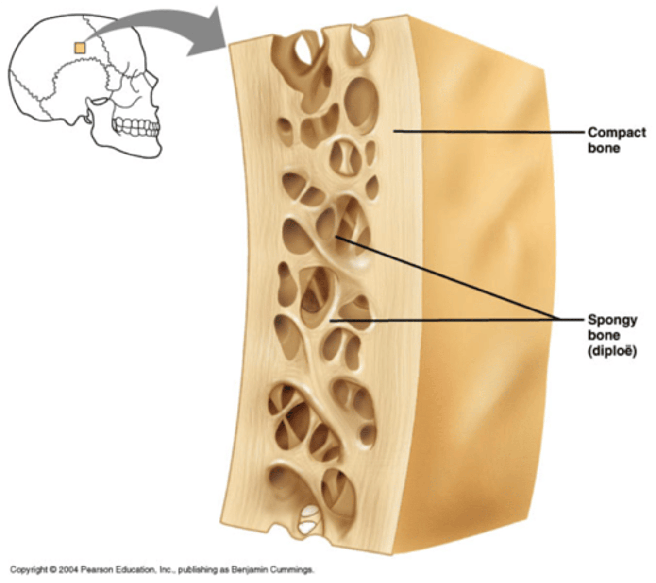
Structure of Short, Irregular, Flat bone
thin plates of spongy bone covered by compact bone; plates sandwiched between connective tissue membranes; periosteum (outer layer) and endosteum; bone marrow throughout spongy bone; inner layer of spongy bone called "diploe"; hyaline cartilage covers articular surfaces
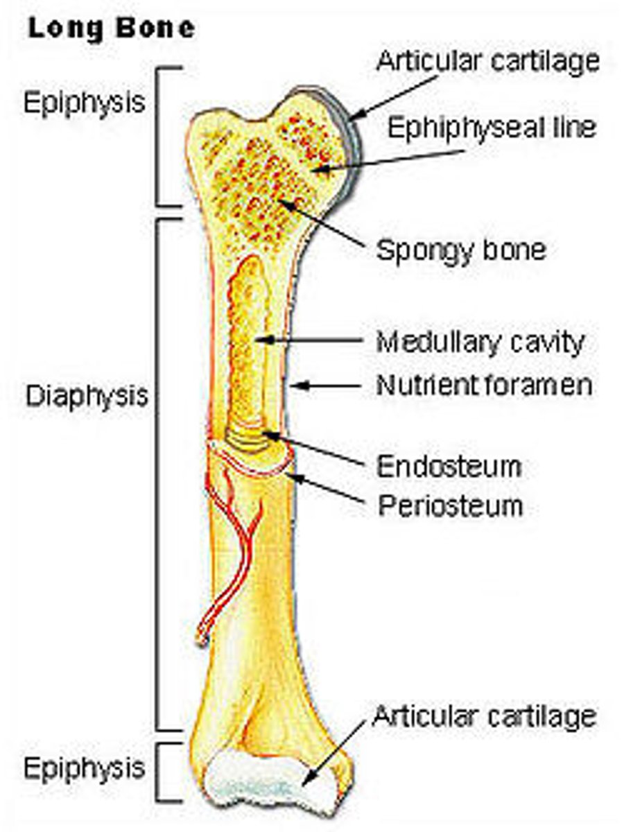
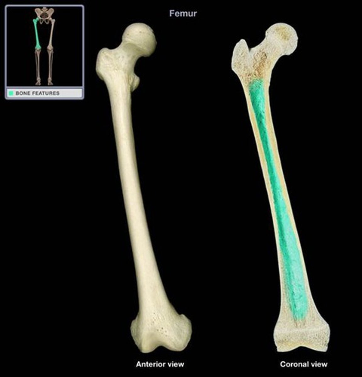
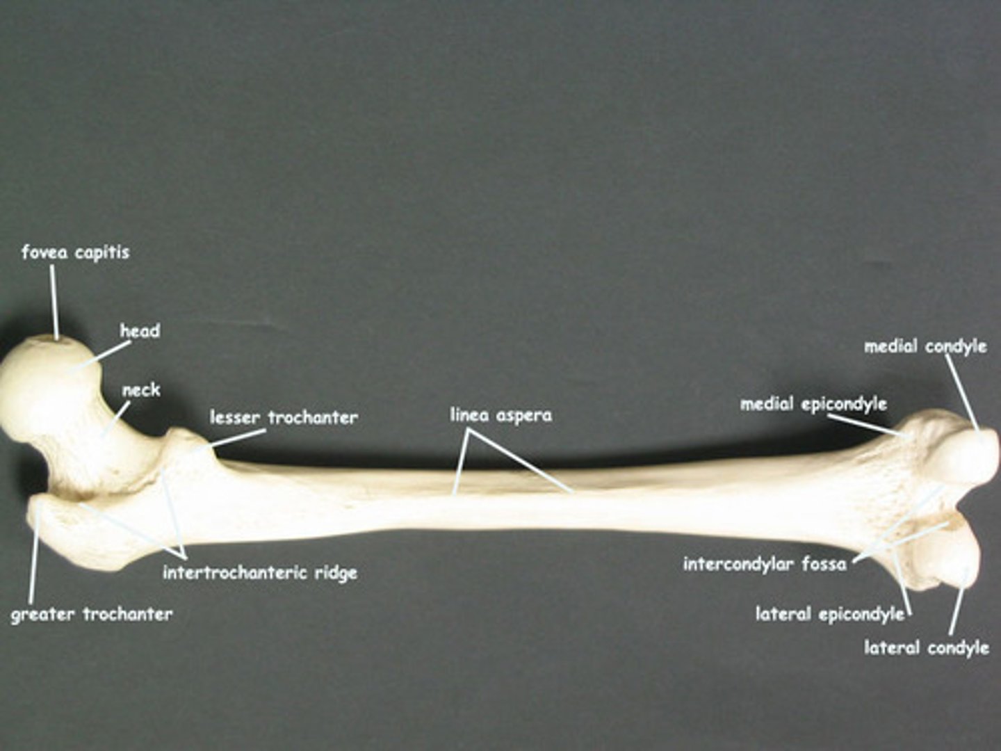
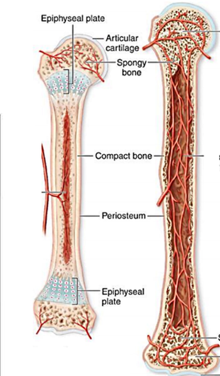
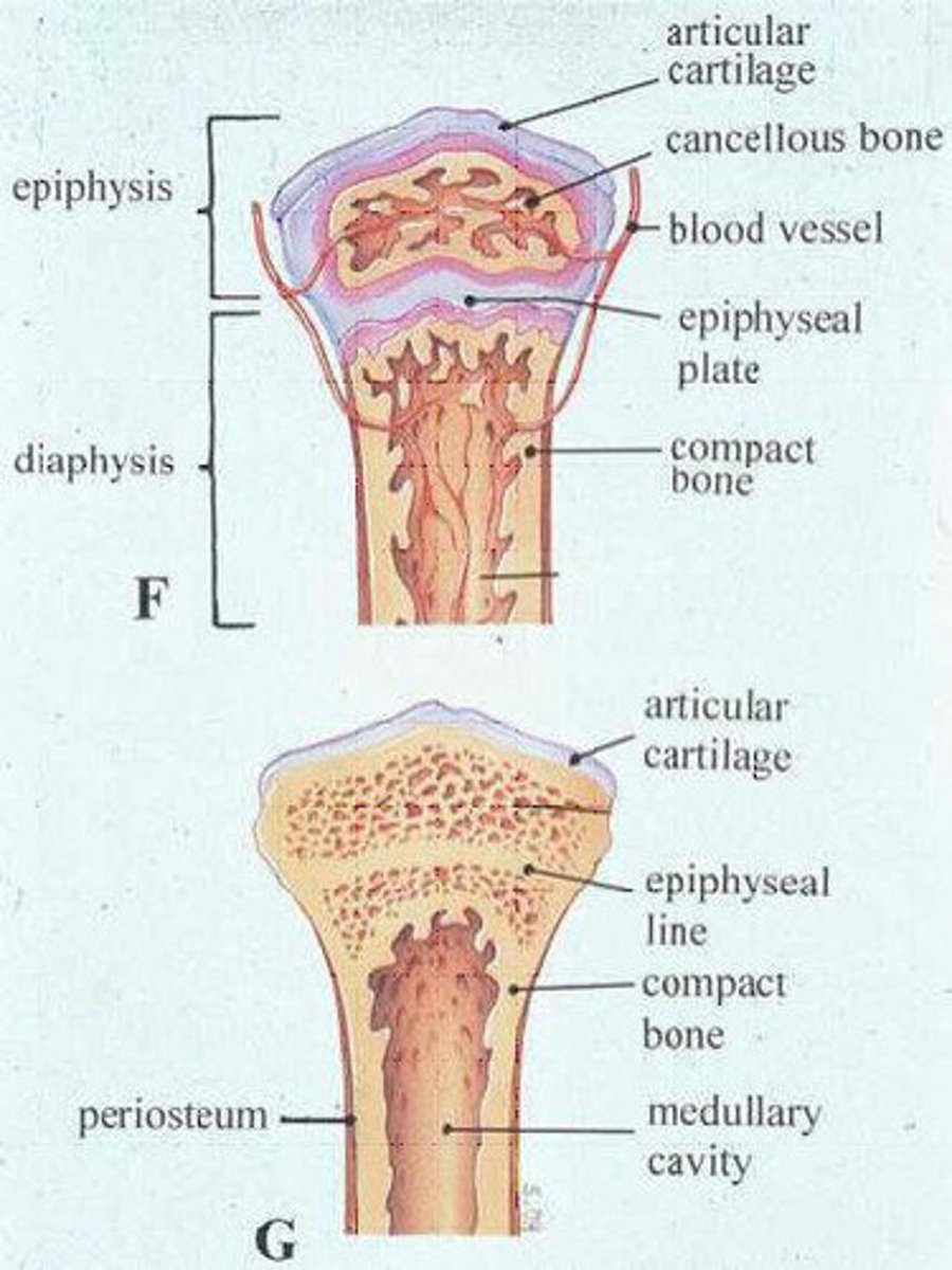
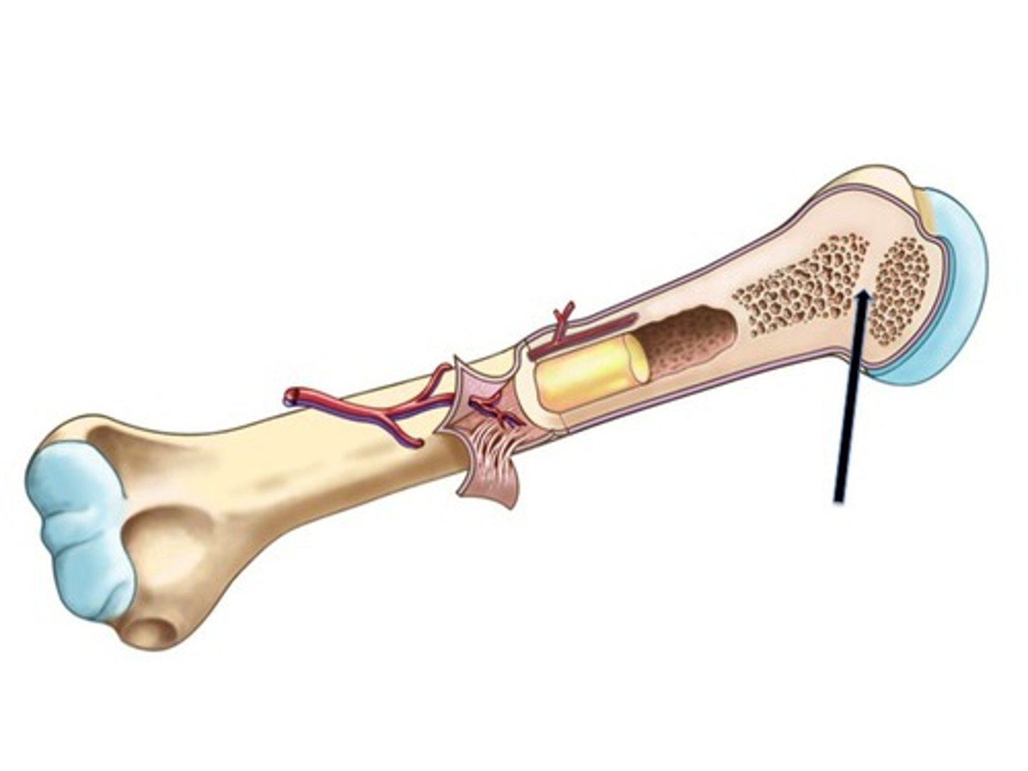
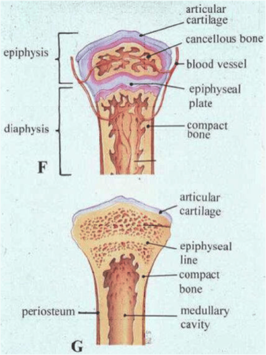
Structure of typical long bone
diaphysis (tubular shaft forms long axis with compact bone surrounding medullary cavity); epiphyses (expanded ends of bone with external compact bone and internal spongy bone); articular cartilage covers articular surfaces; between is epiphyseal line (remnant of childhood bone growth at epiphyseal plate)
TERM
Periosteum
DEFINITION
White, double-layered membrane that covers external surfaces except joint surfaces; outer fibrous layer of dense irregular connective tissue and Sharpey's fibers secure to bone matrix (anchoring points for tendons and ligaments); inner osteogenic layer abuts bone; contains stem cells (osteogenic cells); many nerve fibers and blood vessels
TERM
Endosteum
DEFINITION
Delicate connective tissue membrane covering internal bone surface
Covers trabeculae of spongy bone
Lines canals that pass through compact bone
Contains osteogenic cells that can differentiate into other bone cells
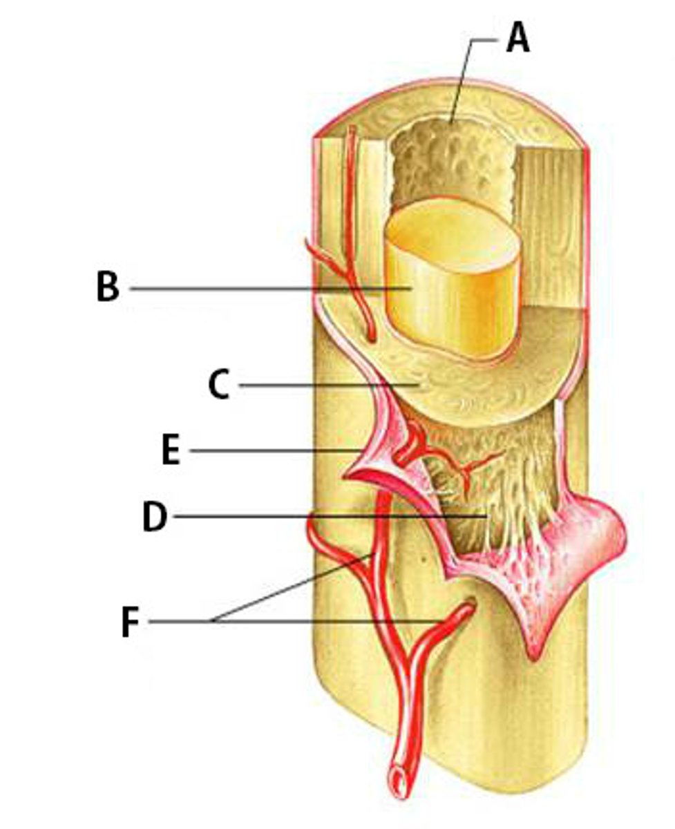
Diaphysis
tubular shaft forms long axis; compact bone surrounding medullary cavity
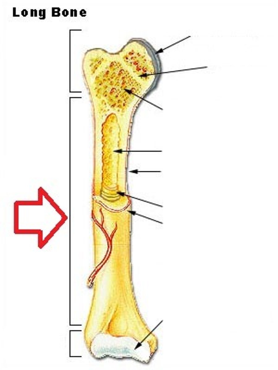
Epiphyses
bone ends; external compact bone; internal spongy bone; articular cartilage covers articular surfaces
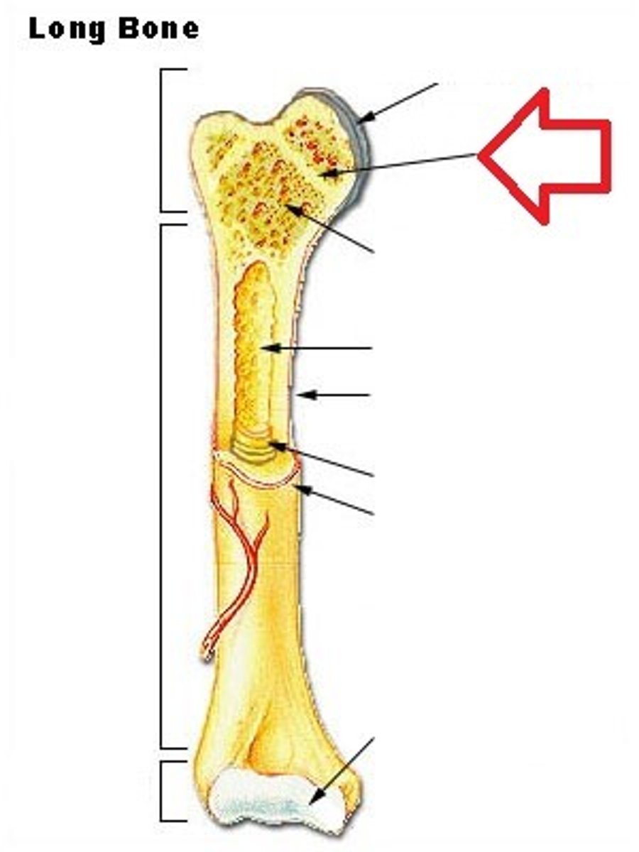
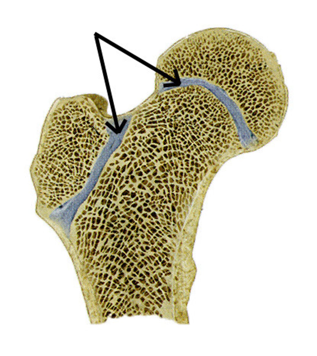
Epiphyseal line
between the epiphyses and diaphysis; remnant of childhood bone growth at epiphyseal plate
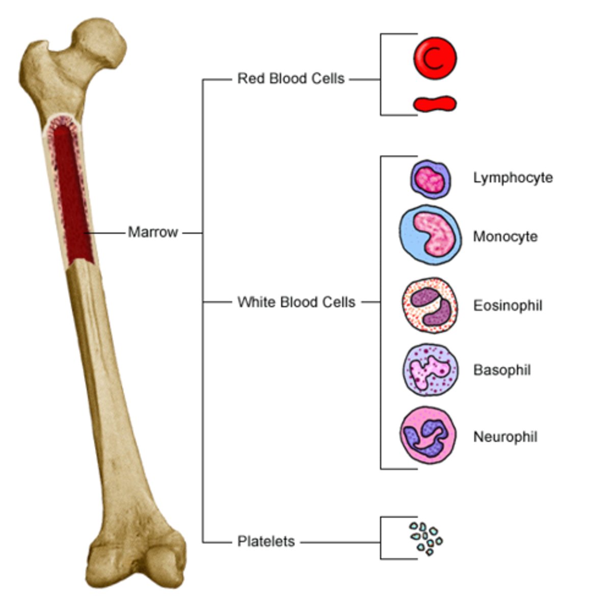
Medullary cavities
site of red bone marrow and hematopoiesis in fetus and newborns; also found in spongy bone
Hemopoiesis
is the formation of blood cellular component; all cellular blood components are derived from hematopoietic stem cells; yellow marrow can convert to red, if necessary; occurs in spongy and diploe bones of adults
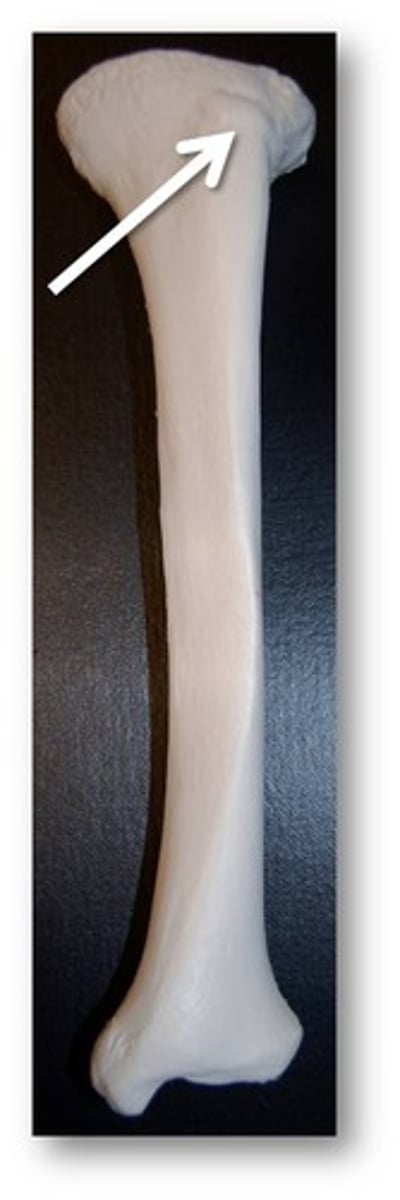
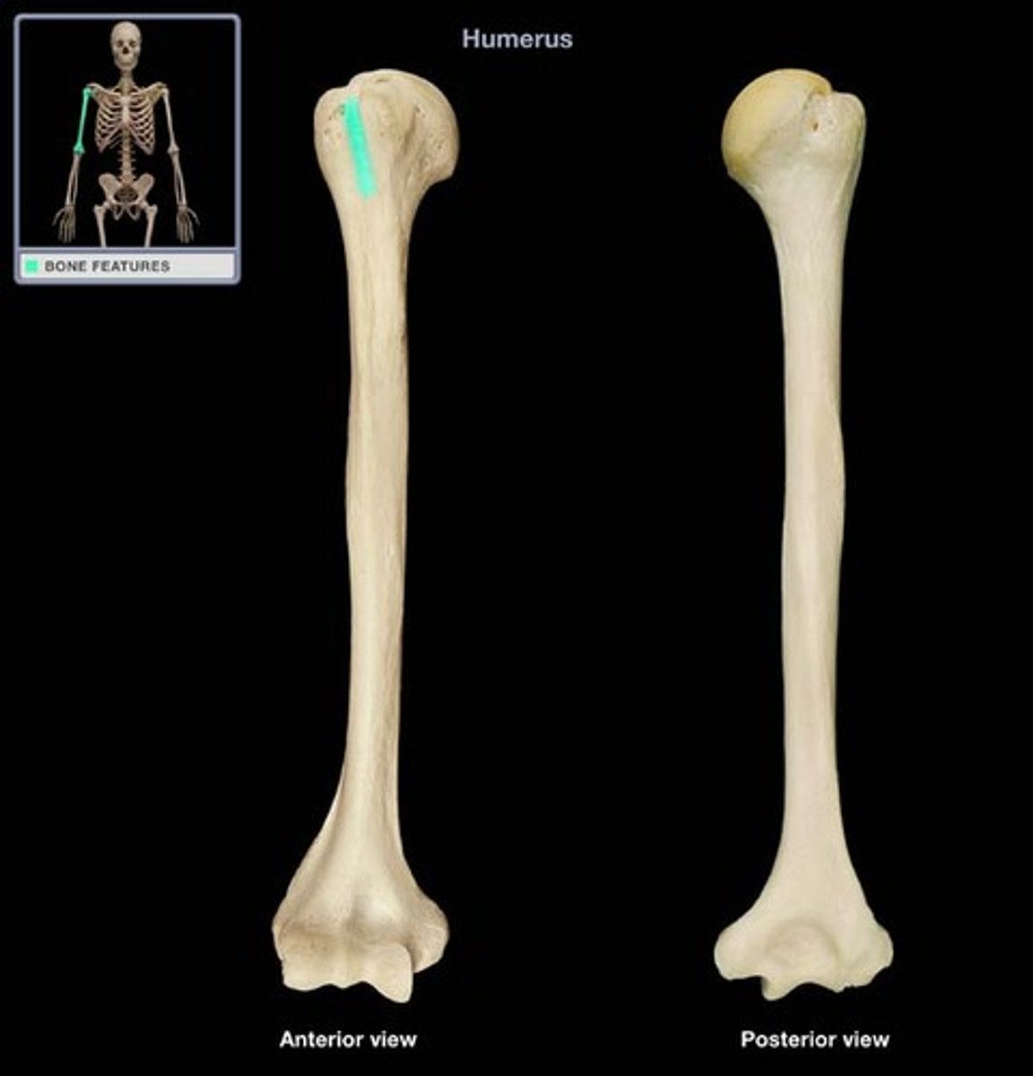
Tuberosity
Large rounded projection; may be roughened
Crest
Narrow ridge of bone; usually prominent

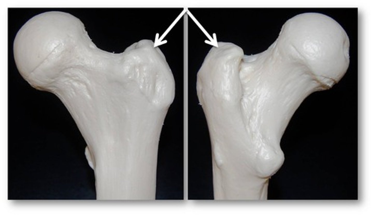
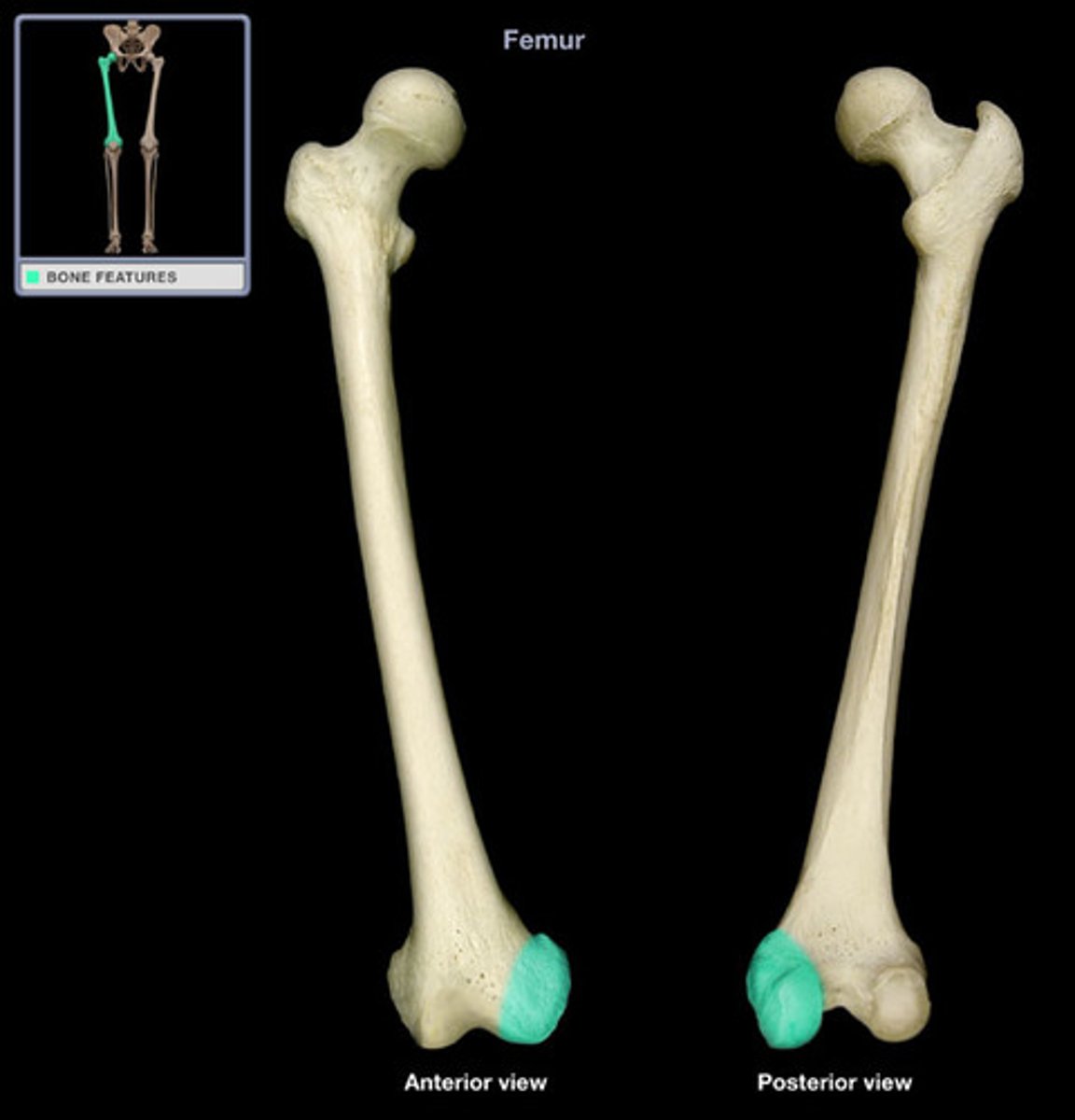
Trochanter
A large, irregularly shaped process (only process is on the femur)
Line
Narrow ridge of bone, less prominent than a crest (Ex: linea aspira)
Tubercle
Small rounded projection or process
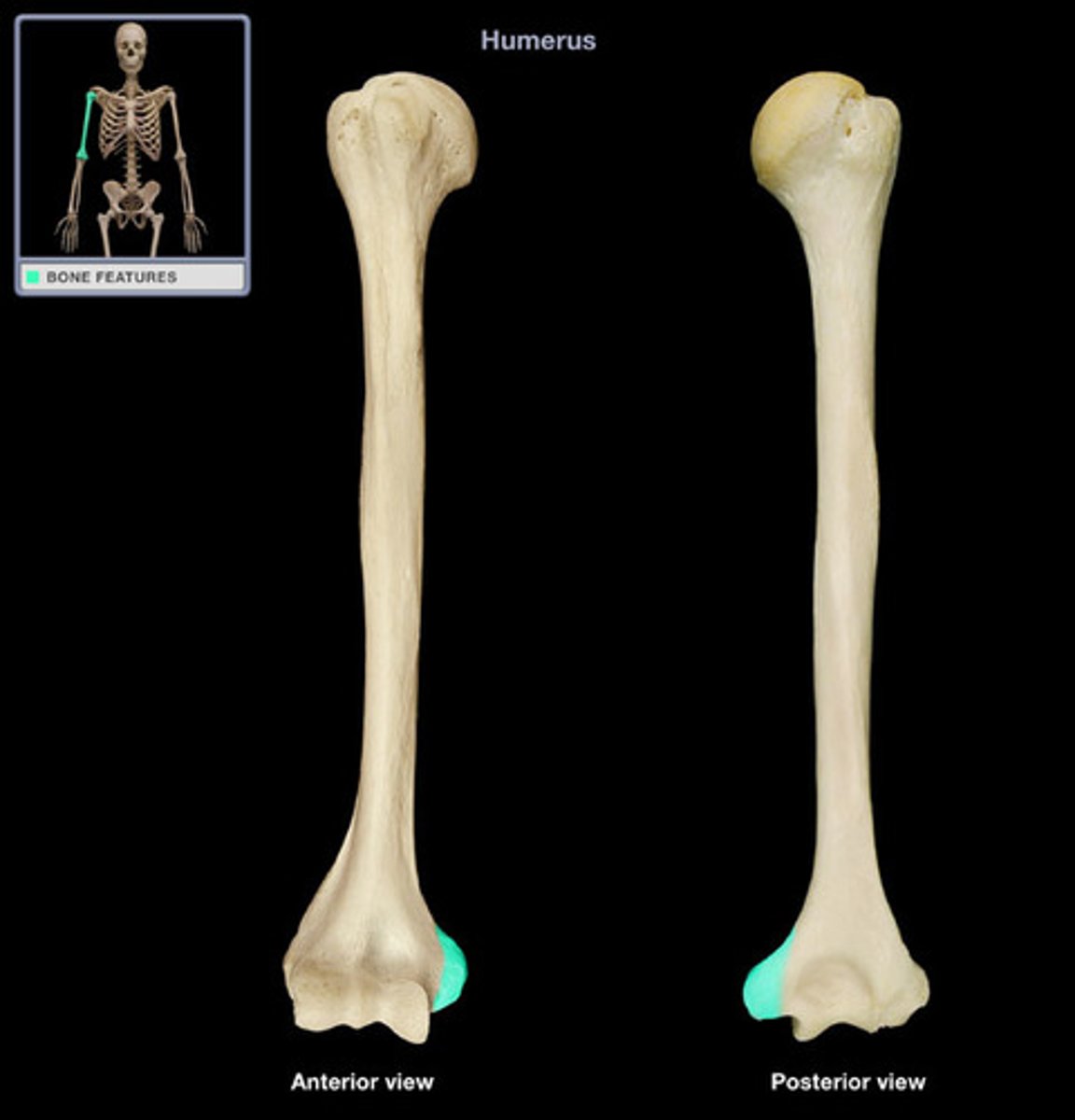
Epicondyle
Raised area on or above a condyle
Spine
Sharp, slender, often pointed projection
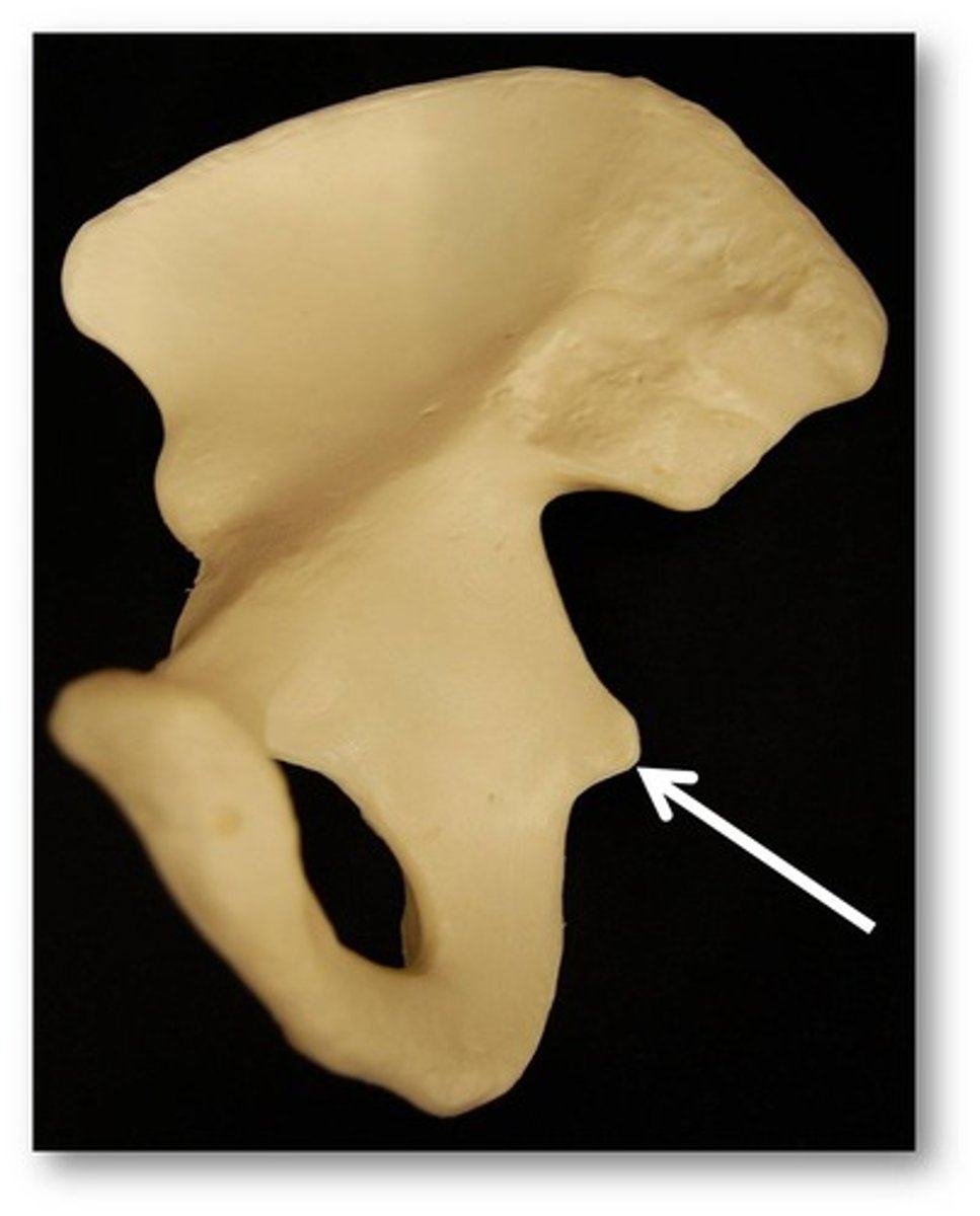
Process
any bony prominence
Projections that are sites of muscle and ligament attachment
Tuberosity, Crest, Trochanter, Line, Tubercle, Epicondyle, Spine, Process
Projections that help to form joints
Head, facet, condyle, ramus
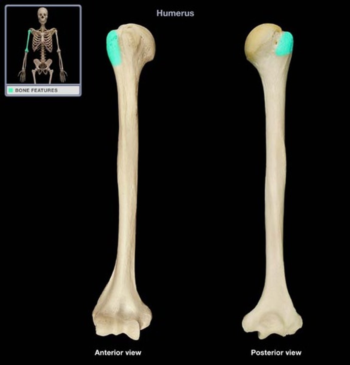
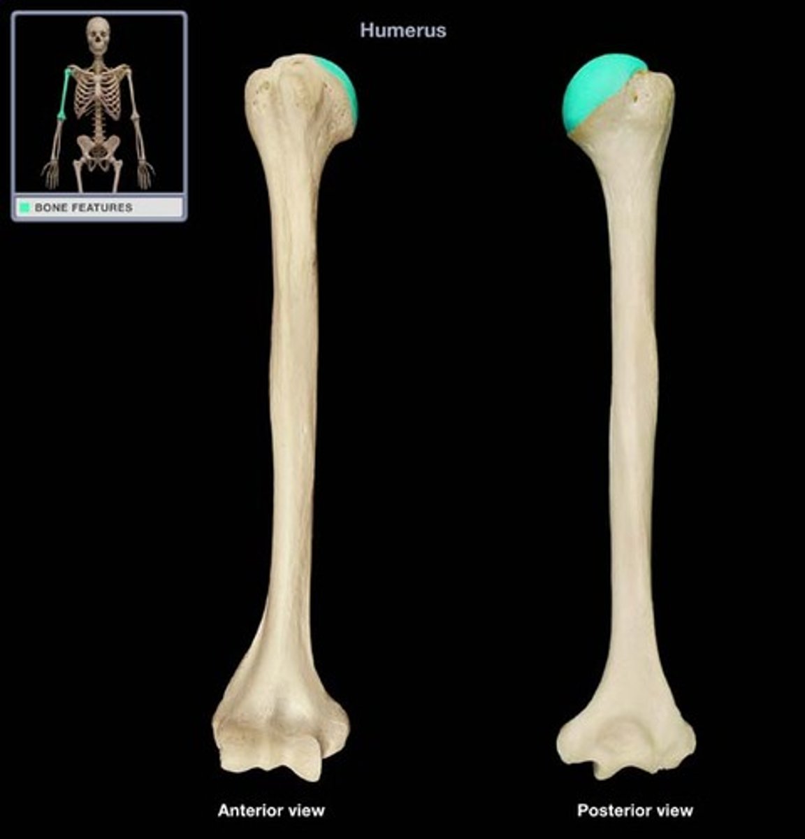
Head
Bony expansion carried on a narrow neck
Facet
Smooth, nearly flat articular surface
Condyle
Rounded articular projection
Ramus
Armlike bar of bone
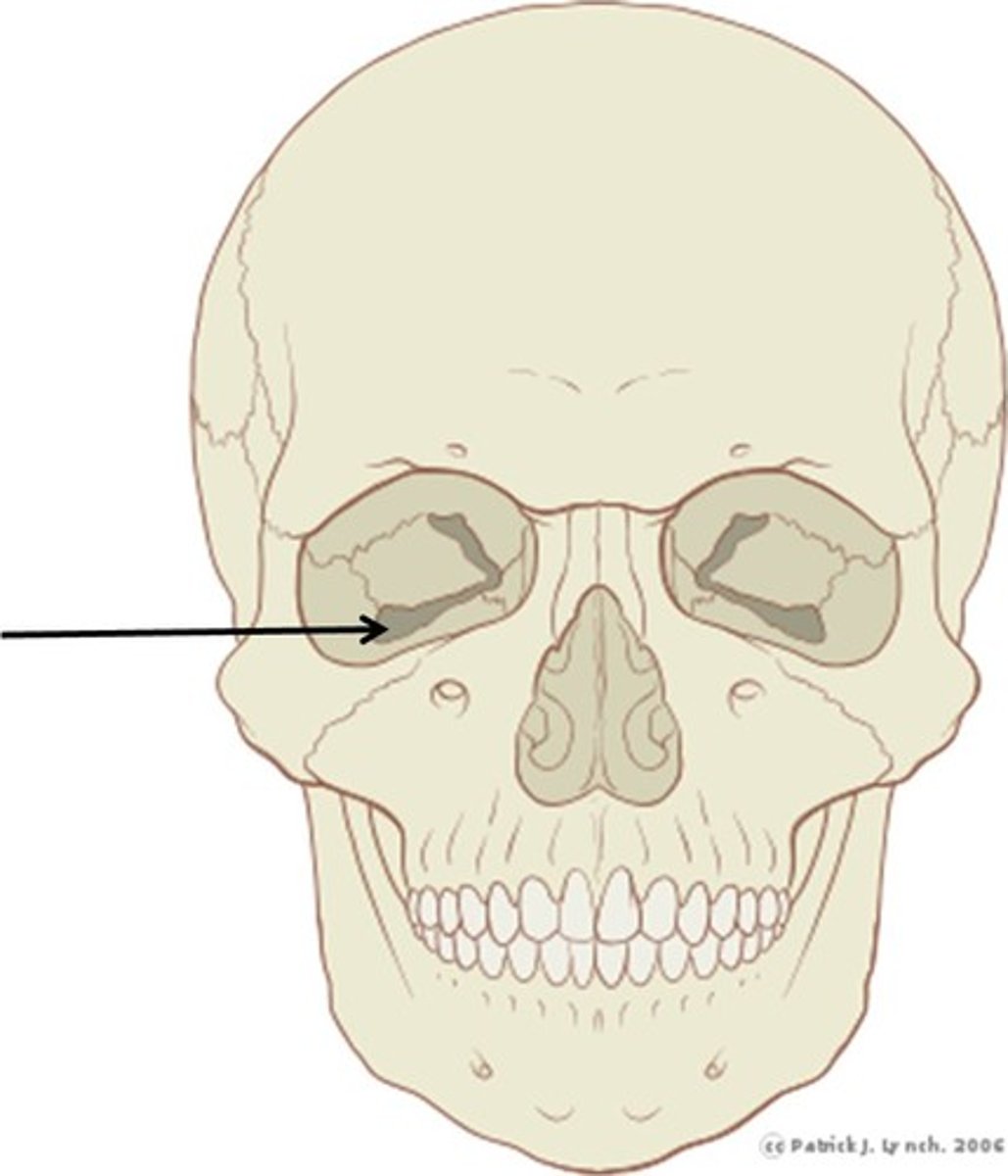
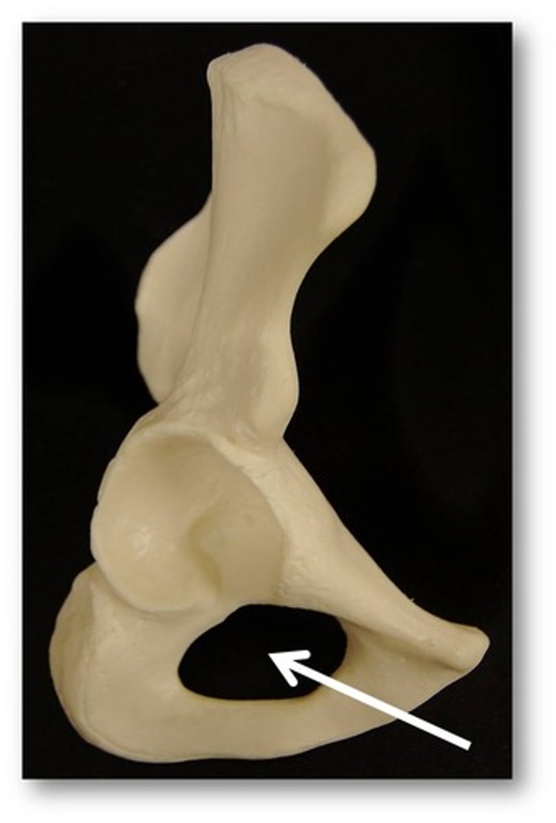
Depression and Openings
(for passage of blood vessels and nerves) groove, fissure, foramen, notch; others- meatus, sinus, fossa
Groove
Furrow
Fissure
Narrow, slitlike opening
Foramen
round or oval opening through a bone
Notch
indentation at the edge of a structure
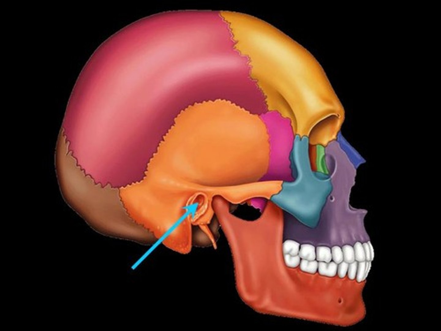
Meatus
canal-like passageway
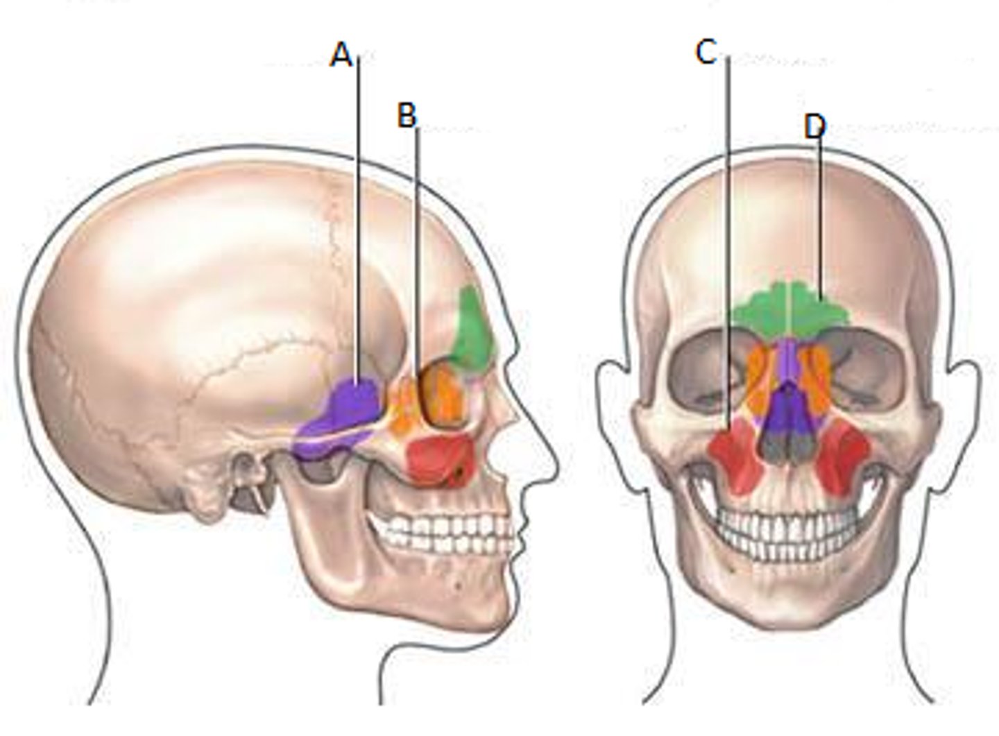
Sinus
Cavity within a bone, filled with air and lined with mucus membrane
Fossa
Shallow; basinlike depression in a bone, often serving as an articular surface
Types of bone cells
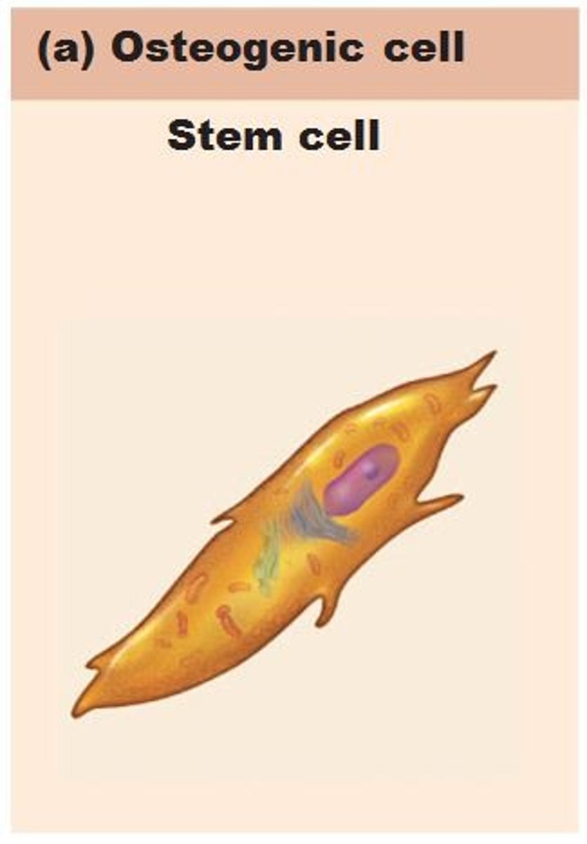
Osteogenic cells
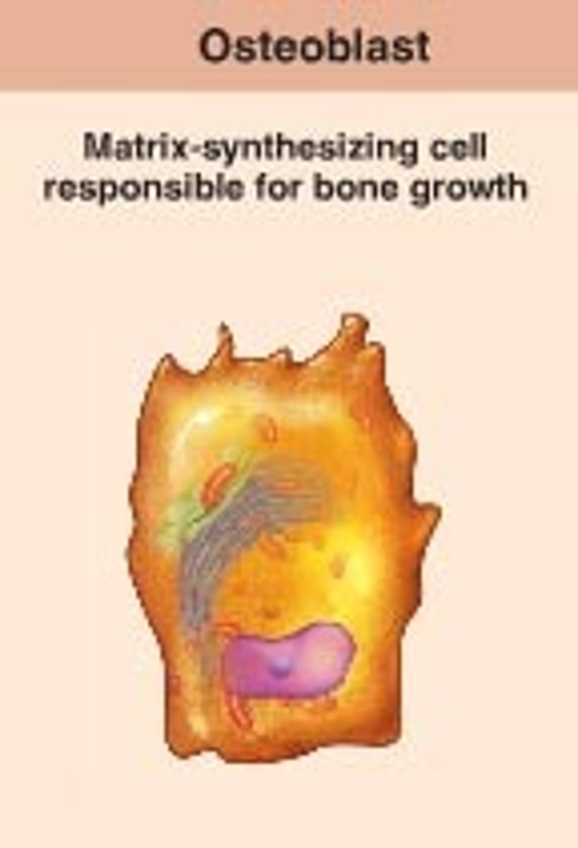
Osteoblasts
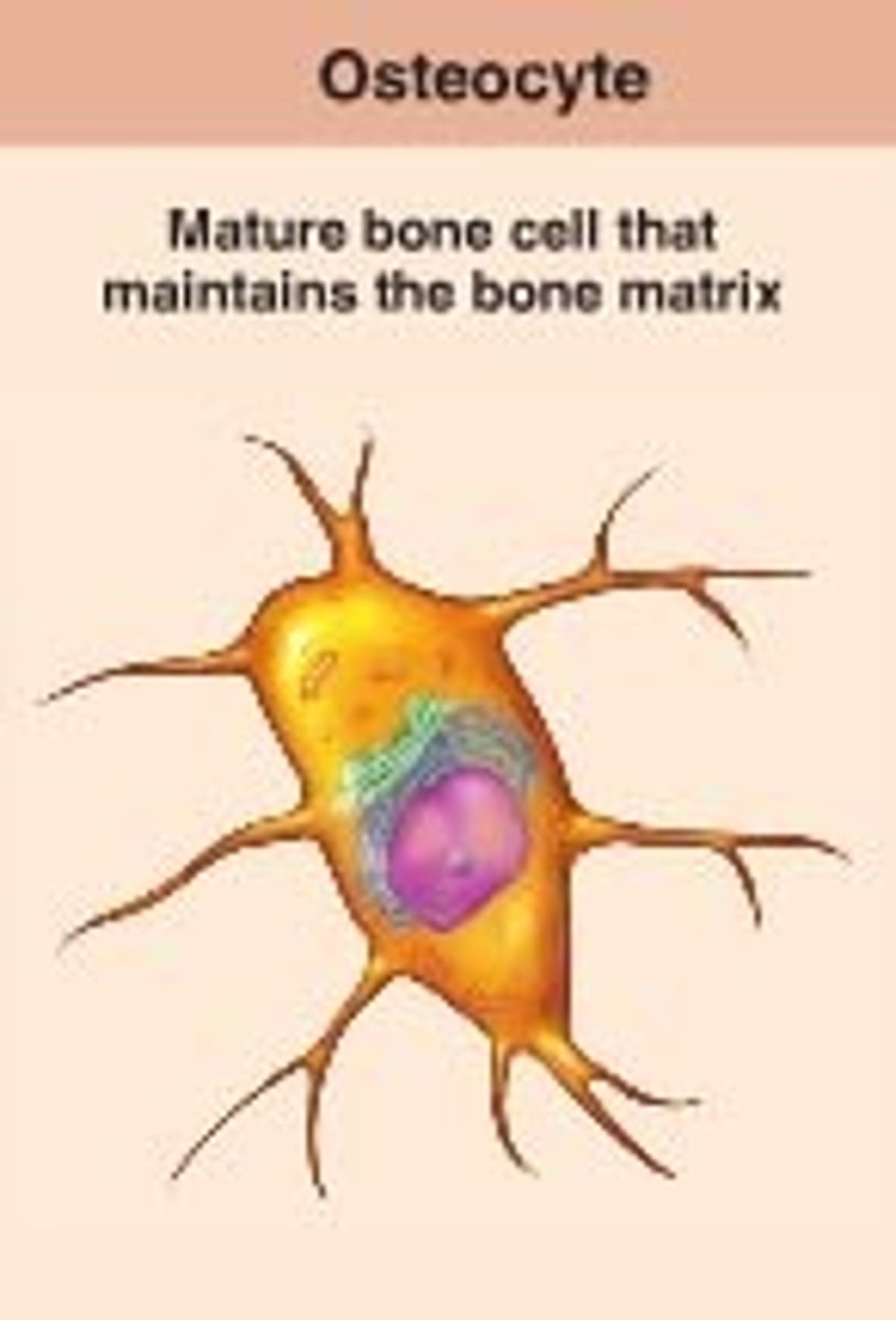
Osteocytes
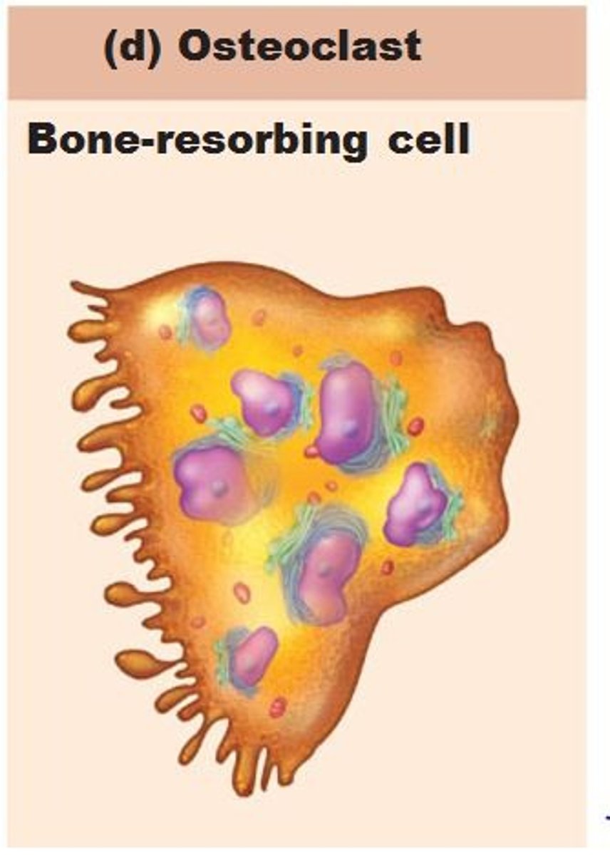
Osteoclasts
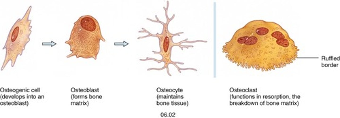
Osteogenic Cells
(also called osteoprogenitor cells); mitotically active stem cells in periosteum and endosteum; when stimulated differentiate into osteoblasts or bone lining cells; some persist as osteogenic cells
Osteoblasts
bone-forming cells; secrete unmineralized bone matrix or osteoid; includes collagen and calcium-binding proteins; collagen = 90% of bone protein; actively mitotic
Osteocytes
mature bone cells in lacunae; monitor and maintain bone matrix; act as stress or strain sensors; respond to and communicate mechanical stimuli to osteoblasts and osteoclasts (cells that destroy bone) so bone remodeling can occur
Osteoclasts
derived from hematopoietic stem cells that become macrophages; giant, multinucleate cells for bone resorption; when active, rest in resorption bay and have ruffled border; ruffled border increases surface area for enzyme degradation of bone and seals off area from surrounding matrix
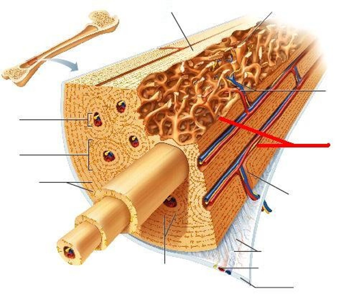
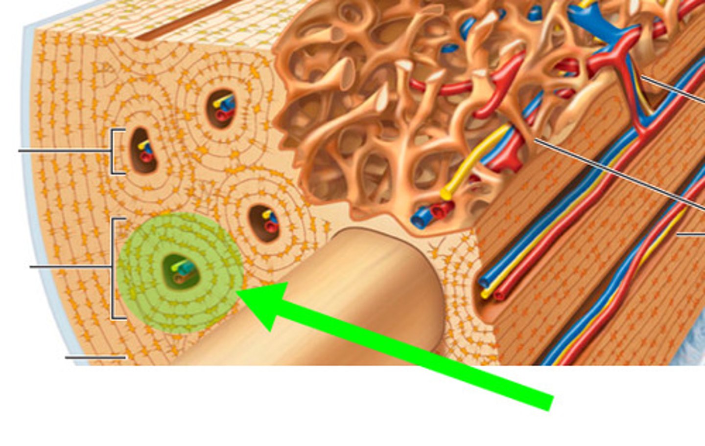
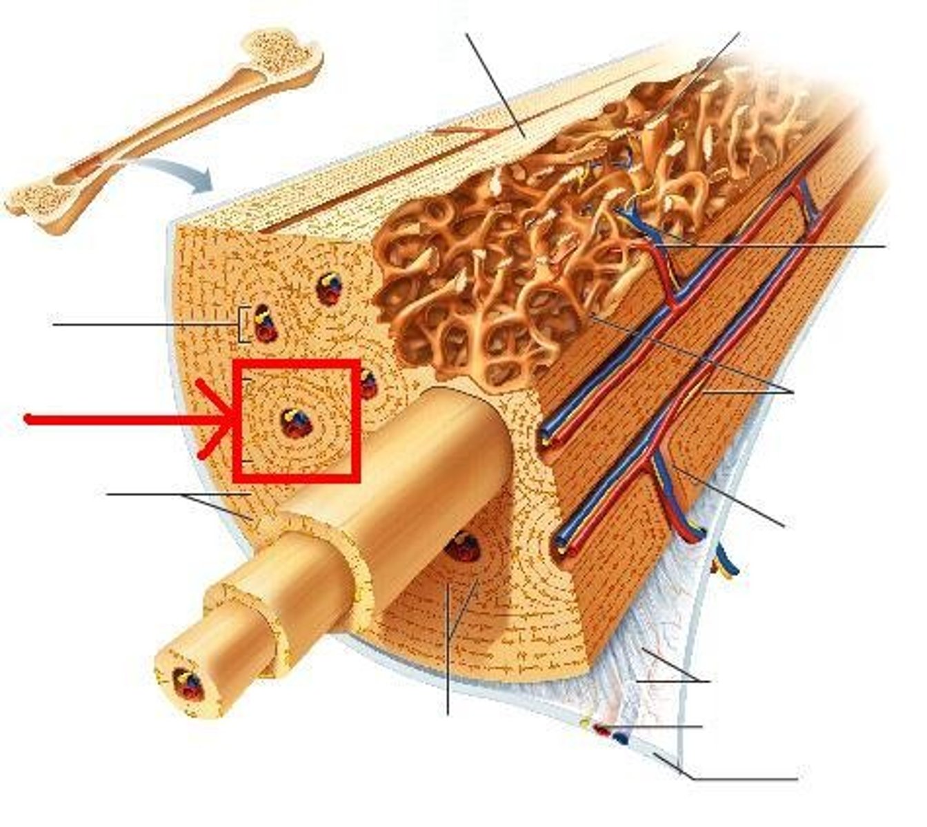
Compact Bone
(also called lamellar bone) : hard, dense bone tissue that is beneath the outer membrane of a bone; withstands stress
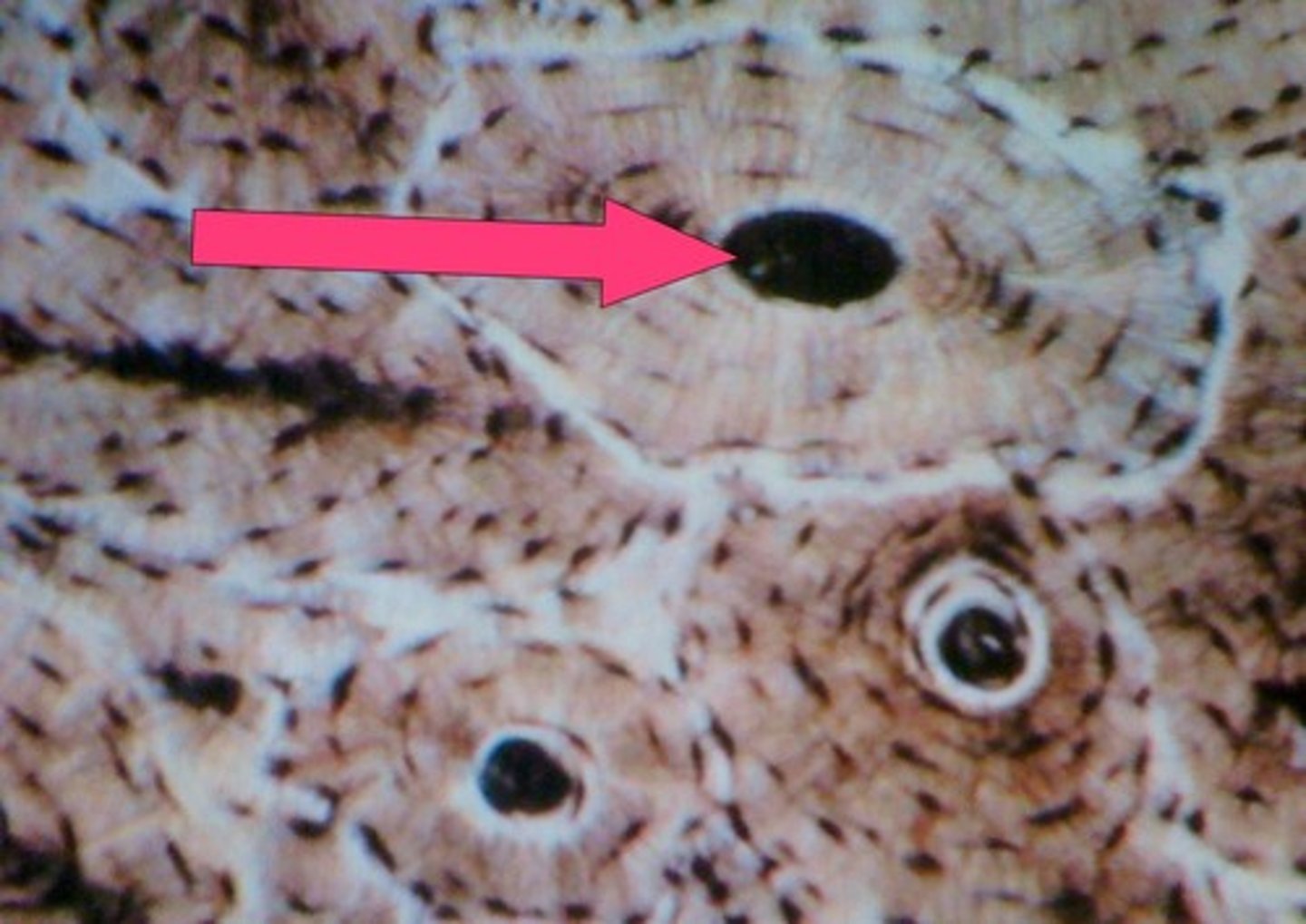
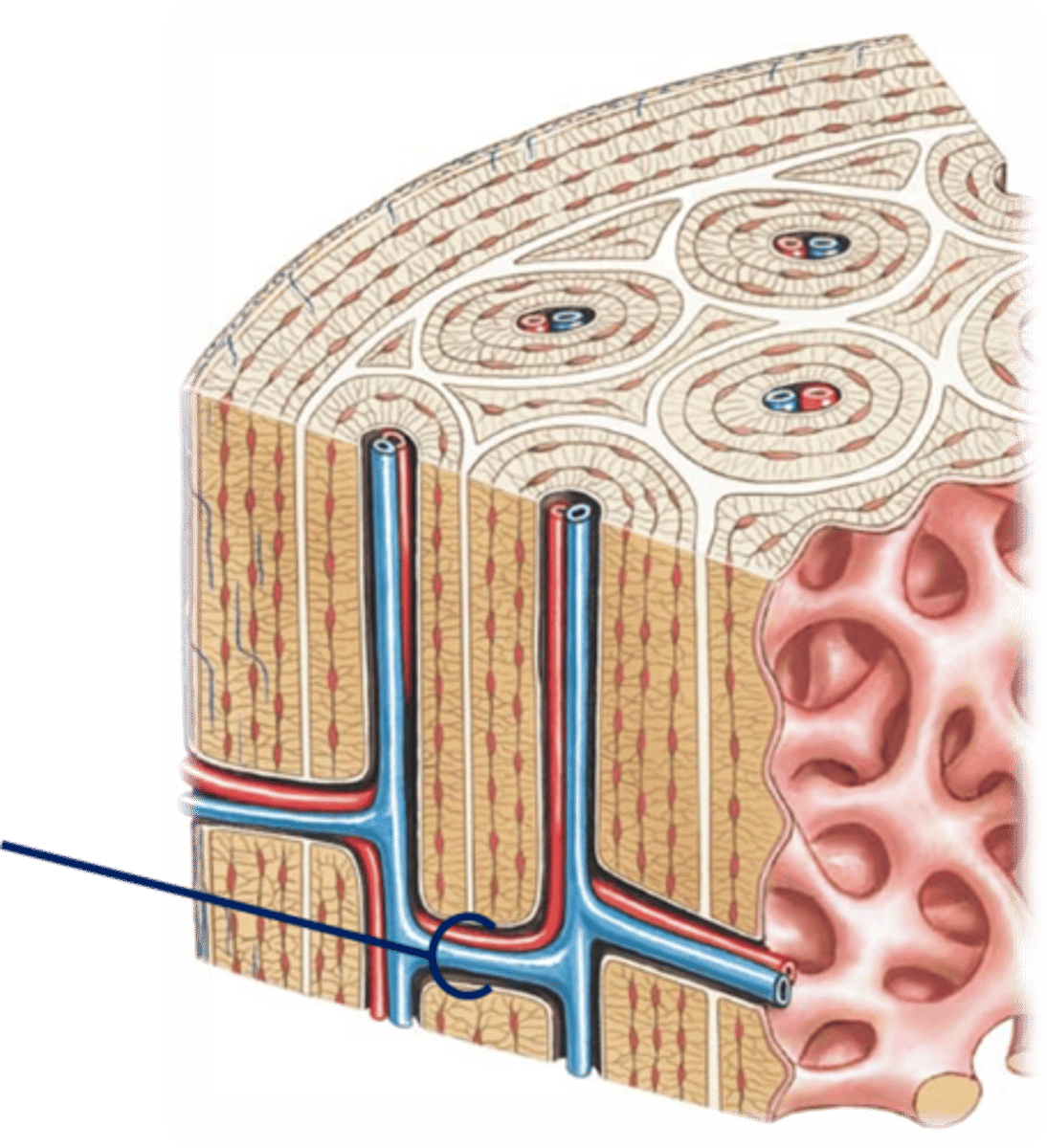
Osteon or Haversian system
structural unit of compact bone; elongated cylinder parallel to long axis of bone
TERM
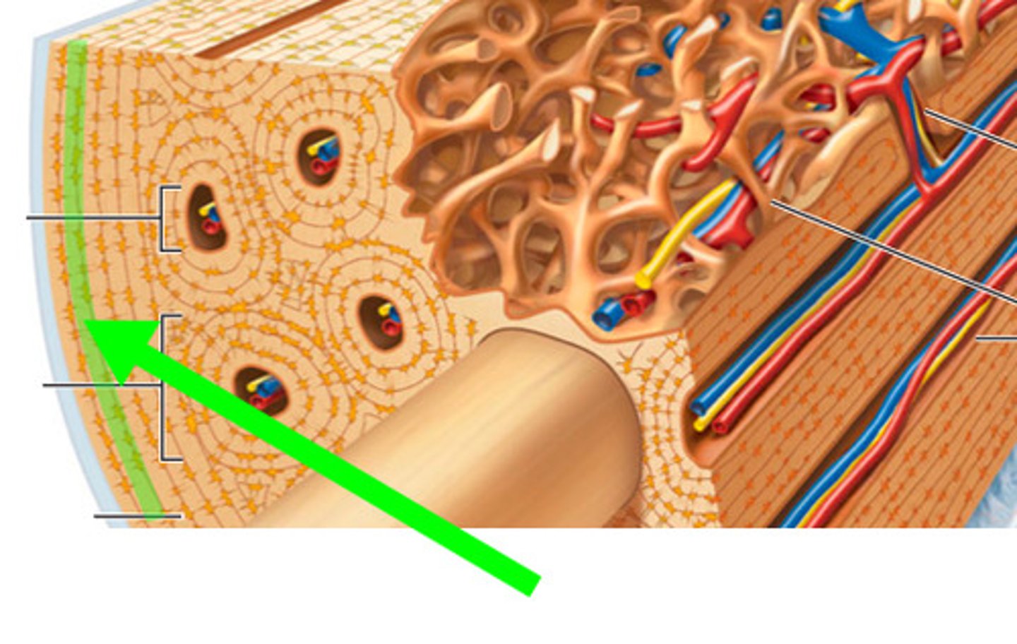
Lamellae
DEFINITION
hollow tubes of bone matrix; collagen fibers in adjacent rings run in different directions; withstands stress - resist twisting
TERM
Osteon
DEFINITION
structural unit of compact bone
TERM
central canal
DEFINITION
haversian canal runs through core of osteon; contains blood vessels and nerve fibers; lined by enosteum
perforating (Volkmann's) canals
canals lined with endosteum at right angles to central canals; connect blood vessels and nerves of periosteum, medullary cavity, and central canal
lacunae
small cavities that contain osteocytes
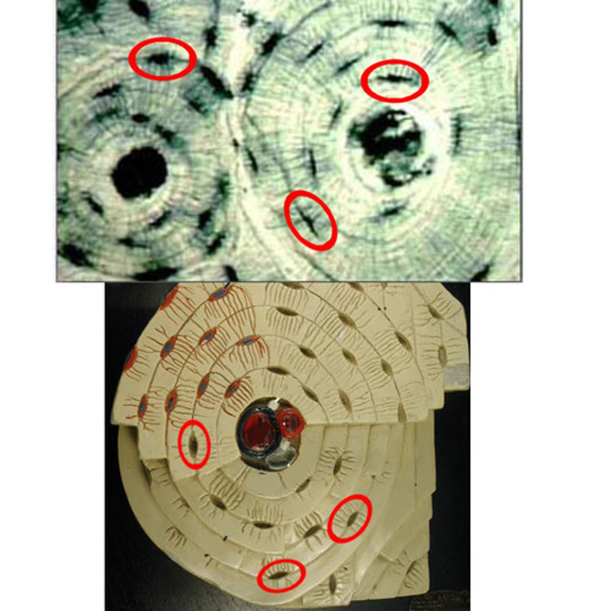
canaliculi
hairlike canals that connect lacunae to each other and central canal
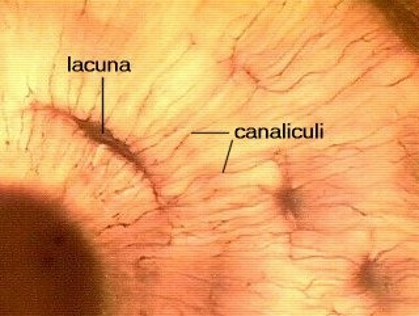
canaliculi formation and function
when matrix hardens and cells are trapped the canaliculi form; allow communication; permit nutrients and wastes to be relayed from one osteocyte to another throughout osteon
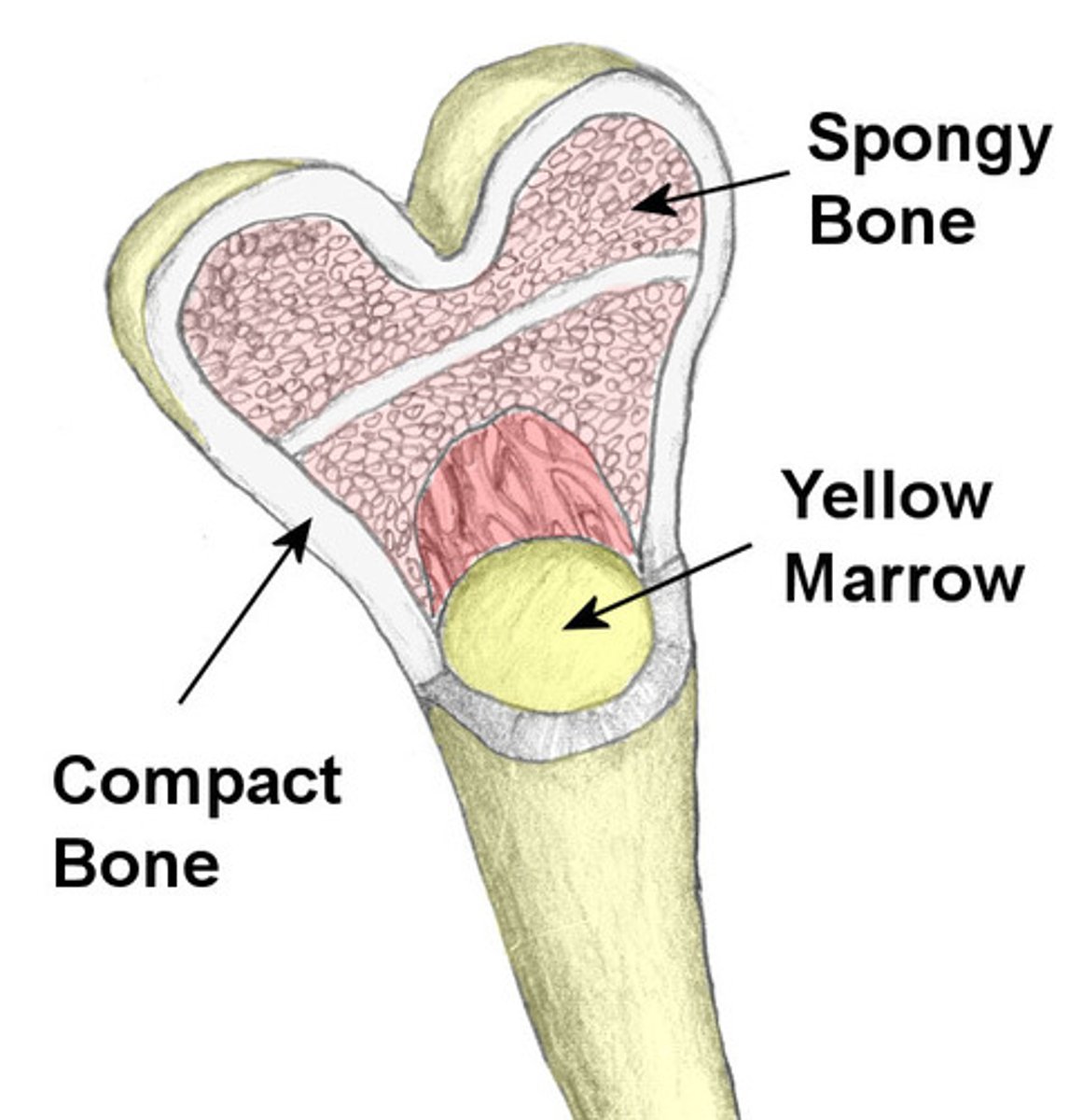
Spongy Bone
located inside compact bones at widened ends of long bones, has spaces in matrix so it looks like a sponge, still rigid, adaptation- "to make bones lighter in weight"; appears poorly organized

Trabeculae
align along lines of stress to help resist it; no osteons; contain irregularly arranged lamellae and osteocytes interconnected by canaliculi; capillaries in endosteum supply nutrients

Organic Components
includes cells and osteoid; made of ground substance (proteoglycans and glycoproteins); collagen fibers; contributes to structure; provides tensile strength and flexibility; stretch and break easily on impact to dissipate energy and prevent fracture; if no addition trauma, bonds re-form
Inorganic Components
hydroxyapatites (mineral salts); 65% of bone by mass; mainly of tiny calcium phosphate crystals in and around collagen fibers; responsible for hardness and resistance to compression
Strength of bone
half as strong as steel in resisting compression; as strong as steel in resisting tension
Ossification (osteogenesis)
process of bone tissue formation; formation of bony skeleton; begins in 2nd month of development; postnatal bone growth until early adulthood; bone remodeling and repair is lifelong
When does ossification occur?
Begins in 2nd month of development; postnatal bone growth until early adulthood
Endochondral
_______________ ossification is when bone forms by replacing hyaline cartilage; forms most of skeleton
Intramembranous ossification
bone develops from fibrous membrane; bones called membrane bones; forms flat bones, e.g. clavicles and cranial bones

Two types of ossification
intramembranous and endochondral
Clavicles
endochondral ossification forms most all bones inferior to base of skull except _______
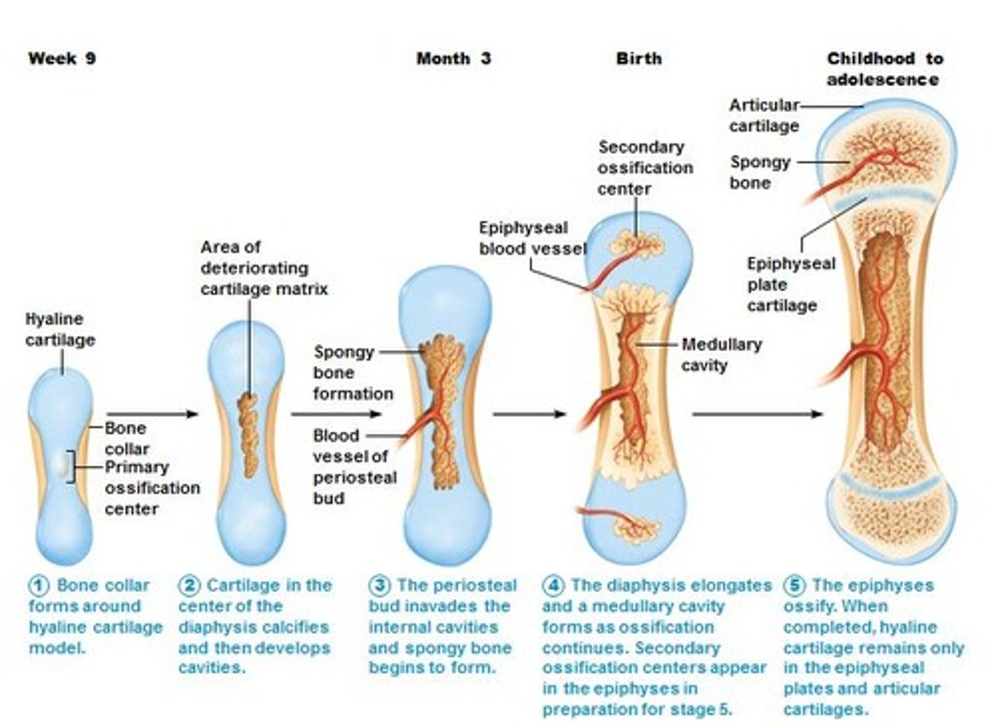
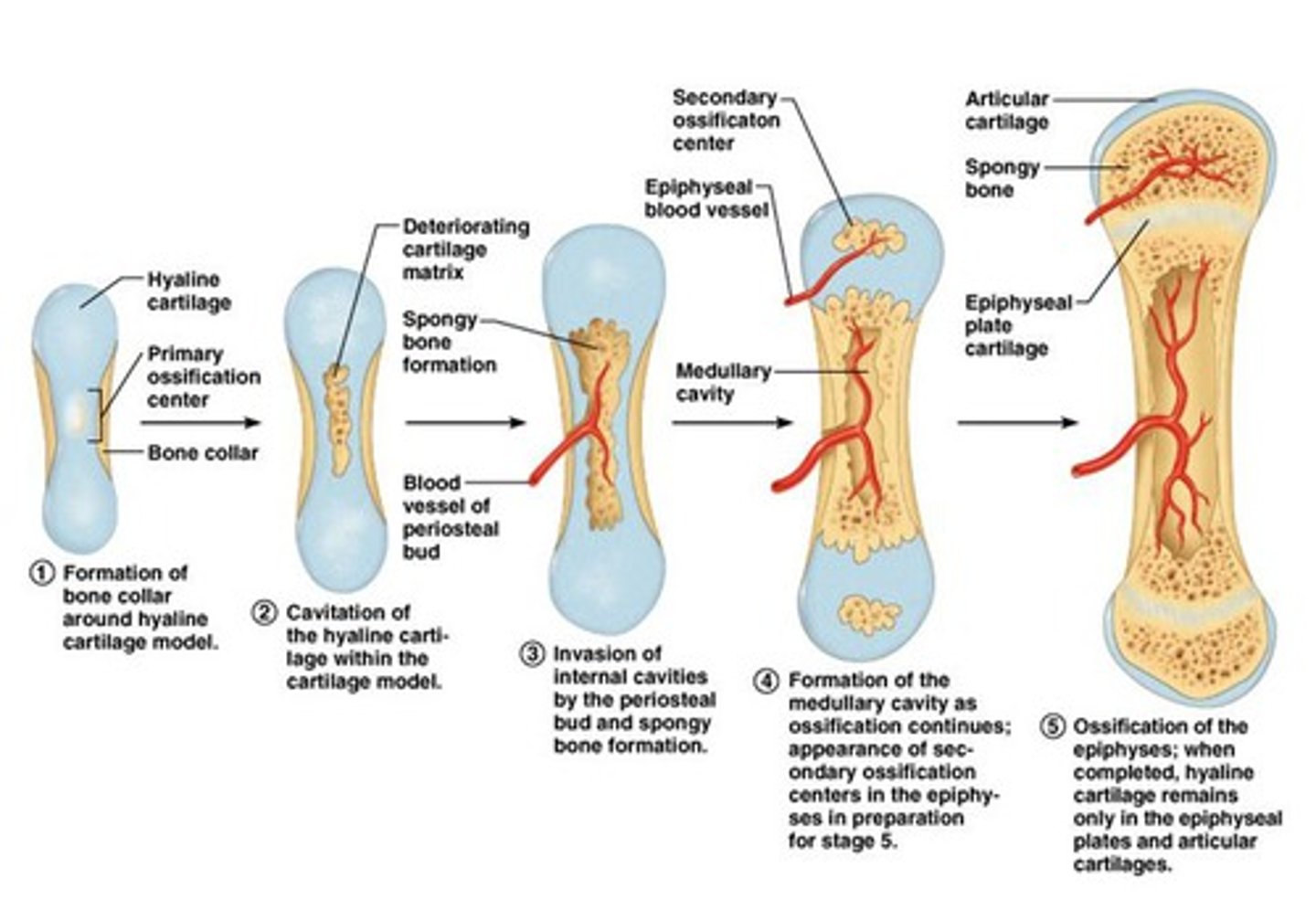
Endochondral ossification in a long bone
1) Bone collar forms around the diaphysis of the hyaline cartilage model.
2)Cartilage in the center of the diaphysis calcifies and then develops cavities.
3)The periosteal bud invades the
internal cavities and spongy bone forms.
4) The diaphysis elongates and a medullary cavity forms. Secondary ossification centers appear in the epiphyses.
5)The epiphyses ossify. When completed, hyaline cartilage remains only in the epiphyseal plates and articular cartilages.
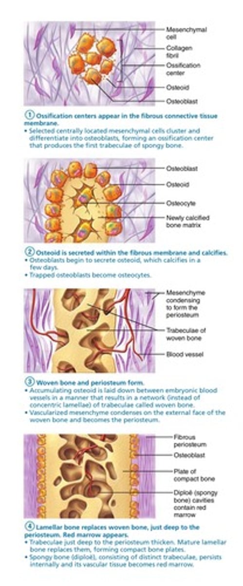
Steps of Intramembranous ossification
1)Ossification centers appear in the fibrous
connective tissue membrane; osteoblasts forming an ossification center that produces the first trabeculae of spongy bone.
2) Osteoid is secreted within the fibrous membrane and calcifies; trapped osteoblasts become osteocytes.
3) Woven bone and periosteum form.
• Accumulating osteoid is laid down in a manner that results in a network of trabeculae called woven bone; external face of the woven bone and becomes the periosteum.
4) Lamellar bone replaces woven bone, just deep to the periosteum. Red marrow appears.
postnatal Bone Growth
interstitial (longitudinal) growth and appositional growth
are both types of ____________
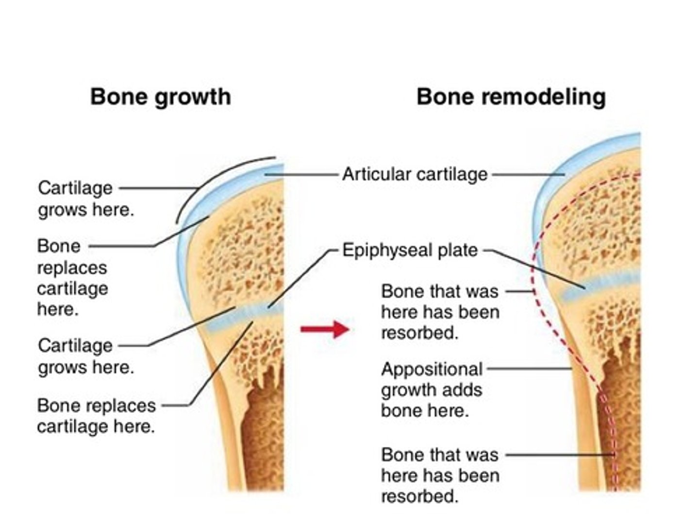
interstitial bone growth
aka longitudinal bone growth- increase in length of long bones-
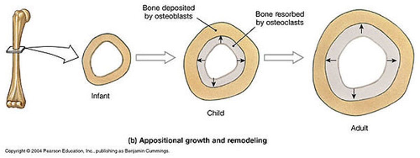
appositional bone growth
increase in bone thickness
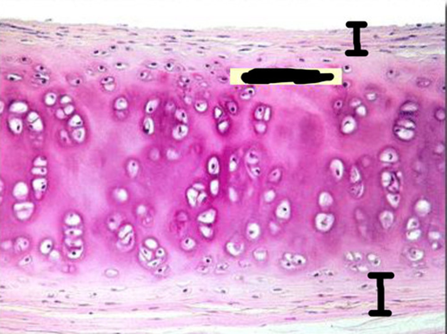
Epiphyseal cartilage
interstitial growth requires presence of ___________; epiphyseal plate maintains constant thickness
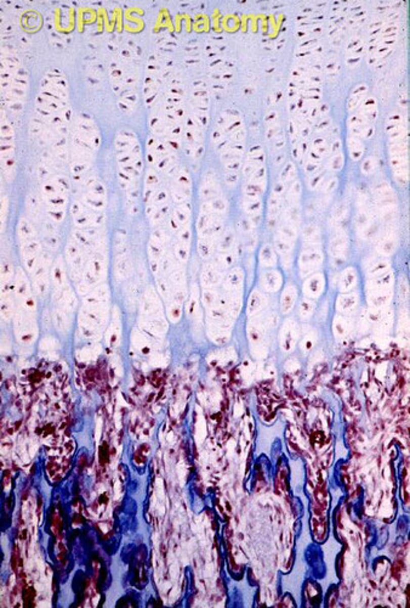
zones interstitial bone growth
resting (quiescent) , proliferation (growth), and hypertrophic zone, calcification, ossification zone
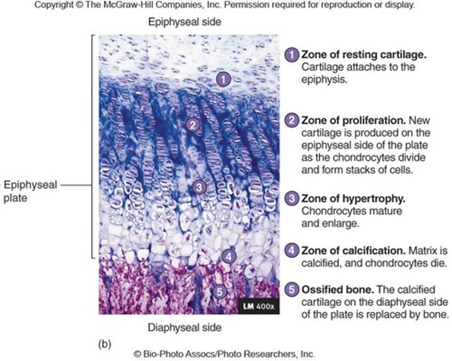
Resting (quiescent) zone
zone where cartilage on epiphyseal side of epiphyseal plate; relatively inactive
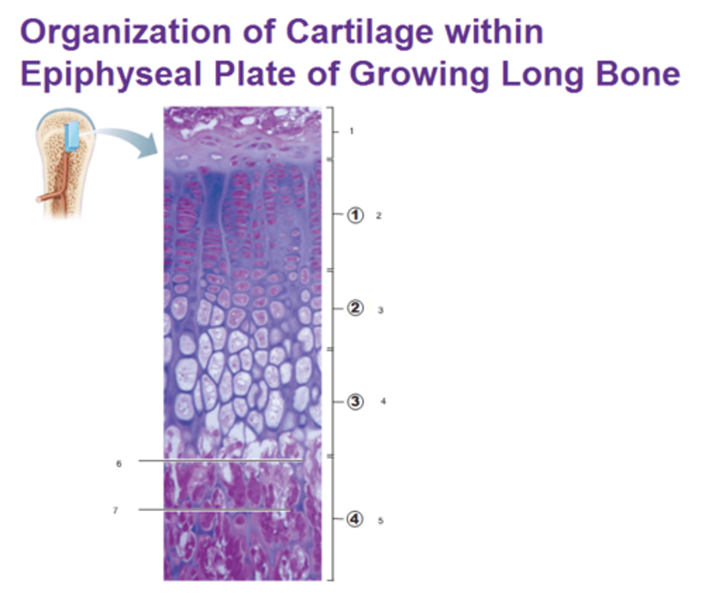
Proliferation (growth) zone
zone where cartilage on diaphysis side of epiphyseal plate; rapidly divide pushing epiphysis away from diaphysis lengthening
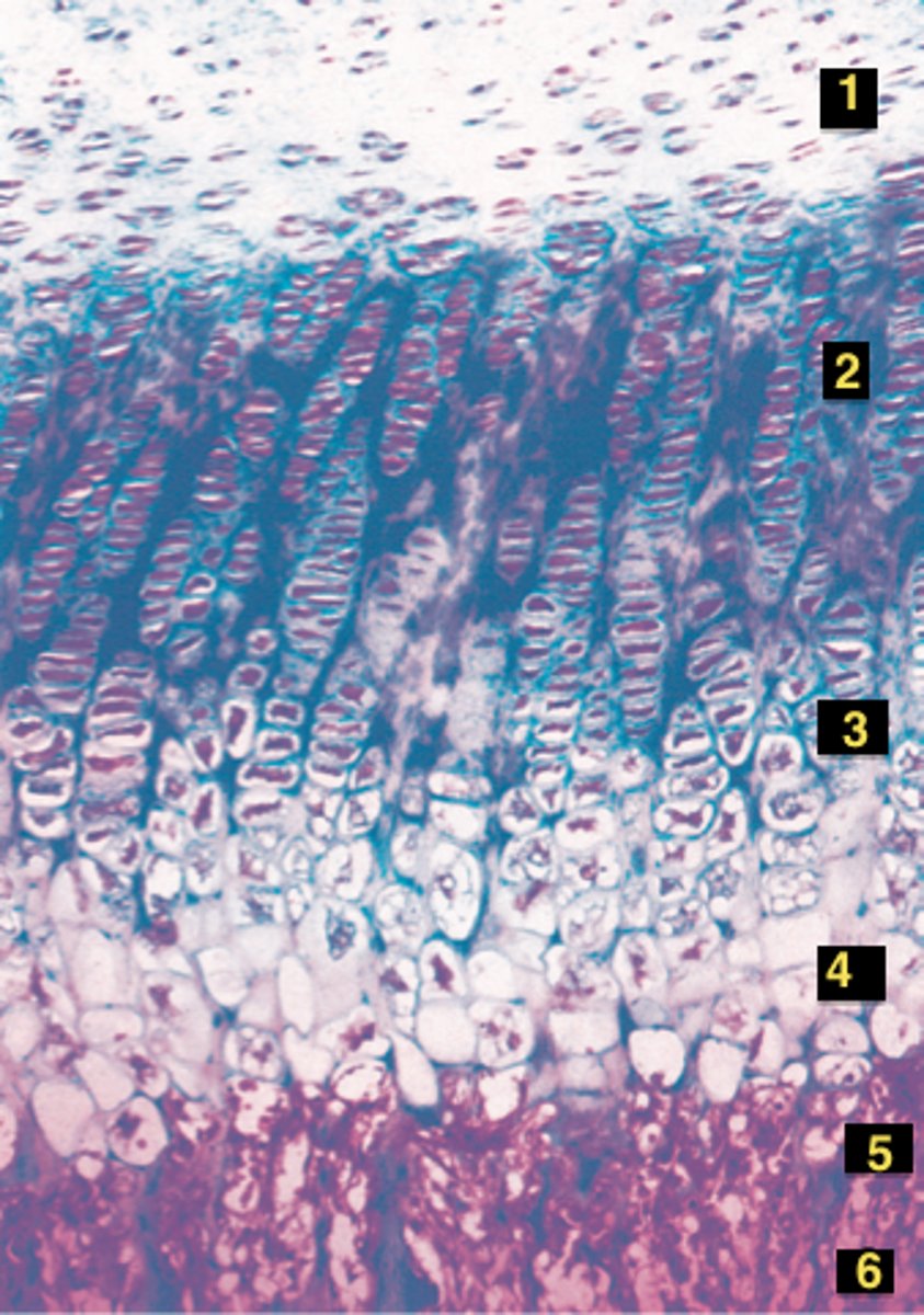
Hypertrophic zone
zone where older chondrocytes closer to diaphysis and their lacunae enlarge and erode interconnecting spaces
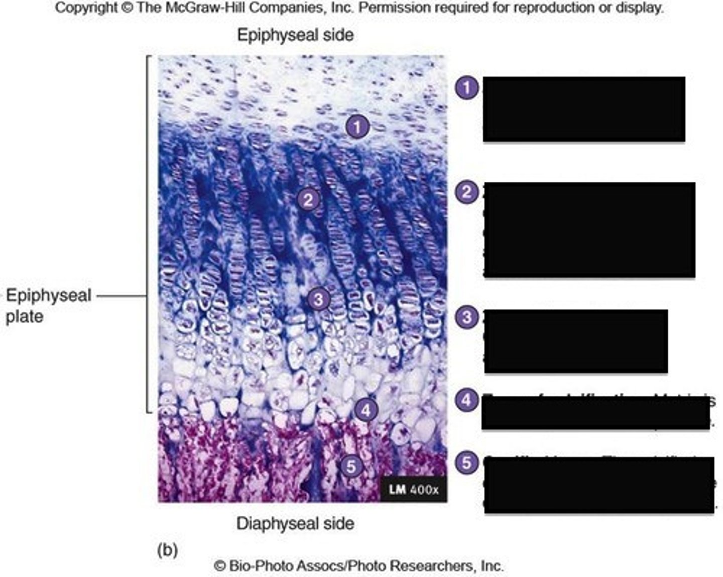
Calcification zone
surrounding cartilage matrix calcifies, chondrocytes die and deteriorate
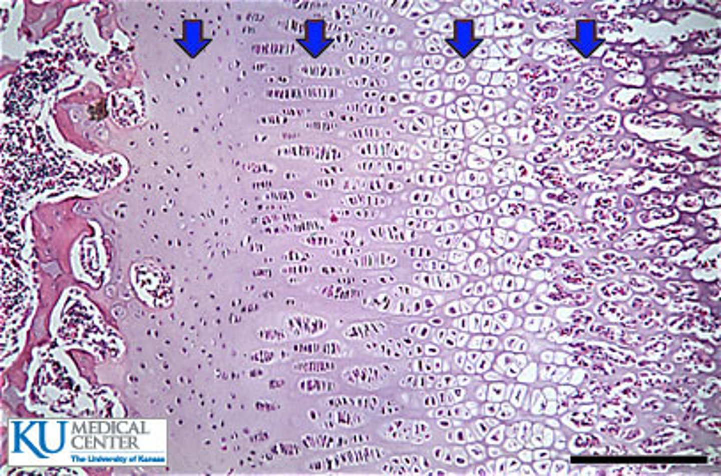
Ossification zone
zone where chondrocyte deterioration leaves long spicules of calcified cartilage at epiphysis-diaphysis junction; spicules eroded by osteoclasts; covered with new bone by osteoblasts; ultimately replaced with spongy bone
Epiphyseal plate
Near end of adolescence, chondroblasts divide less often
the __________________ thins then is replaced by bone
Epiphyseal plate closure
when ________ happens, bone lengthening ceases because requires presence of cartilage; bone of epiphysis and diaphysis fuses; females - about 18 years; males - about 21 years
Widen
in appositional growth it allows lengthening bone to ___________; usually more building up than breaking down; thicker, stronger bone but not too heavy
Thins
when the epiphyseal plate becomes a line the epiphyseal plate ________then is replaced by bone
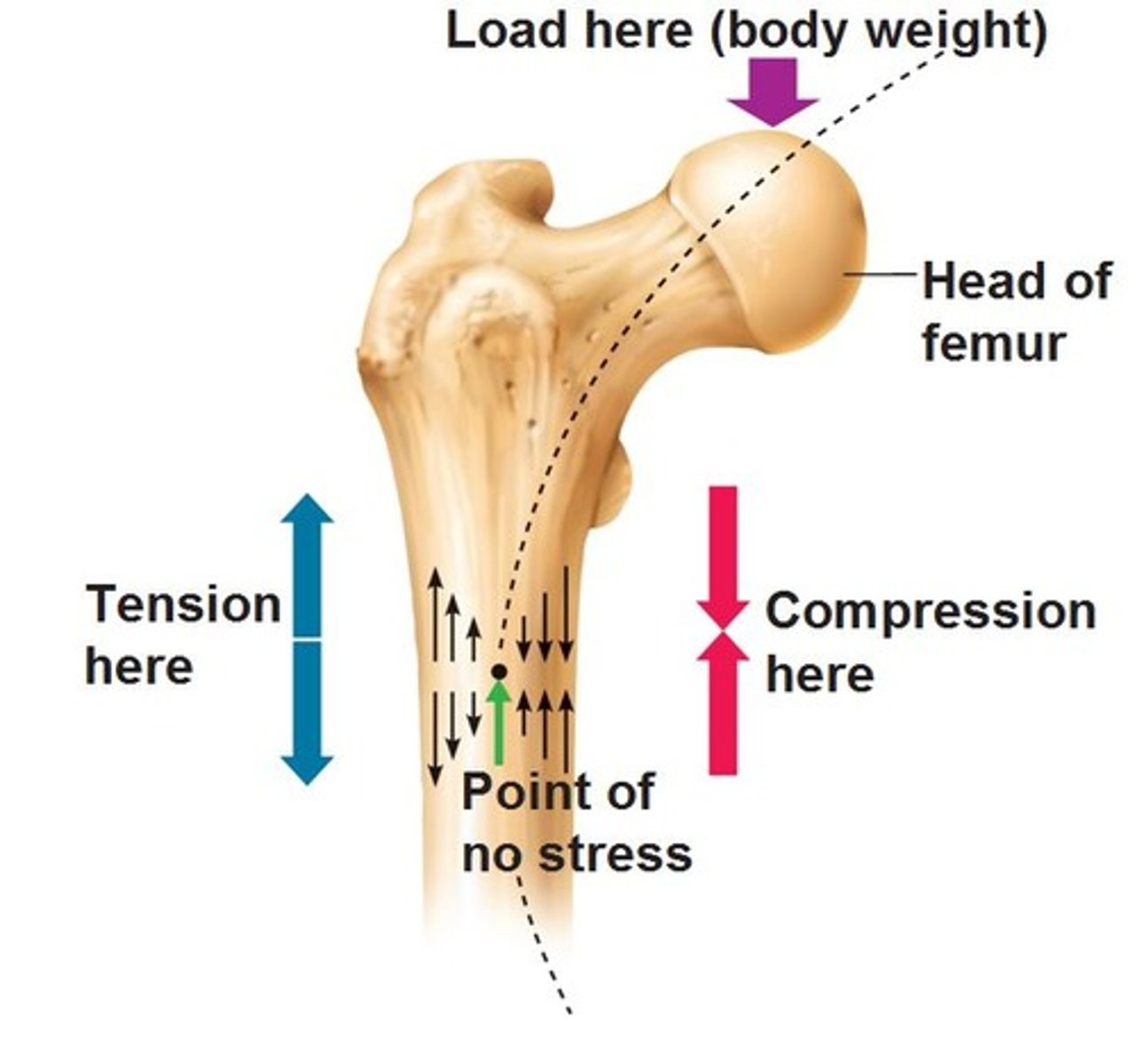
Stresses
Bones reflect ________ they encounter
How do bones respond to mechanical stress?
Bones stress when weight bears on them or muscles pull on them
Wolff's Law of Bone
Bones grow or remodel in response to demands placed on it; explains how handedness (right or left handed) results in thicker and stronger bone of that upper limb; curved bones thickest where most likely to buckle; trabeculae form trusses along lines of stress; large, bony projections occur where heavy, active muscles attach; bones of fetus and bedridden featureless
Fracture Classification
displaced, non-displaced, complete, incomplete, closed, and opened are possible fractures
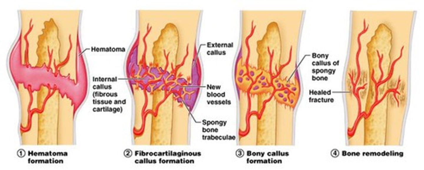
healing of a bone fracture
(1) hematoma forms (2) fibrocartilage callus forms (3) bony callus forms (4) bone remodeling
Osteomalcia
bones poorly mineralized; calcium salts not adequate; soft, weak bones but bone density may be normal; pain upon bearing weight
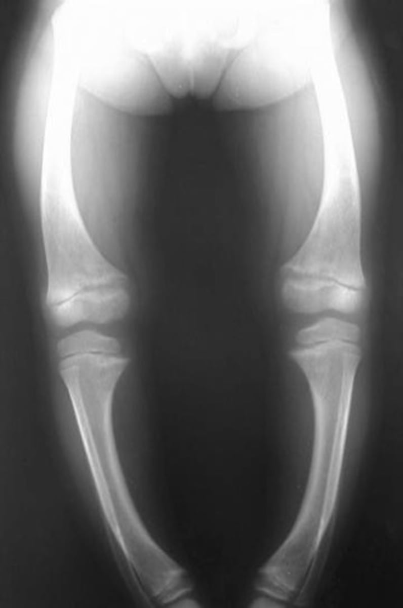
Rickets
osteomalacia of children; bowed legs and other
bone deformities; bones ends enlarged
and abnormally long; cause: vitamin D
deficiency or insufficient dietary calcium
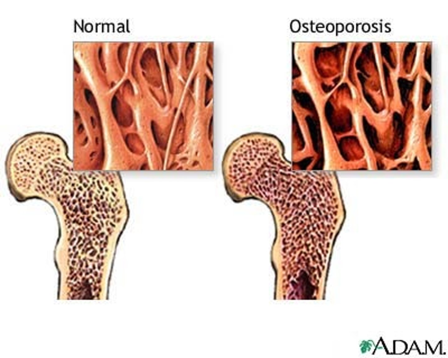
Osteoporosis
group of diseases where bone resorption outpaces deposit; spongy bone of spine and neck of femur most susceptible; vertebral and hip fractures common
Pagets disease
excessive and haphazard bone deposit and resorption; bone made fast and poorly; very high ratio of spongy to compact bone and reduced mineralization; usually in spine, pelvis, femur, and skull; rarely occurs before age 40; cause unknown - possibly viral
Yellow bone marrow
contains stored fat
Red bone marrow
found in cancellous bone; site of hematopoiesis