Chapter 20: Herat Part 2
 Call Kai
 Learn
Learn Practice Test
 Spaced Repetition
 Match
Match Flashcards
 Knowt Play
1/32

Earn XP
Description and Tags
Gibrous skeleton of heart, heart valve, cardiac conduction system, cardiac cycle, heart sounds
Name | Mastery | Learn | Test | Matching | Spaced | Call with Kai |
|---|
No analytics yet
Send a link to your students to track their progress
33 Terms
Fibrous skeleton of the Heart
dense CT 4 rings sorround valves
structural foundations for valves (anchor valve with dense CT), insertion for myocardia, electrically insulates atria and ventricles separately
pump 2 step process
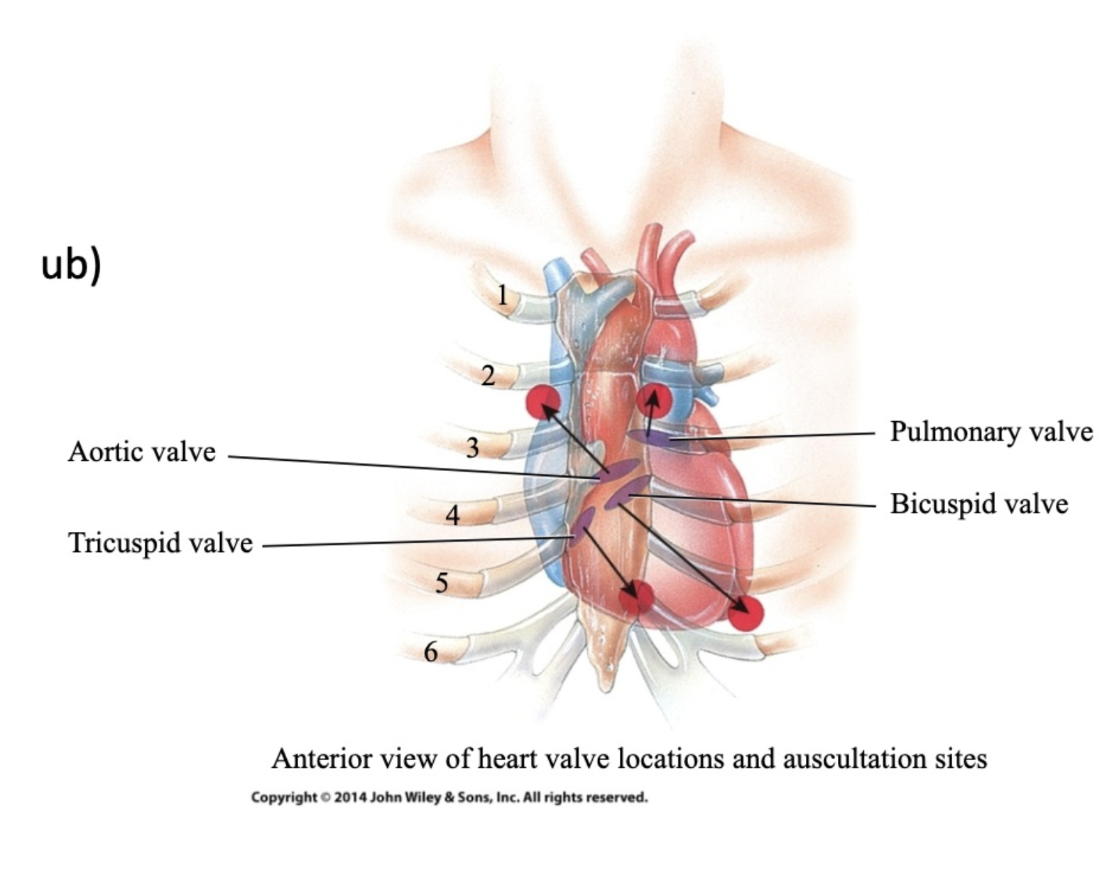
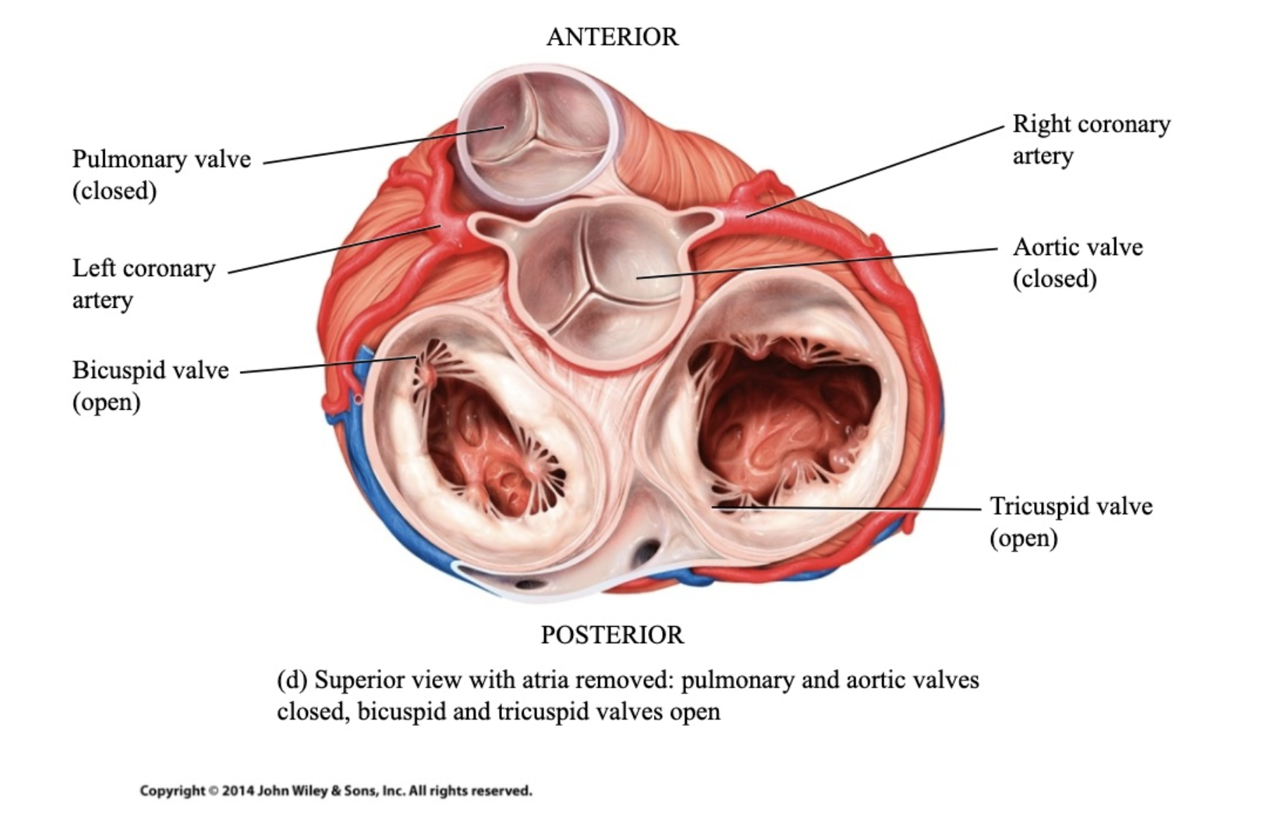
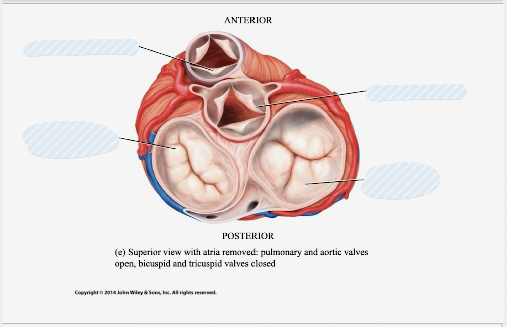
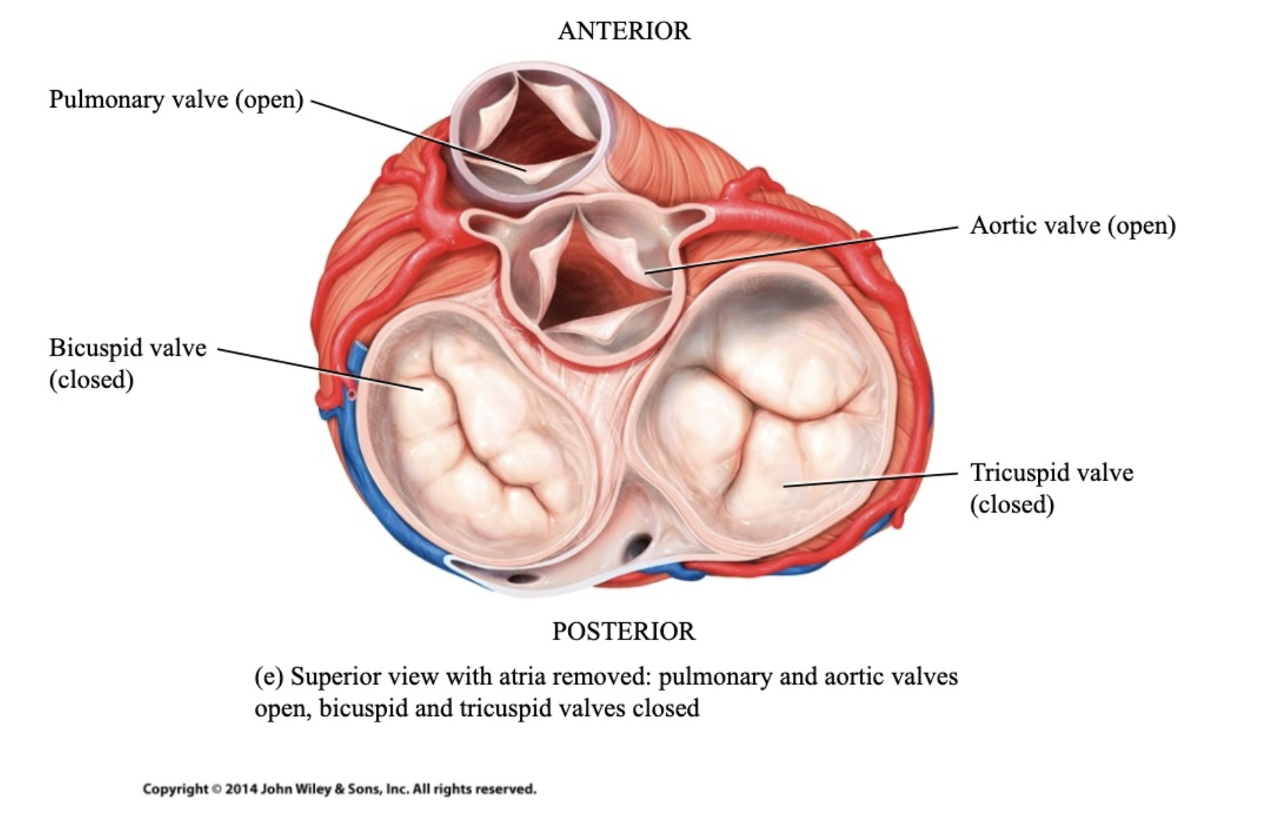
Av valves
in arota
When open, cusps project into ventricles
blood flow from atrium into ventricle
Blood moves from atria to ventricles
valve open when Atrial pressure > ventricular pressure
When closed, edges of cusps meet and create a barrier
so blood doesnt flow backwards
Chordae tendineae prevent cusps from opening backwards into atria
valves closes: Atrial pressure < ventricular pressure
Semilunar Valves
in pulmonary vlave
3 crescent moon cusps
made og thicker CT
Open for blood to pass from ventricles into arteries
valve open when Ventricular pressure > arterial pressure
force cusps apart allowing blood to be ejected from ventricle to artery
close when arterial pressure > Ventricular pressure
Autorhythmic cells
repeatedly and rhythmically generate action potentials
doesnt need a stimulus
Pacemaker
sets rhythm of the heart
What are the cardiac muscle cells connected by in cardiac conjunction?
Gap junction
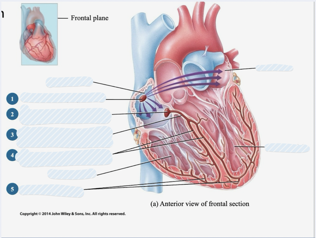
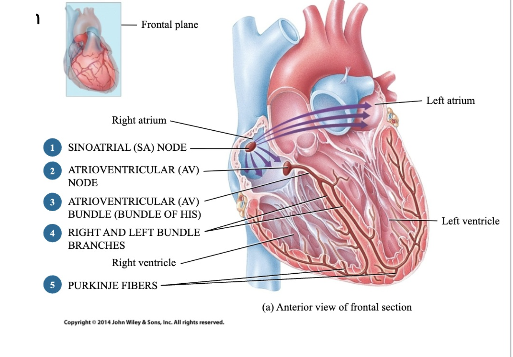
cardiac conjection order and what they do
SINOATRIAL (SA) NODE - in charge of generating AP is a pacemaker cell
ATRIOVENTRICULAR (AV) NODE - helps transmit impulse to contract from atrium to ventricles
ATRIOVENTRICULAR (AV) BUNDLE (BUNDLE OF HIS) - only place electrical actviity can cross atrium to ventricle
RIGHT AND LEFT BUNDLE BRANCHES
PURKINJE FIBERS - activate cardiac muscle of ventricles to initiate ventricular contraction
why is it important that there is a delay between the contraction of the atria and contraction of ventricles?
If no dely dont expect blood to nove if all 4 chambers contract at the same time. delay is necessary to give blood somehwere to go when chambers contract. The ventricles have to be relaxed to recieve blood when atria is contracting.
What initiates the contraction of the heart?
SA node
What transmits AP across the fibrous skeleton after short delay?
AV node
What carries AP to the apex of the heart?
His/ AV bundle and bundle branches
What delivers action potetnial to ventricular cardiac muscle cells
Purkenjie fibers
Membrane potential
difference in electrical potential (mV) on either side of the membrane
every cell has it but cant maniupulate it
muscle and neurons can alter MP in response to stimulus what makes them excitable
Resting membrane potential
difference of inside and outside of an unstimulated cell
Negative charges in cytosol, positive charges in ECM = polarized
inside of cell more negative than outside (polarized)
neg pole = inside
pos pole = outside
Depolarization
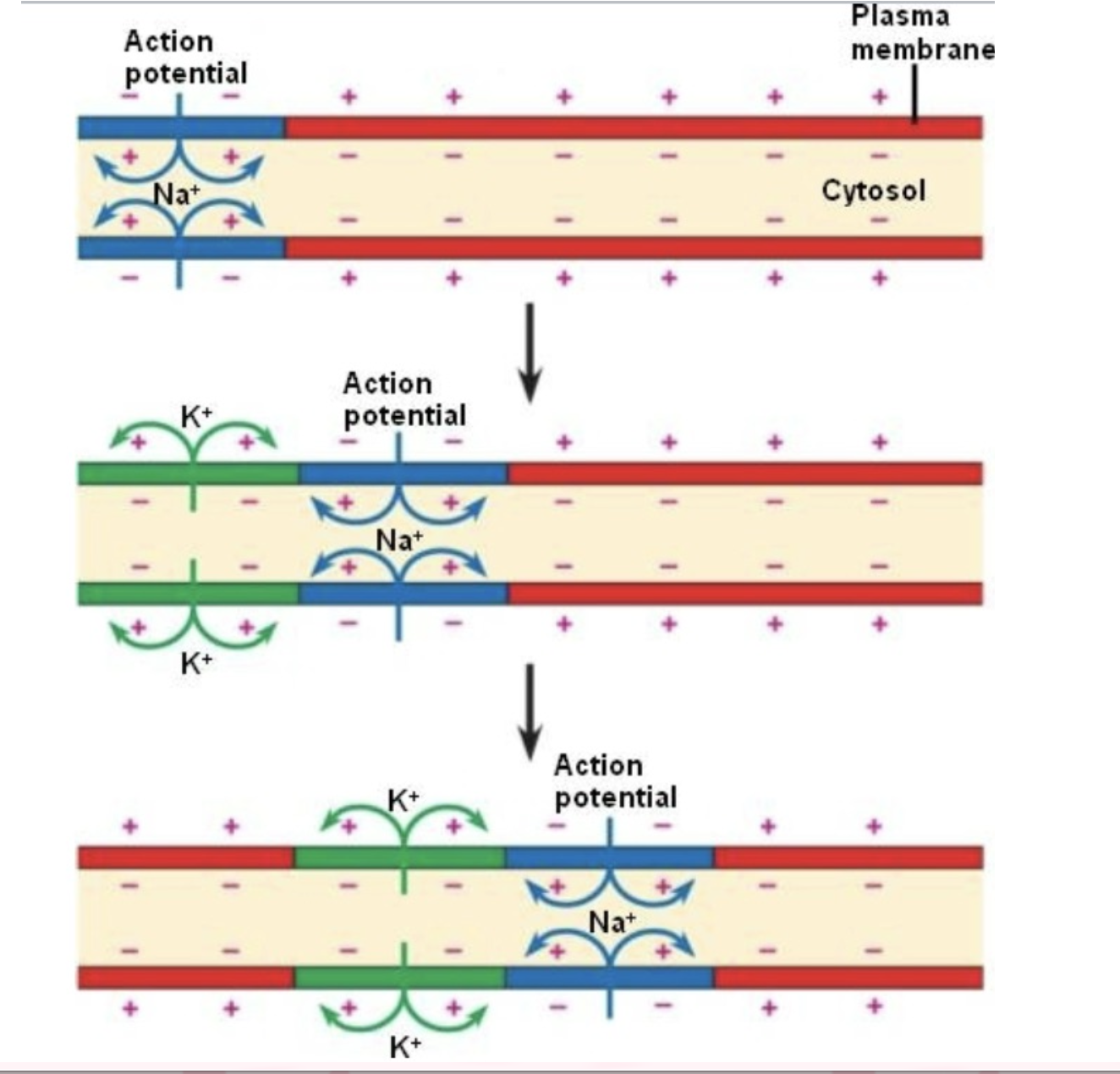
decrease in difference between outside and inside cell (let sodium in)
in excitable cells if stimulated can respond to that by depolarizing which is a decrease in polarization between inside and outside of a cell
example is letting more positive enter the cell which will reduce the difference less polarized
have to go to Resting can't stay depolarized forever
let positive into depolarize to repolarize take positive out of the cel
Repolarization
returning membrane to resting membrane potential after a disturbance
let potassium out
Action potential
rapid depolarization and repolarization
Happens in one discrete part of membrane
influx of + ion charge (sodium) causes adjacent mebrane to depoloarize
depolarize happens in 1 part of membrane depolarizing and AP then cause next part of membrane to have it
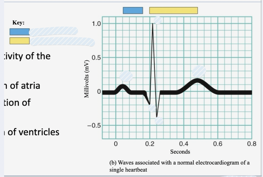
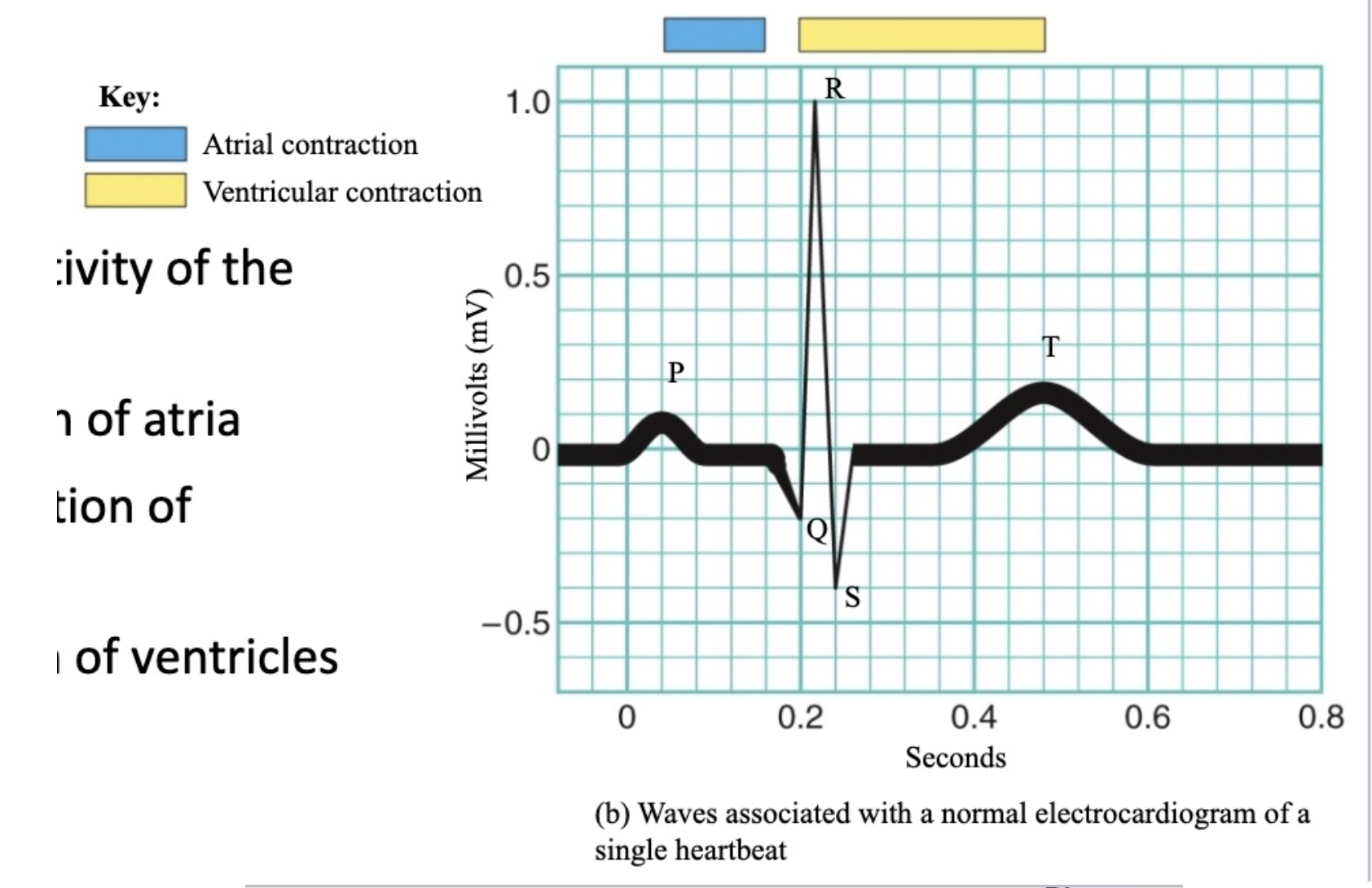
ECG
tells us the pattern of electrical activity of the heart
3 waves of ECG
P wave – depolarization of atria
QRS wave – depolarization of ventricles
stringer signal because lots of cardiac muscle in ventricles
T wave – repolarization of ventricles
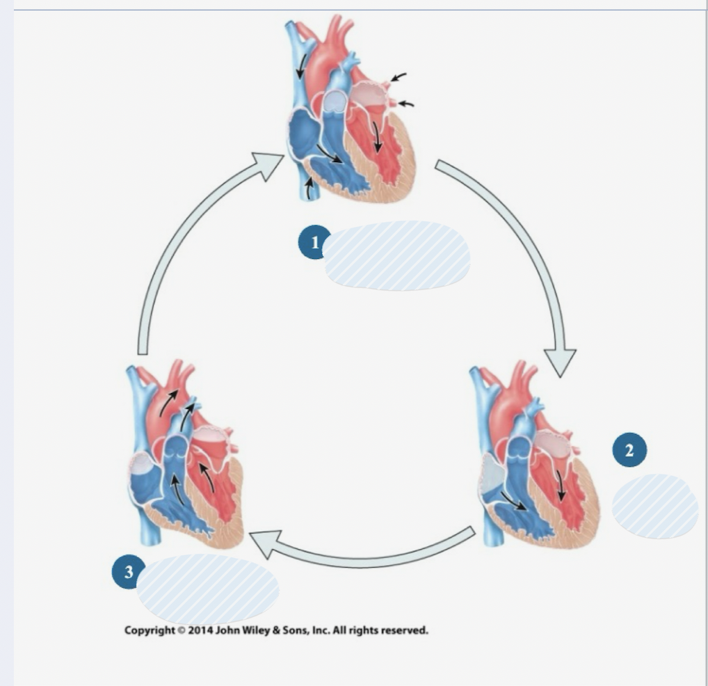
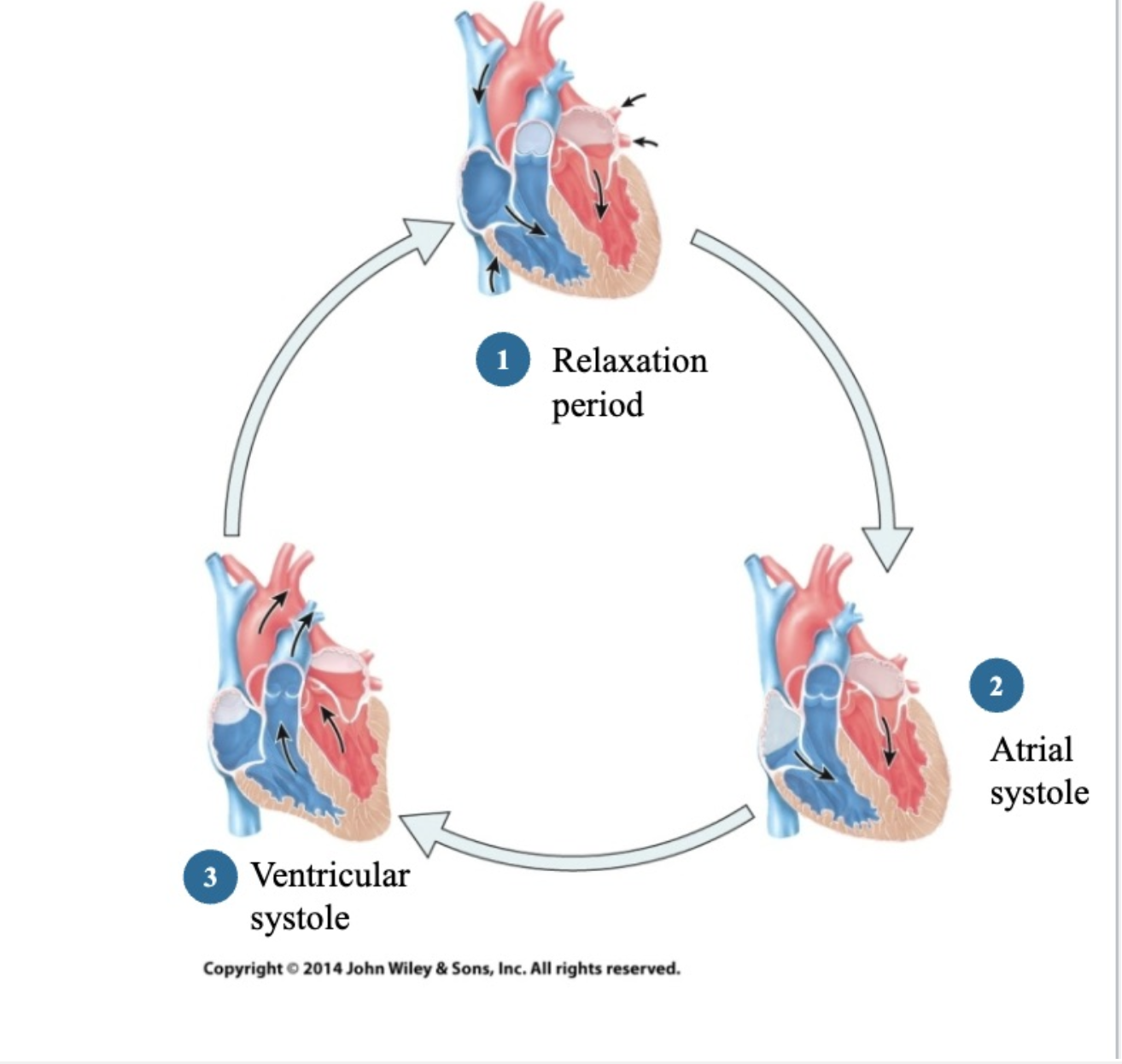
Cardiac Cycle steps
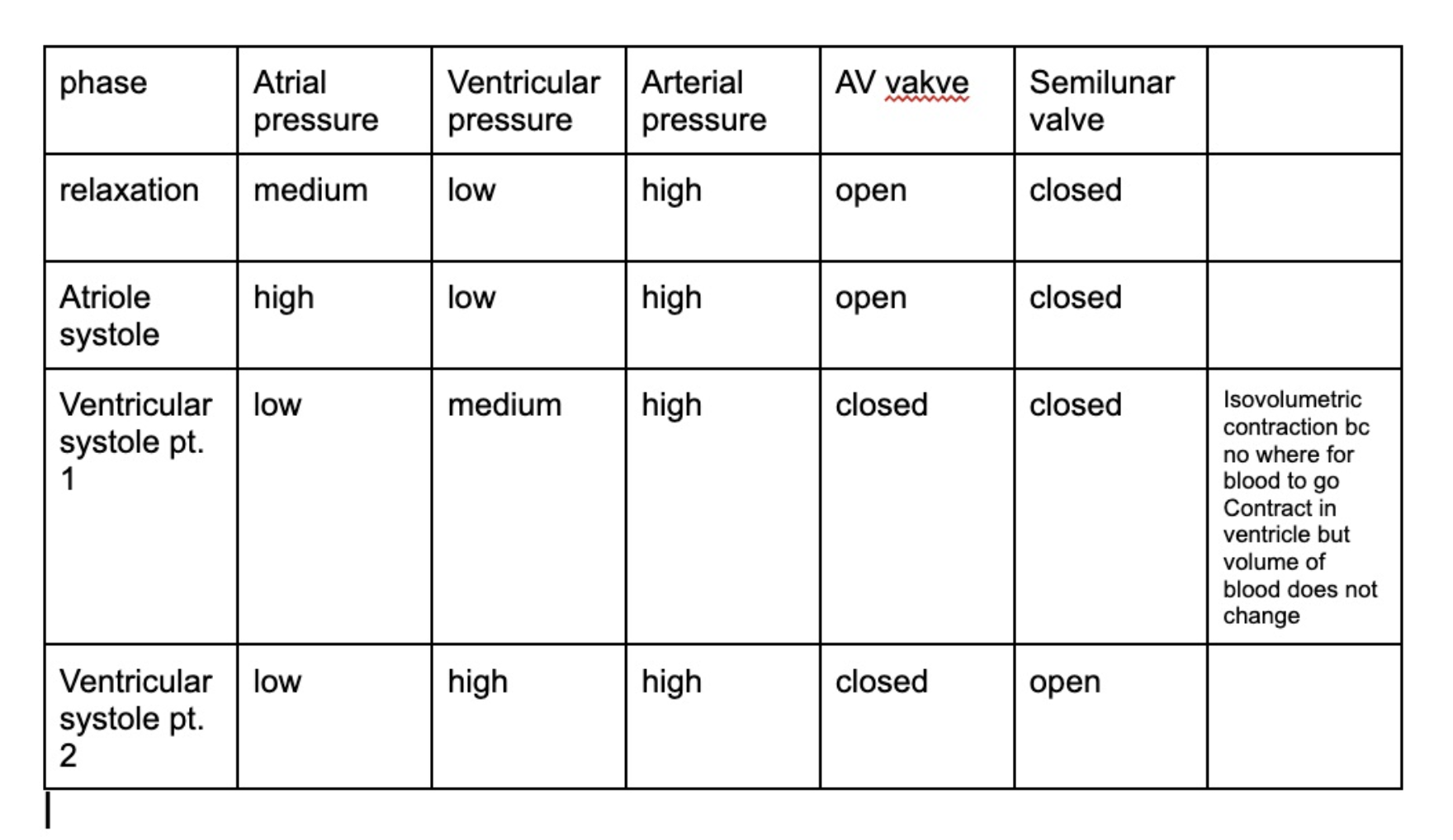
relaxation period
atrial systole
ventricular systole
Relaxation period
All 4 chambers in diastole – relaxation
Atrial pressure > ventricular pressure < arterial pressure
Atrial systole
Atria contract
Atrial pressure > ventricular pressure < arterial pressure
Ventricular systole
Ventricles contract
1st part of ventricular systole:
Atrial pressure < ventricular pressure < arterial pressure – all valves closed
2nd part of ventricular systole:
Atrial pressure < ventricular pressure > arterial pressure – semilunar valves open, blood is ejected
How many heart sounds
4 sounds caused by turbulence of blood
Sounds
S1 – AV valves closing (lub)
S2 – Semilunar valves closing (dub)
S3 – ventricular filling in relaxation (not heard)
S4 – atrial systole (not heard)