Anatomy test 2 - Axial Skeleton GVSU
1/85
Earn XP
Description and Tags
BMS 208
Name | Mastery | Learn | Test | Matching | Spaced | Call with Kai |
|---|
No analytics yet
Send a link to your students to track their progress
86 Terms
Orbit
a bony passageway in the skull that allows for the passage of nerves and blood vessels to and from the eye, connecting the orbit to the cranial cavity.
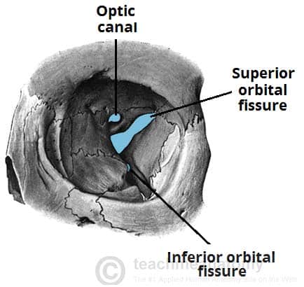
superior orbital fissure
a slit-like opening located in the posterior part of the orbit that serves as a pathway for cranial nerves and blood vessels entering or exiting the orbit.

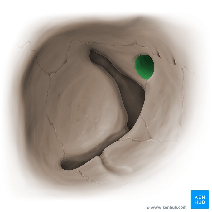
Inferior orbital fissure
a gap in the floor of the orbit that connects the orbit with the pterygopalatine fossa and the maxillary sinus, allowing the passage of nerves and blood vessels.

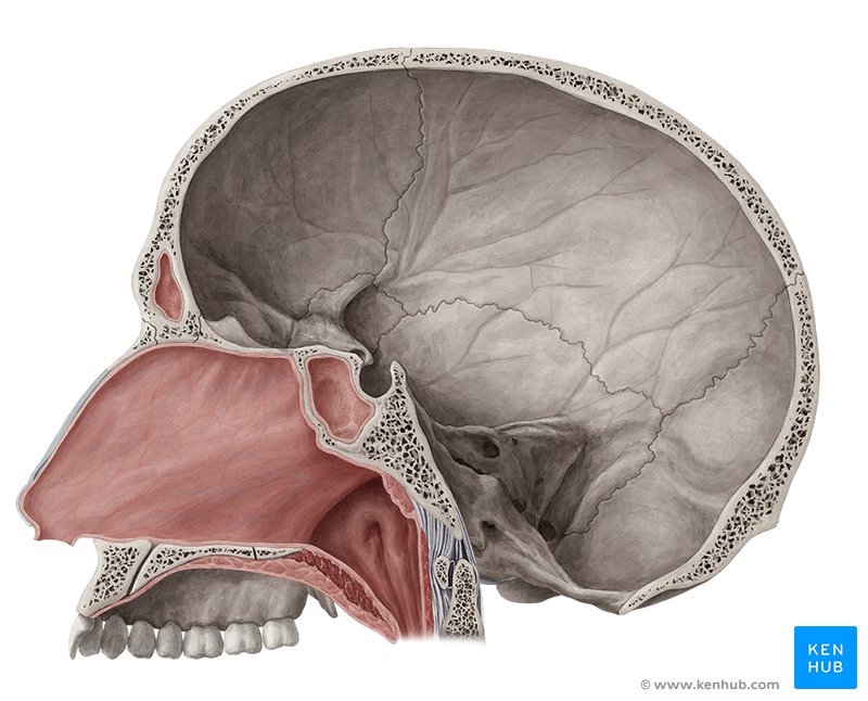
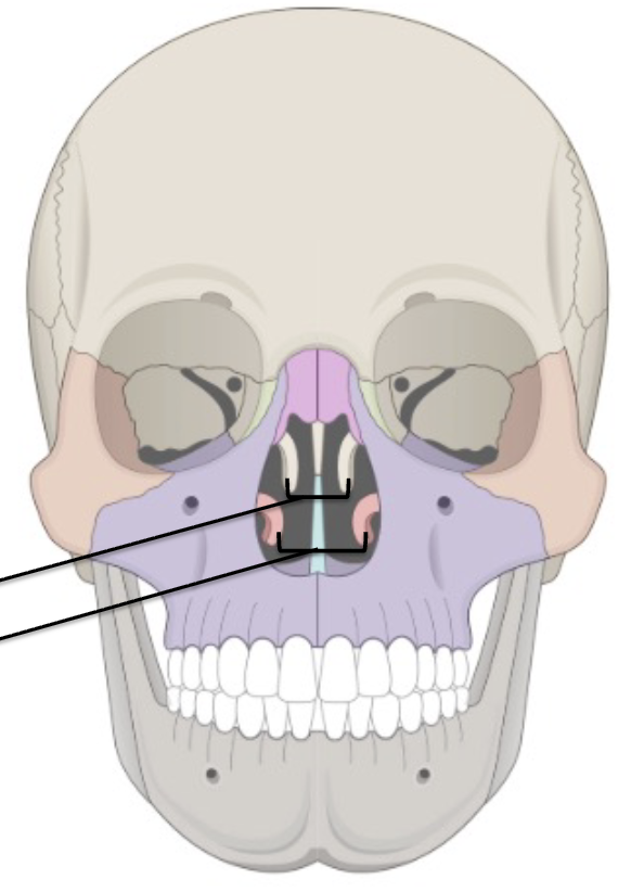
nasal cavity
the space behind the nose that is lined with mucous membrane, allowing for the passage of air and filtration of particles before reaching the lungs.
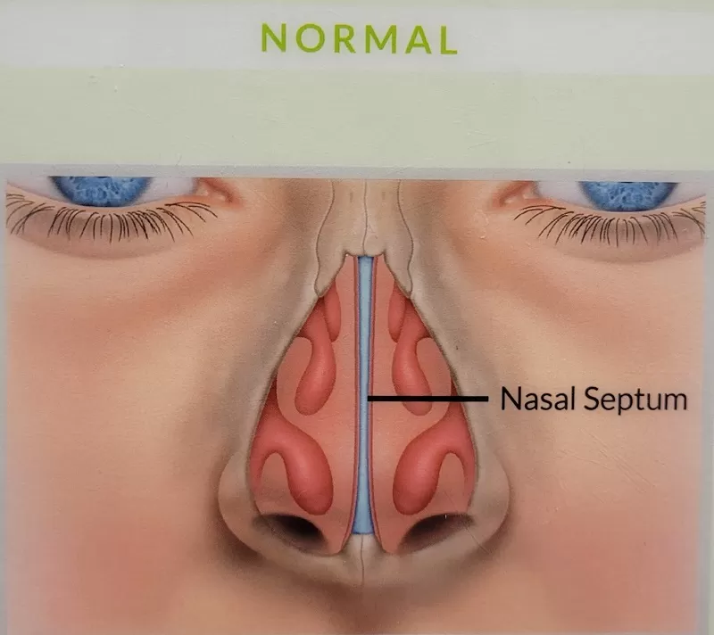
nasal septum
wall of cartilage and bone that divides the nasal cavity into two halfs
oral cavity
the hollow space within the mouth that contains the teeth, gums, tongue, and the opening of the salivary glands, playing a crucial role in digestion and speech
zygomatic arch
the bony arch that forms the prominence of the cheek, made up of the zygomatic bone and part of the temporal bone, serving as an attachment point for facial muscles and contributing to the structure of the skull.

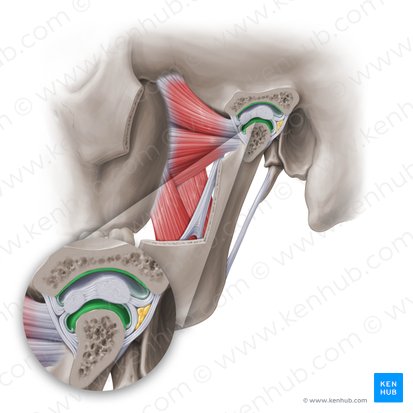
Temporomandibular joint
the joint that connects the jawbone (mandible) to the skull (temporal bone), allowing for movement of the jaw during activities such as chewing and speaking
Hard palate
the bony front part of the roof of the mouth, separating the oral cavity from the nasal cavity, playing a role in speech and the eating process.
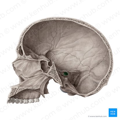
Jugular foramen
an opening in the skull located at the base of the skull that allows for the passage of the internal jugular vein and cranial nerves, playing a crucial role in the venous drainage of the brain.
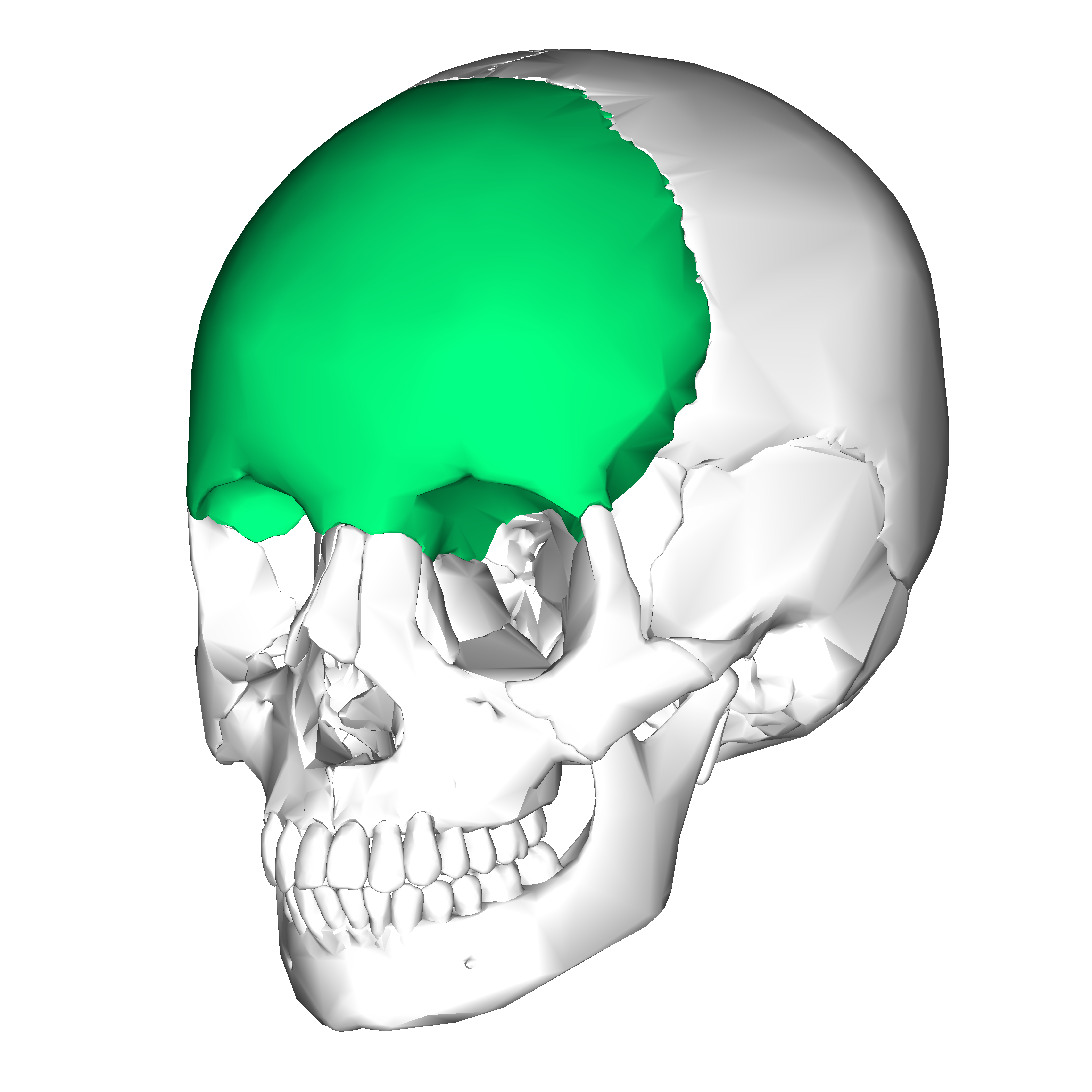
Frontal bone
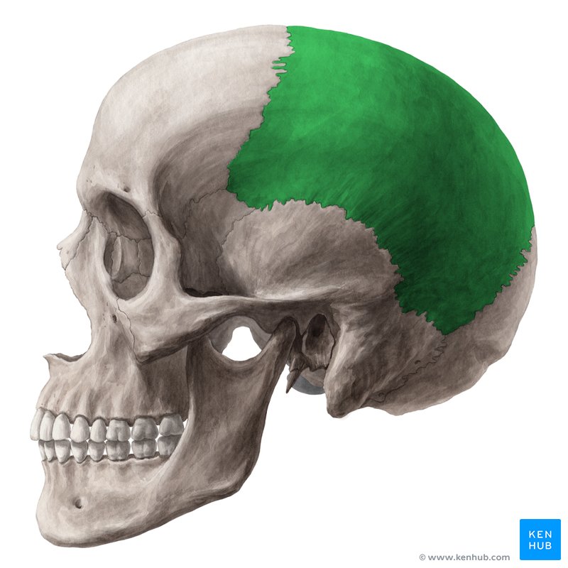
Parietal
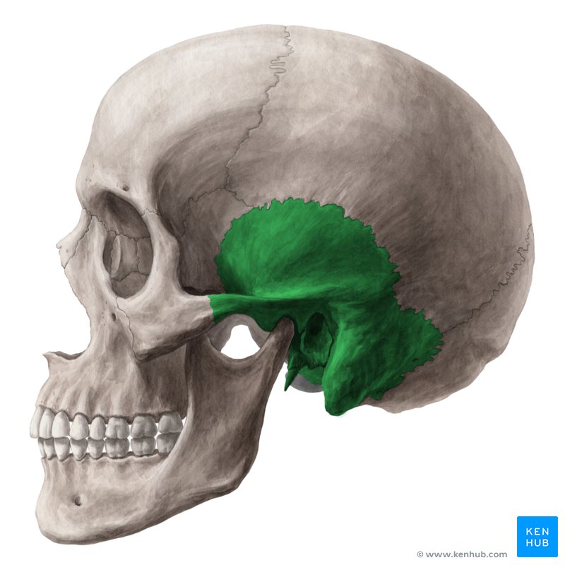
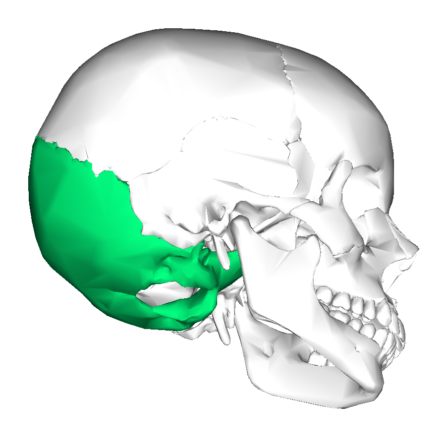
Temporal
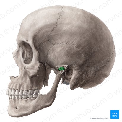
External acoustic (auditory) meatus
an S-shaped tube conducting sound waves from the outer ear (auricle) to the tympanic membrane (eardrum)
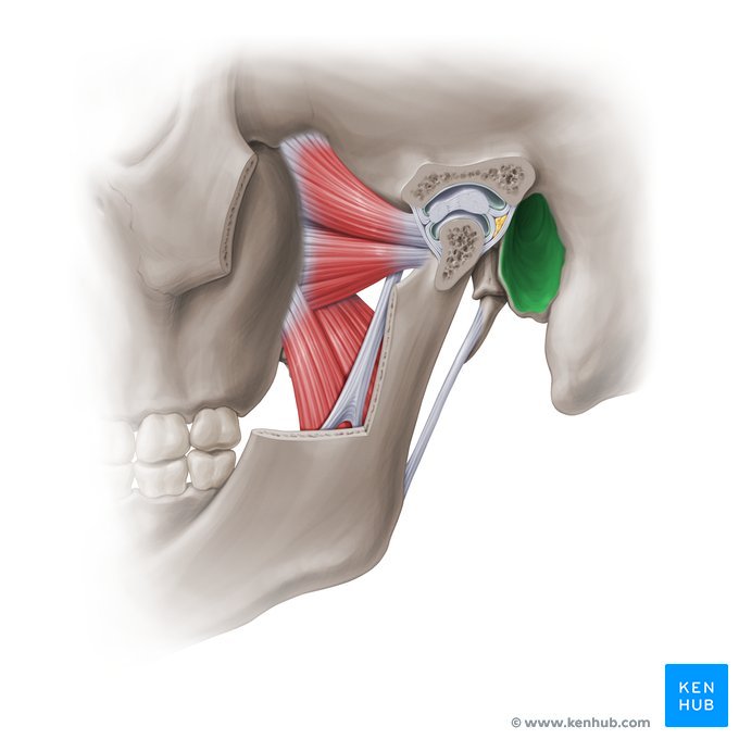
Mandibular fossa
a deep, oval-shaped depression in the squamous part of the temporal bone of the skull, located just in front of the external acoustic meatus
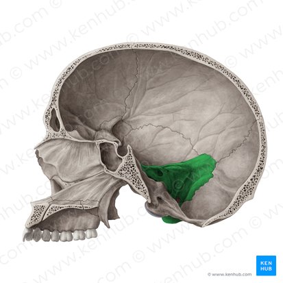
Petrous portion
dense, pyramid-shaped part of the temporal bone located at the base of the skull between the sphenoid and occipital bones
Internal acoustic (auditory) meatus
bony canal within the petrous portion of the temporal bone, acting as a passageway between the posterior cranial fossa and the inner ear
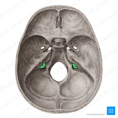
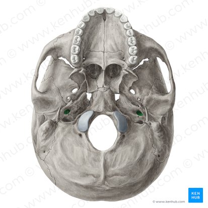
Carotid canal
a bony, L-shaped passage located within the petrous part of the temporal bone at the base of the skull. It allows the internal carotid artery and the sympathetic plexus to travel from the neck into the middle cranial fossa, providing a protected pathway for blood supply to the brain
Ear ossicles
three tiny bones in the middle ear that form a chain to transmit and amplify sound vibrations from the eardrum to the inner ear
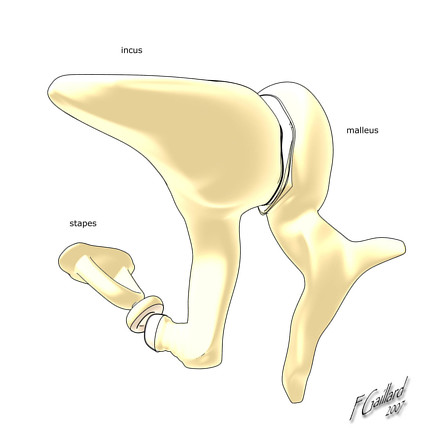
Malleus
the outermost and largest of the three small bones (ossicles) in the mammalian middle ear, connected to the eardrum and the incus

Incus
a tiny anvil-shaped bone (ossicle) in the middle ear of mammals

Stapes
the smallest bone in the human body, located in the middle ear

Occipital
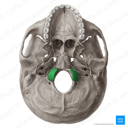
Occipital condyles
paired, oval-shaped bony protuberances on the inferior surface of the occipital bone at the base of the skull, located on either side of the foramen magnum
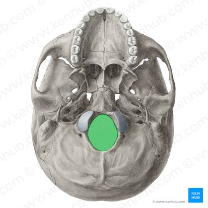
Foramen magnum
Ethmoid
Olfactory foramina
Superior and middle nasal conchae
thin, curved bony plates projecting from the ethmoid bone into the lateral walls of the nasal cavity
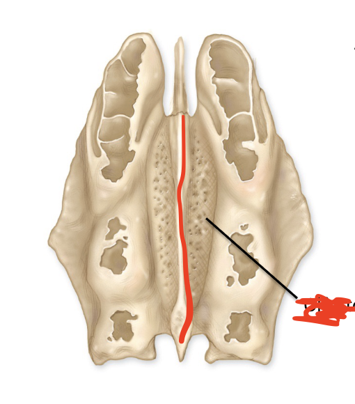
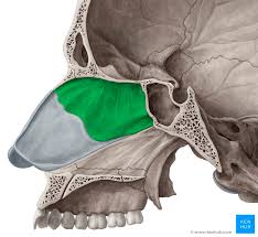
Perpendicular plate
a thin, flat, vertical bone located in the skull's median plane, forming the upper, posterior part of the nasal septum
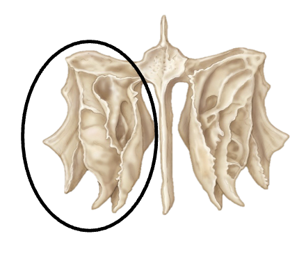
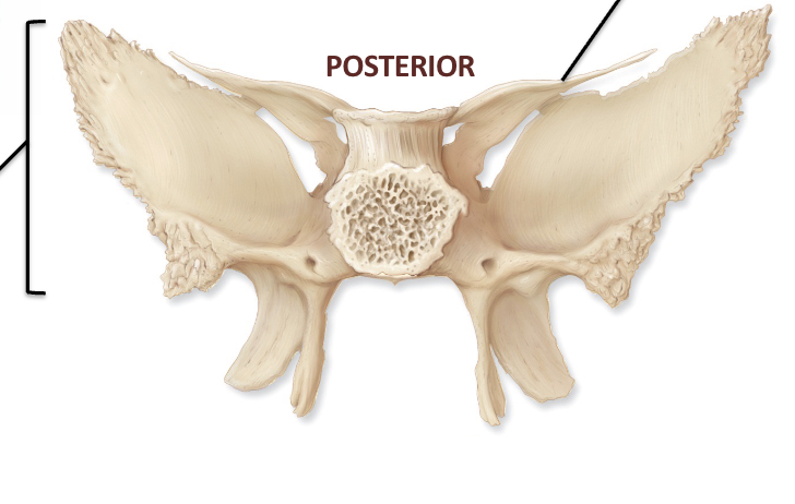
Sphenoid
a complex, butterfly-shaped unpaired bone situated at the base of the skull, acting as a "keystone" that connects the neurocranium to the facial skeleton
Greater wing

lesser wing
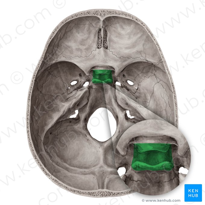
Sella turcica
bony depression located in the sphenoid bone at the base of the skull
Optic canal
funnel-shaped bony passage located in the lesser wing of the sphenoid bone at the orbital apex
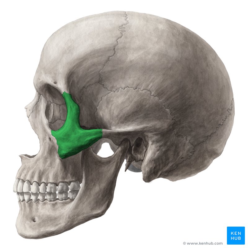
Zygomatic
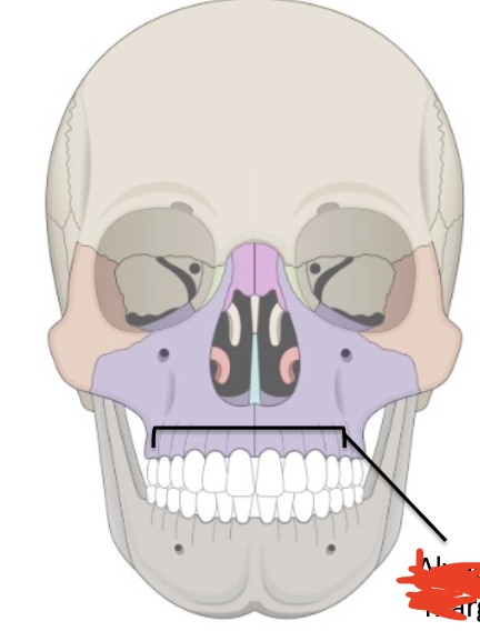
Maxilla
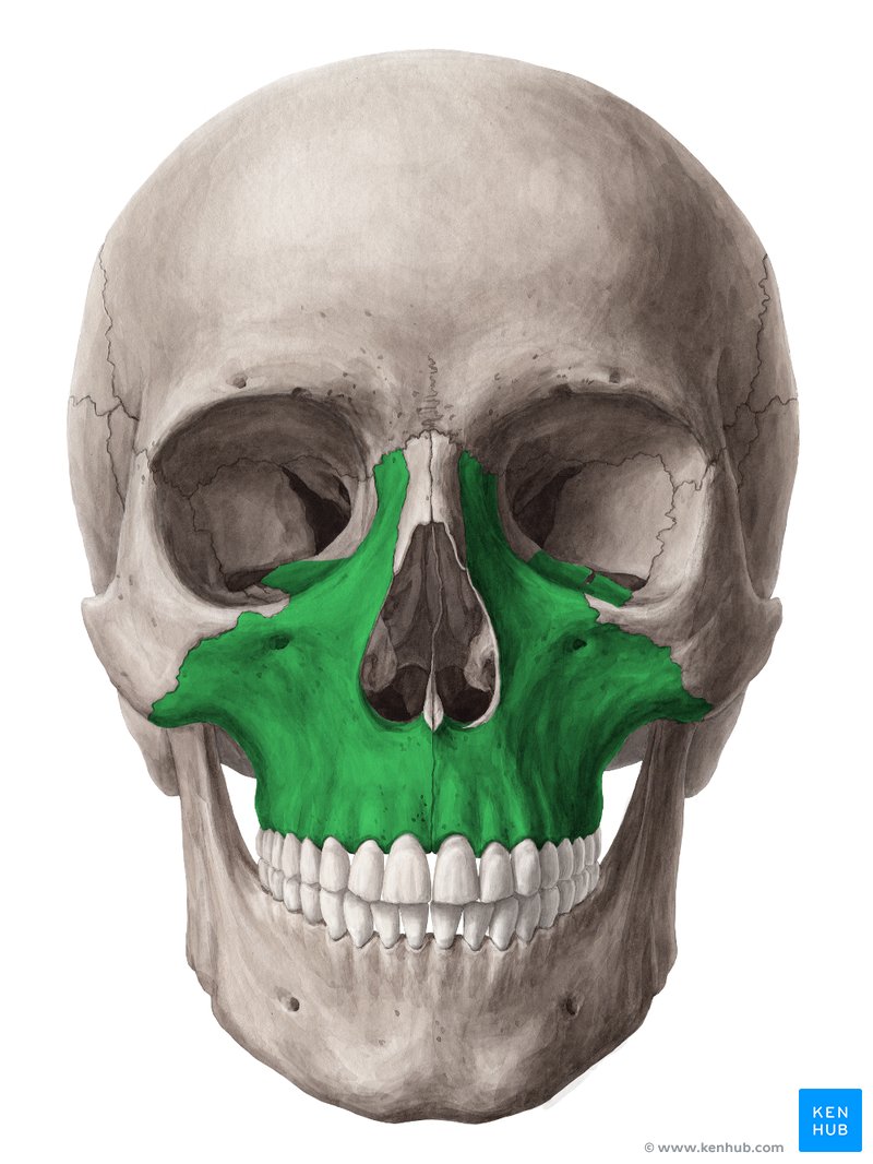
Alveolar margin
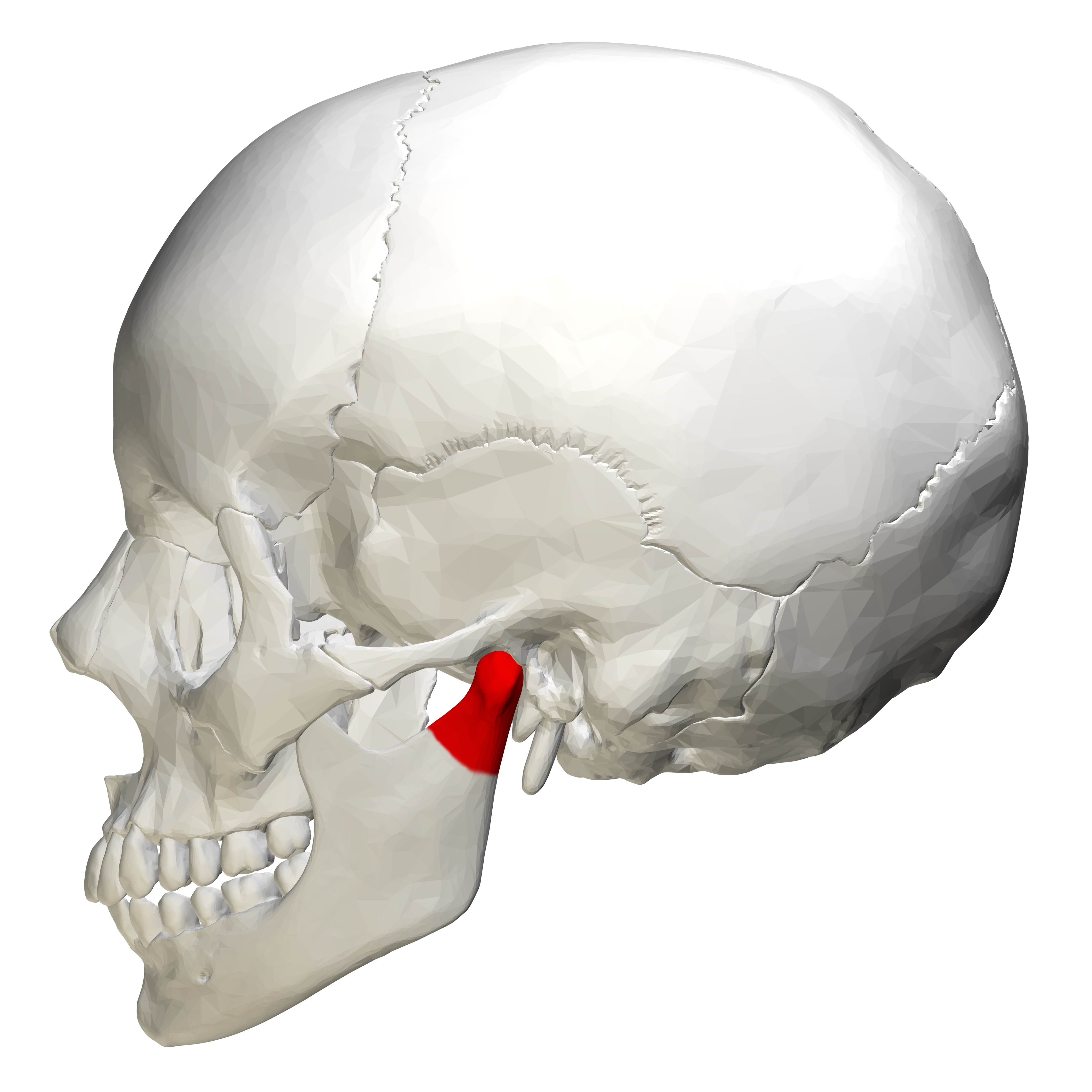
Mandible
mandibular condyle
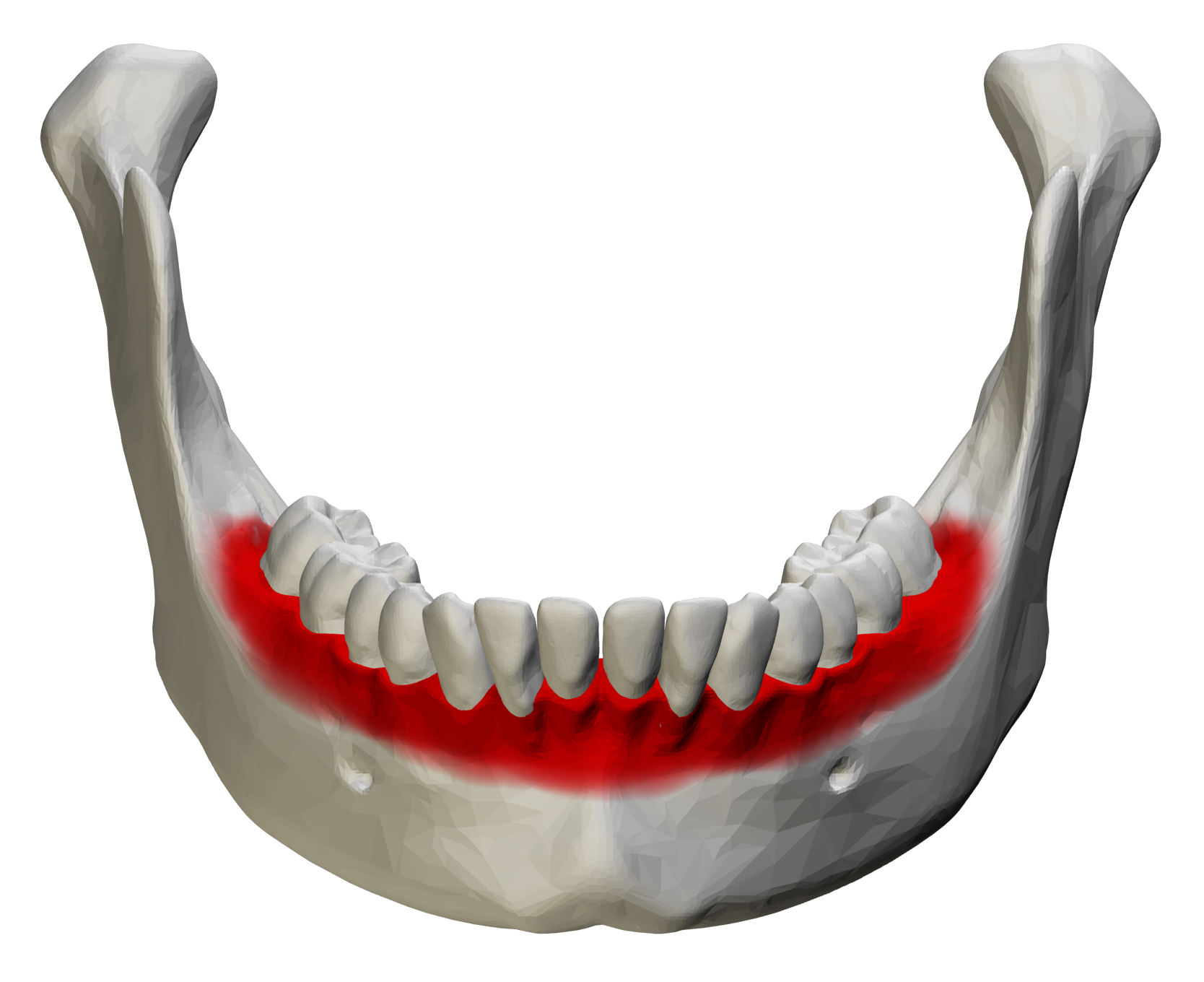
Alveolar margin
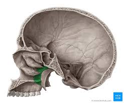
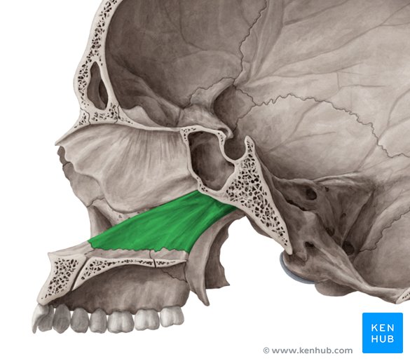
Palatine
pair of L-shaped bones located at the back of the nasal cavity, crucial for forming the posterior hard palate, nasal floor, and part of the eye socket
Vomer
a small, thin, plow-shaped unpaired facial bone located in the midsagittal plane of the skull
Inferior nasal concha
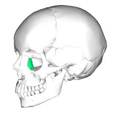
Lacrimal
the structures associated with producing and draining tears
Nasal
nose
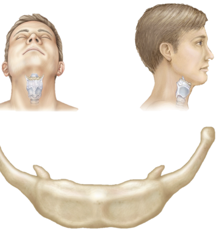
Hyoid
a small, U-shaped "floating" bone in the front of the neck between the chin and thyroid cartilage, crucial for anchoring tongue, larynx, and throat muscles
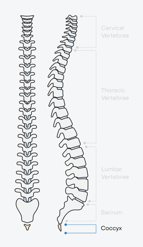
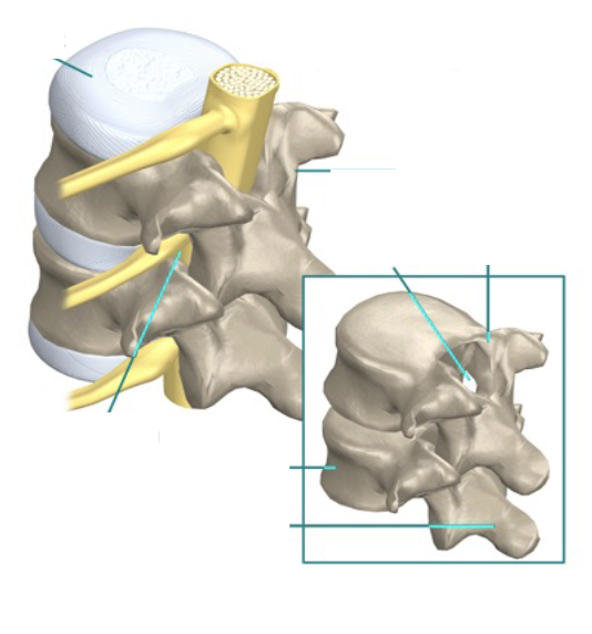
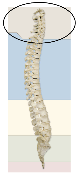
vertebral column
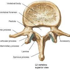
Body (centrum)
the thick, cylindrical, anterior (ventral) portion of a vertebra, serving as the primary, weight-bearing, solid component
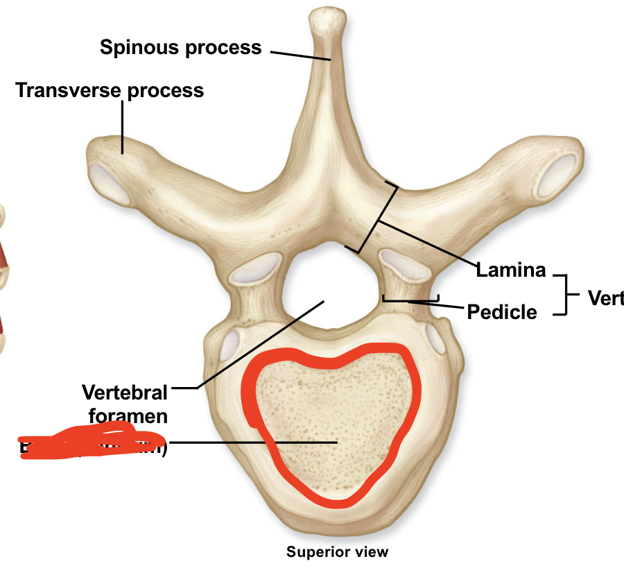
Vertebral (neural) arch
the posterior, bony structure of a vertebra that, along with the vertebral body, forms the vertebral foramen to protect the spinal cord

Pedicle
a small stalk-like structure that supports, connects, or anchors an organ, tissue, or body part

Lamina
two flat, arched plates of bone that form the posterior (back) roof of the vertebral arch

Spinous process
Transverse process
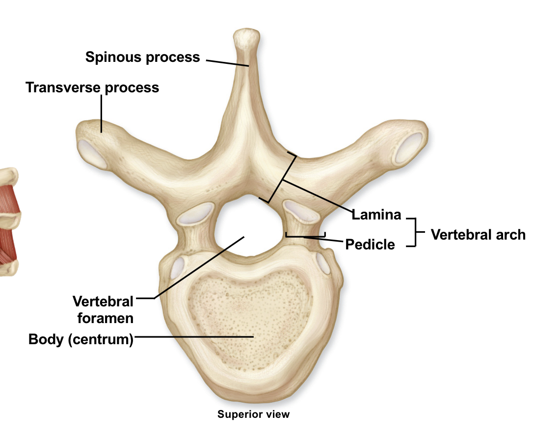
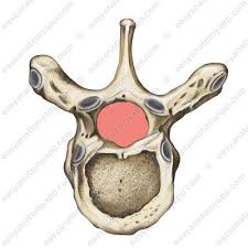
Vertebral foramen
Vertebral canal
an elongated, anatomically sterile, bony tunnel formed by the stacked vertebral foramina of the spine, extending from the skull's foramen magnum down to the sacrum
Superior articular process (purple)
a paired bony projection on the upper surface of a vertebra that extends upward to form a facet joint
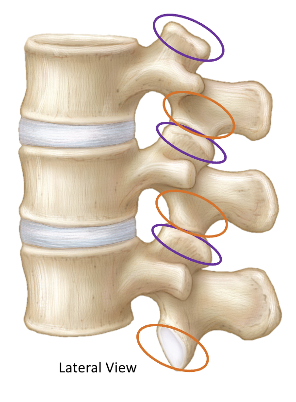
Inferior articular process (orange)
a paired bony projection on the lower side of a vertebra, extending downward from the junction of the lamina and pedicle

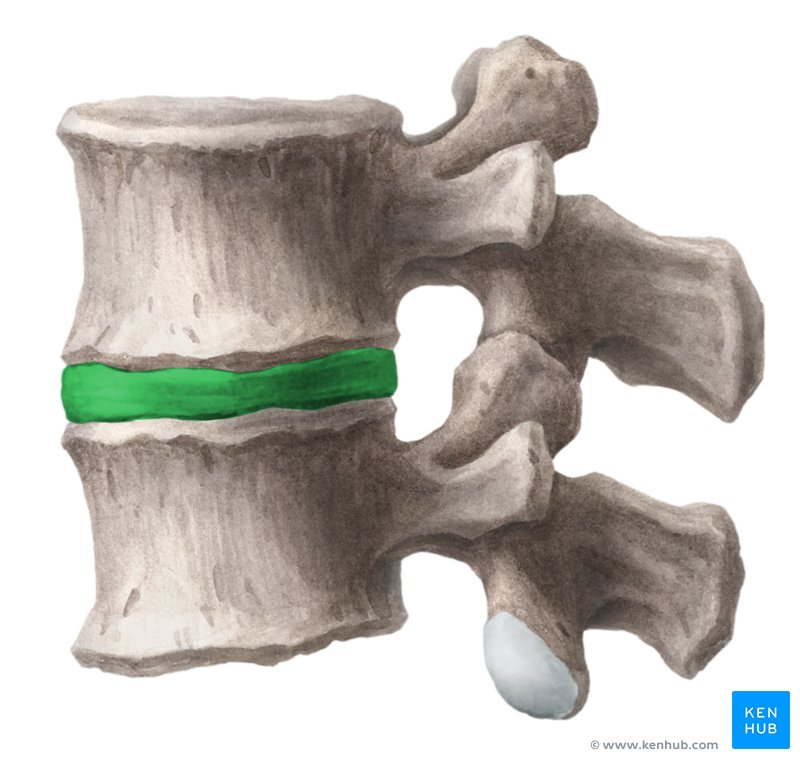
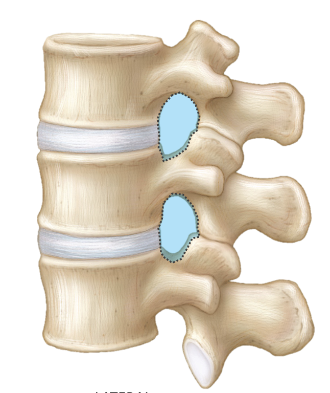
Intervertebral disc
fibrocartilaginous, shock-absorbing cushions located between the bony vertebrae of the spinal column
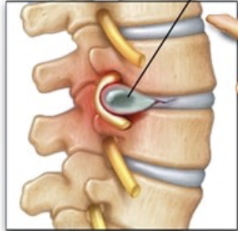
Herniated (“slipped”) disc
occurs when the soft, jelly-like center of a spinal disc pushes through a tear in its tough, outer layer. This material presses on nearby spinal nerves, causing pain, numbness, or weakness in the back, neck, or limbs
Intervertebral foramen
a lateral opening or "doorway" between two adjacent vertebrae, located on both sides of the spinal column, which acts as a passageway for spinal nerves and blood vessels to exit the spinal cord
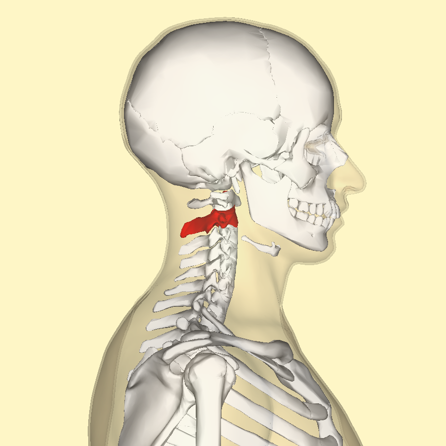
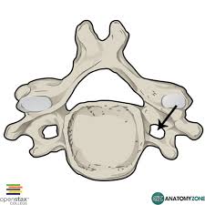
Cervical vertebrae
the seven stacked bones (C1–C7) forming the bony column in the neck, extending from the base of the skull to the top of the thoracic spine
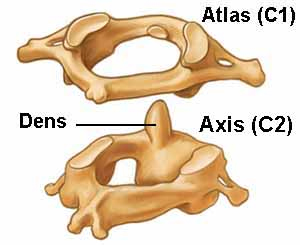
Atlas
the topmost vertebra in the spine, located directly below the skull, responsible for supporting the head's weight and enabling nodding and tilting motions
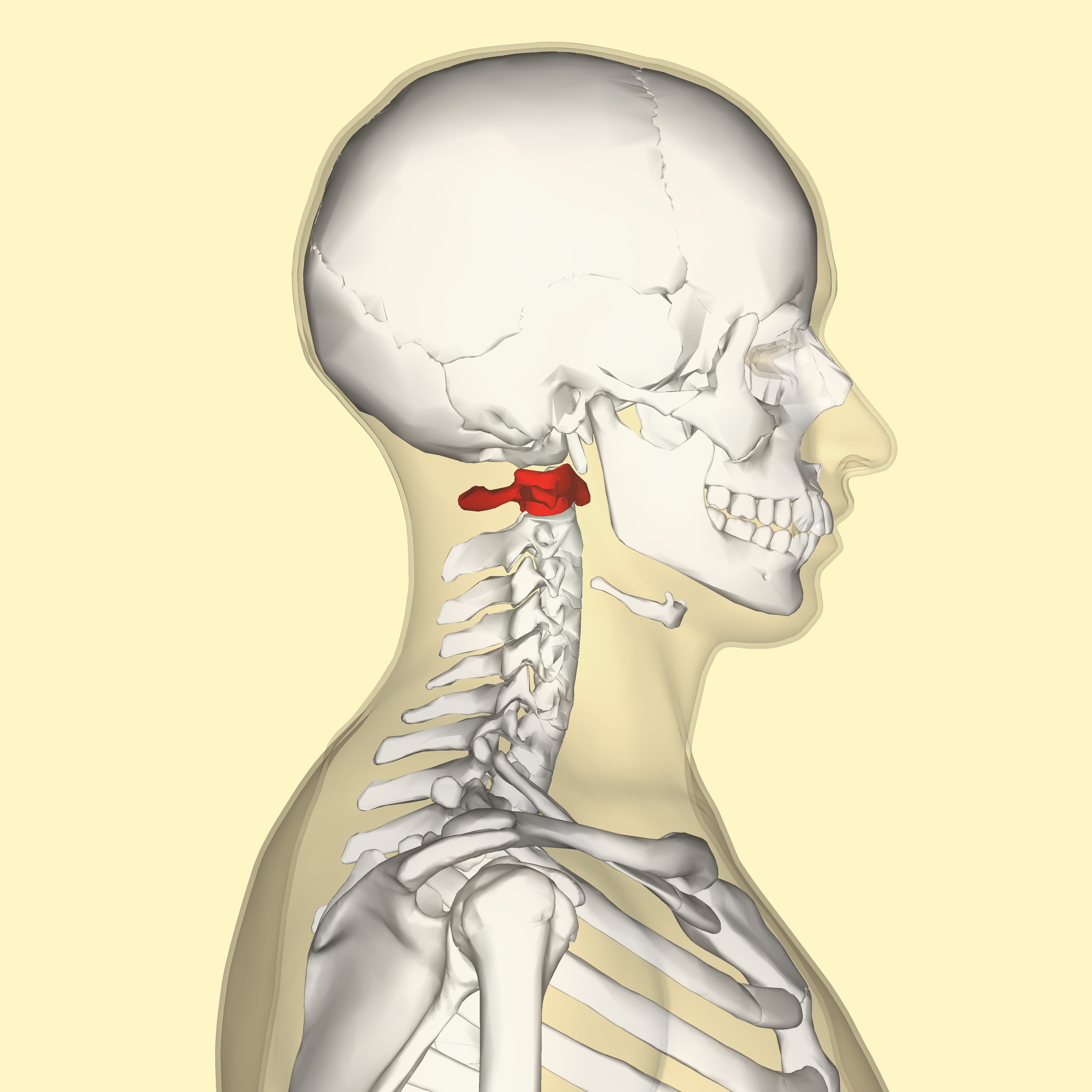
Axis
the second cervical vertebra of the spine, located directly below the atlas (C1) and above C3
Dens (odontoid process)
a peg-like bony projection extending upward from the second cervical vertebra (C2 or axis) into the ring of the first cervical vertebra (C1 or atlas). It acts as a pivot point for the atlanto-axial joint, allowing significant neck rotation.
Transverse foramen
a small, distinct hole located on either side of the transverse processes of the cervical vertebrae (neck bones)
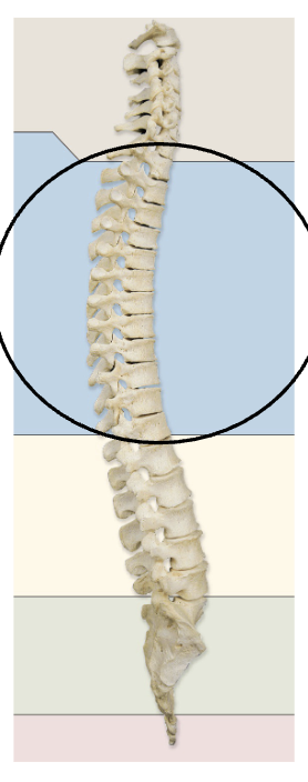
Thoracic vertebrae
the 12 individual bones (T1–T12) forming the middle segment of the vertebral column, situated between the cervical (neck) and lumbar (lower back) spine
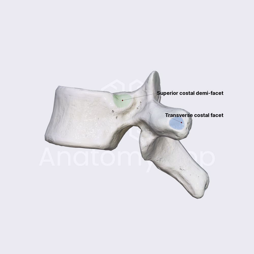
Costal facets
small, smooth, concave surfaces on the sides and transverse processes of thoracic vertebrae that act as attachment points for the ribs
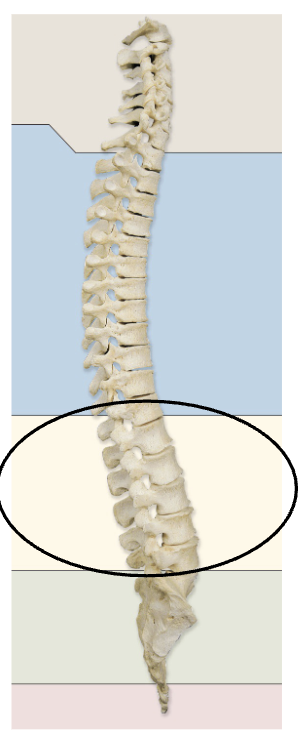
Lumbar vertebrae
the five largest, heaviest segments of the spinal column (designated L1–L5) located in the lower back between the thoracic spine and the sacrum
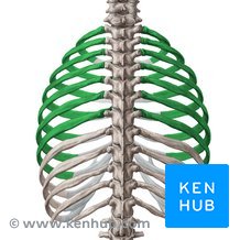
Ribs
24 long, curved, flattened bones (12 pairs) that form the thoracic cage in humans, protecting vital organs like the heart and lungs
Rib shaft
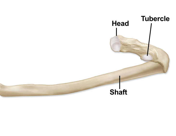
rib head

rib tubercle

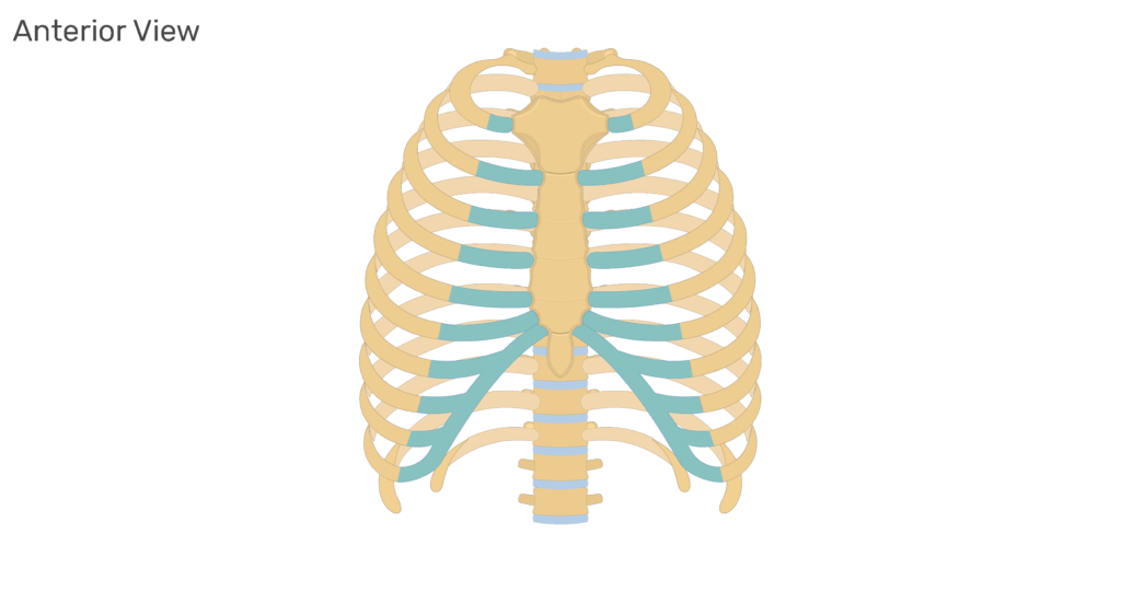
Vertebrosternal ribs
true ribs 1-7 - connect directly to the sternum via their own individual costal cartilages
Vertebrochondral ribs
false ribs 8-10 - any of the three false ribs that are located above the floating ribs and that are attached to each other by costal cartilages
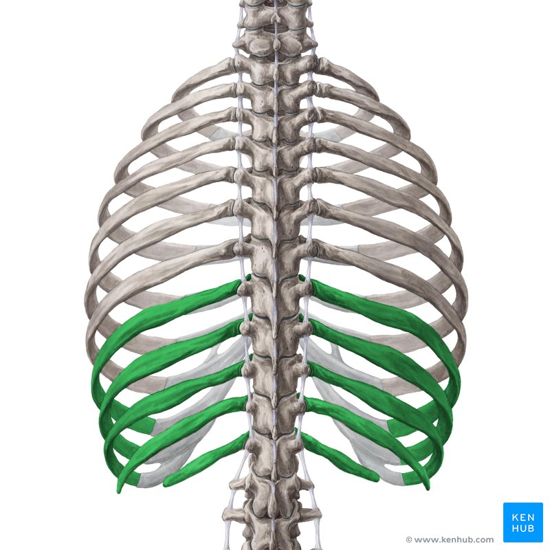
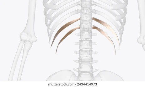
Floating (vertebral) ribs
the last two pairs of ribs (11th and 12th) that attach only to the spine in the back and have no anterior connection to the sternum (breastbone) or other ribs
Costal cartilage
Costal cartilage consists of bars of hyaline cartilage that attach the anterior ends of the ribs to the sternum (breastbone)
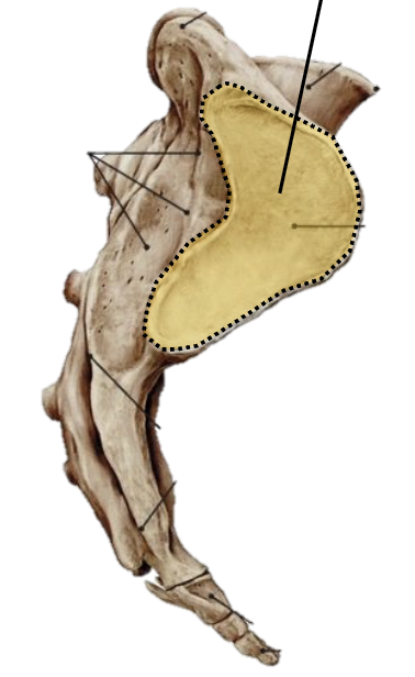
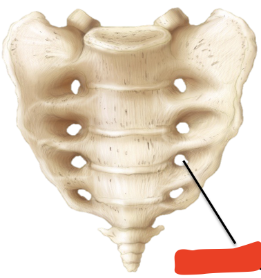
Sacrum
a shield-shaped, triangular bone located at the base of the lumbar spine and the back of the pelvis, formed by five fused vertebrae (S1-S5). It connects the spine to the hip bones, supports upper body weight, and stabilizes the pelvic girdle. It also protects pelvic organs and houses sacral nerves.
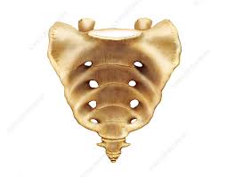
Auricular surface
an L-shaped, ear-shaped articular area located on the lateral surfaces of the upper three sacral vertebrae
Anterior sacral foramina
Posterior sacral foramina
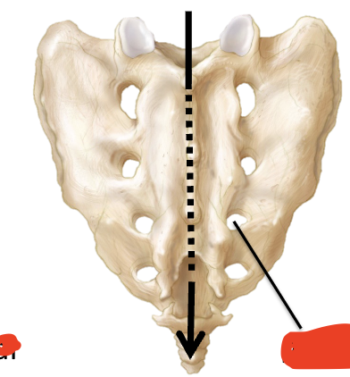
Sacral canal

Sternum
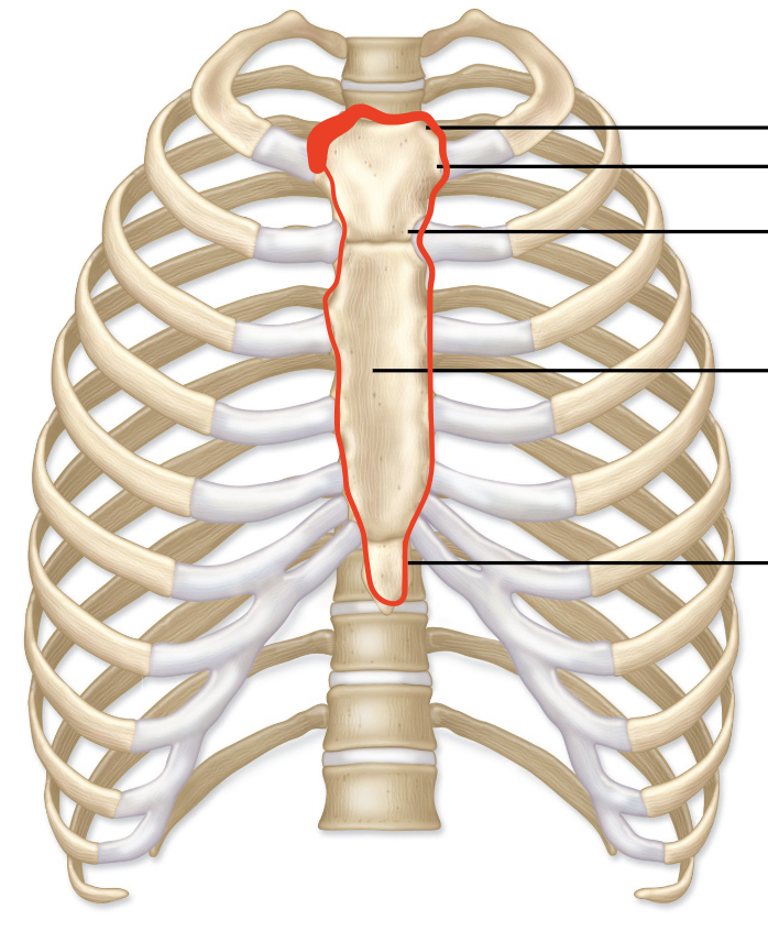
Manubrium
green
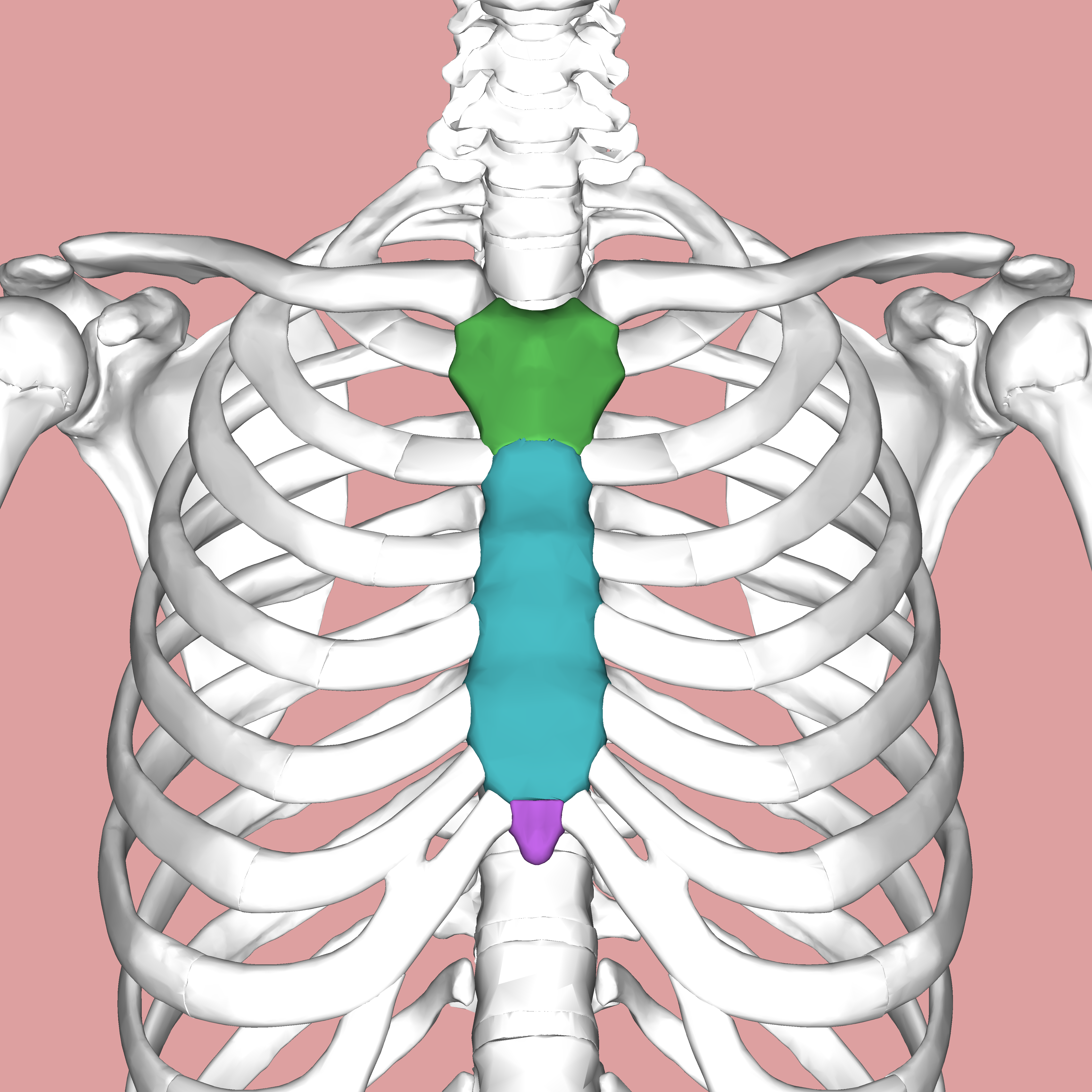
sternum body
blue

Xiphoid process
purple

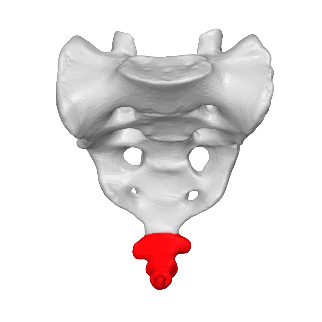
coccyx