Hand Complex
1/46
There's no tags or description
Looks like no tags are added yet.
Name | Mastery | Learn | Test | Matching | Spaced | Call with Kai |
|---|
No analytics yet
Send a link to your students to track their progress
47 Terms
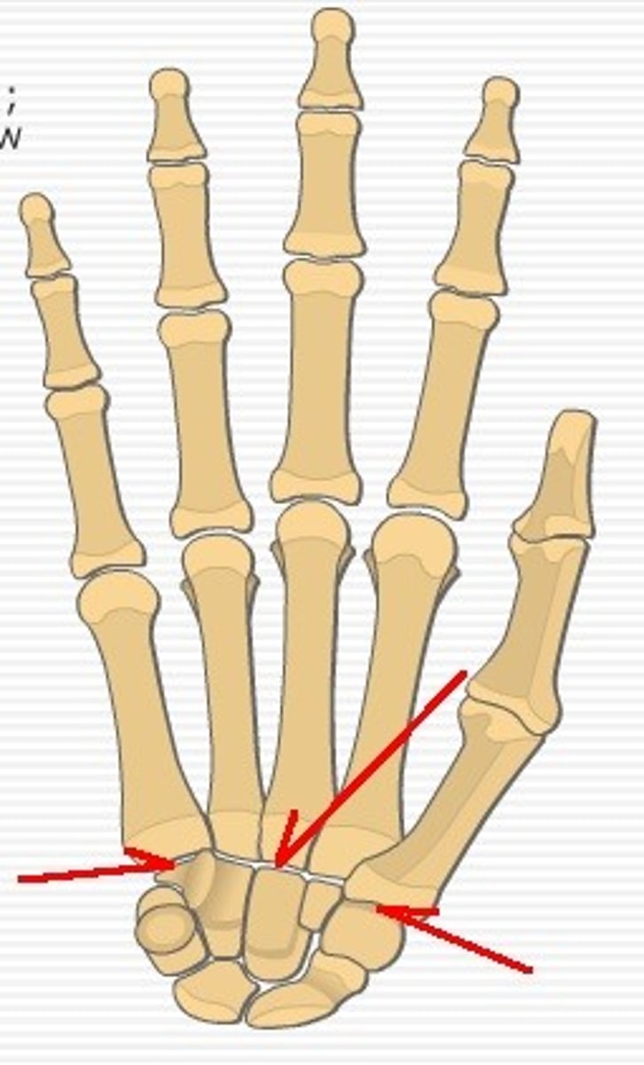
Carpometacarpal (CMC) Joints (2nd-5th)
Joints between carpals and metacarpals.
- Inconsistent classification: debate on plane vs. saddle joint
- 4th/5th joints more mobile than 2nd/3rd joints
1st Carpometacarpal (CMC) joint
saddle joint
2 degrees of freedom:
- Flexion/extension
- Abduction/adduction
more mobile than 2nd & 3rd joint
allows opposition motion
Location: Articulation between 1st metacarpal and
trapezium
Biomechanics:
Most mobile and complex of the CMC joints owing to
its saddle-shaped design
Loose joint capsule to accommodate large motion
Relies on ligaments and tendons for stability
1lb of force at tip of the thumb = 12lbs at the base
Volar Anterior Oblique Ligament (AOL)
Variable anatomy; curtain-like structure covering the volar joint surface
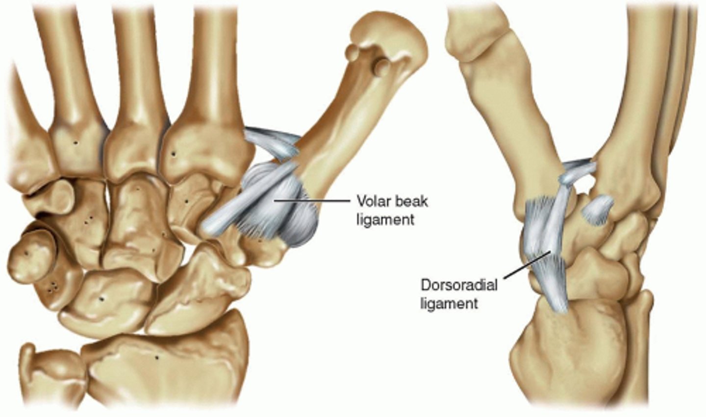
Dorsal Deltoid Ligament
Primary stabilizers of the thumb CMC joint
- Consists of 3 stout bands (fan-shaped like the deltoid): dorsoradial, dorsal central and posterior oblique ligaments
- Originate from the dorsal tubercle of the triquetrum and insert onto the dorsal base of the 1st metacarpal
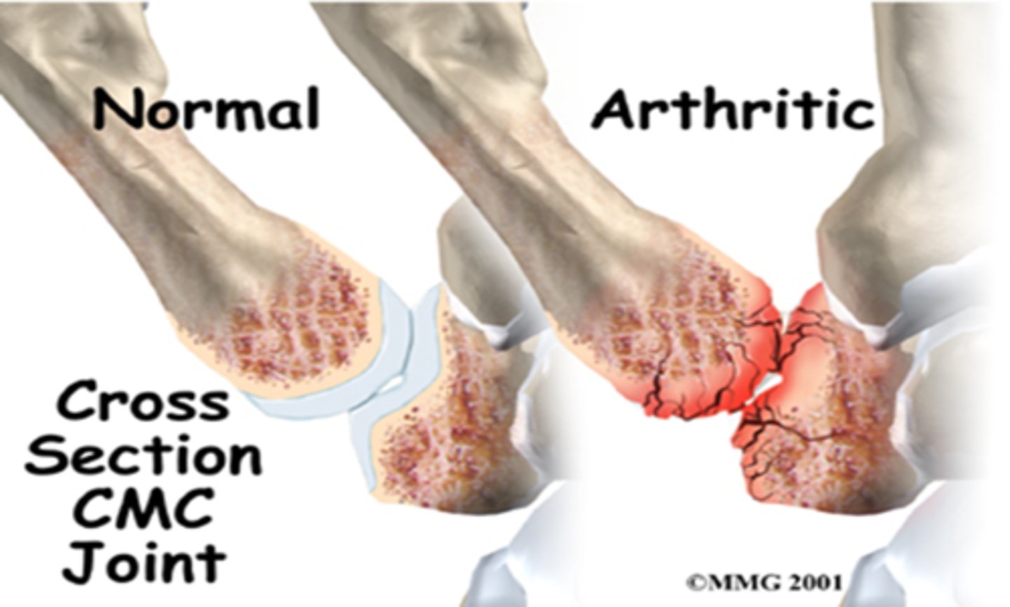
Thumb CMC Joint Osteoarthritis
Pathophysiology:
Degenerative attenuation of stabilizing ligaments followed by increased mechanical stress
of the CMC joint
- 40% and 25% of incidence in women and men over 75 years of age respectively
- Causes debilitating pain and weakness in pinching and loading activities
Considering that CMC OA comes from stress on ligaments and mechanical load on the joint surface, should patients with CMC OA be positioned in its close-packed (maximizing congruency/ligament tautness) or loose-packed (minimizing compression) joint position?
Close-packed position (most congruent): Full opposition
Open-packed position (least congruent): Mid-range
Loose packed position to minimize compression
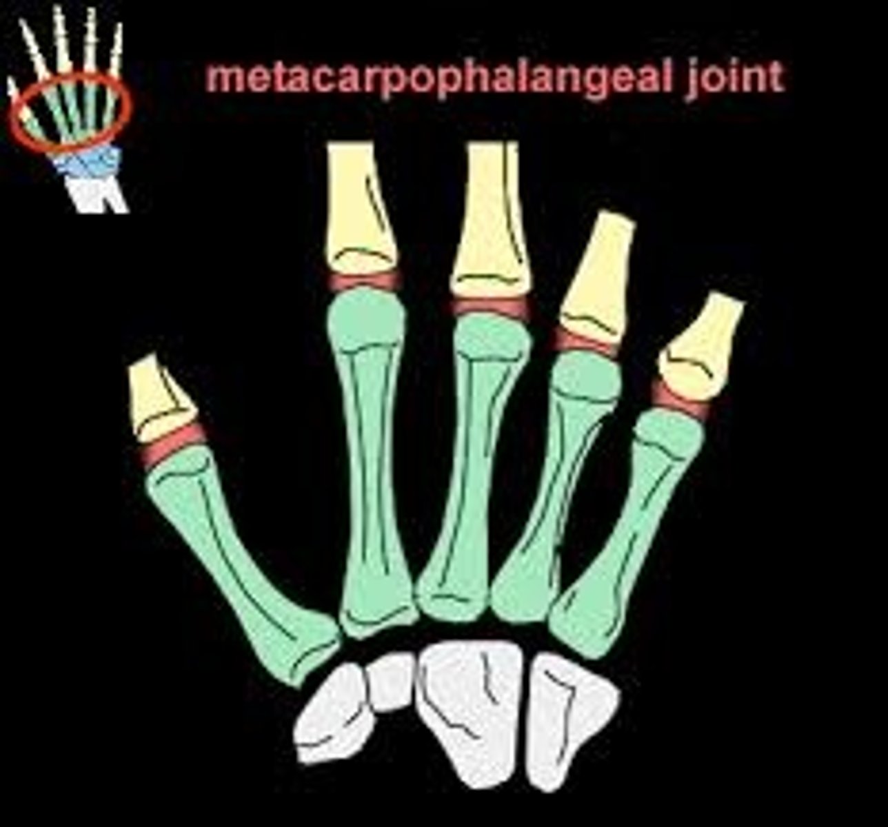
1st Metacarpophalangeal (MCP) joint
Condyloid joint
- Functions more like a "hinge" joint with very limited side-
to-side motion
Metacarpophalangeal (MCP) joint
Joint between metacarpals and phalanges.
Condyloid joint
- 2 degrees of freedom:
- Flexion/extension and abduction/adduction
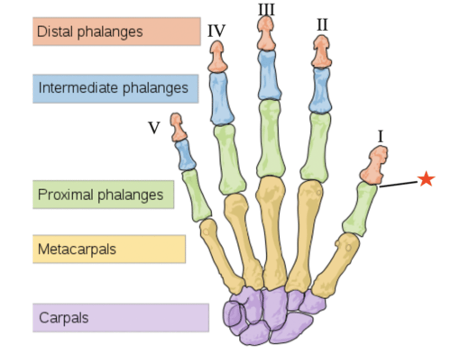
Proximal Interphalangeal (PIP) Joint
Joint between the proximal and middle phalanges
hinge joint: flexion/extension
Distal Interphalangeal (DIP) Joint
joint between middle and distal phalanges
hinge joint: flexion/extension
Interphalangeal (IP) Joint
joint between proximal and distal phalanges of the first digit
hinge joint
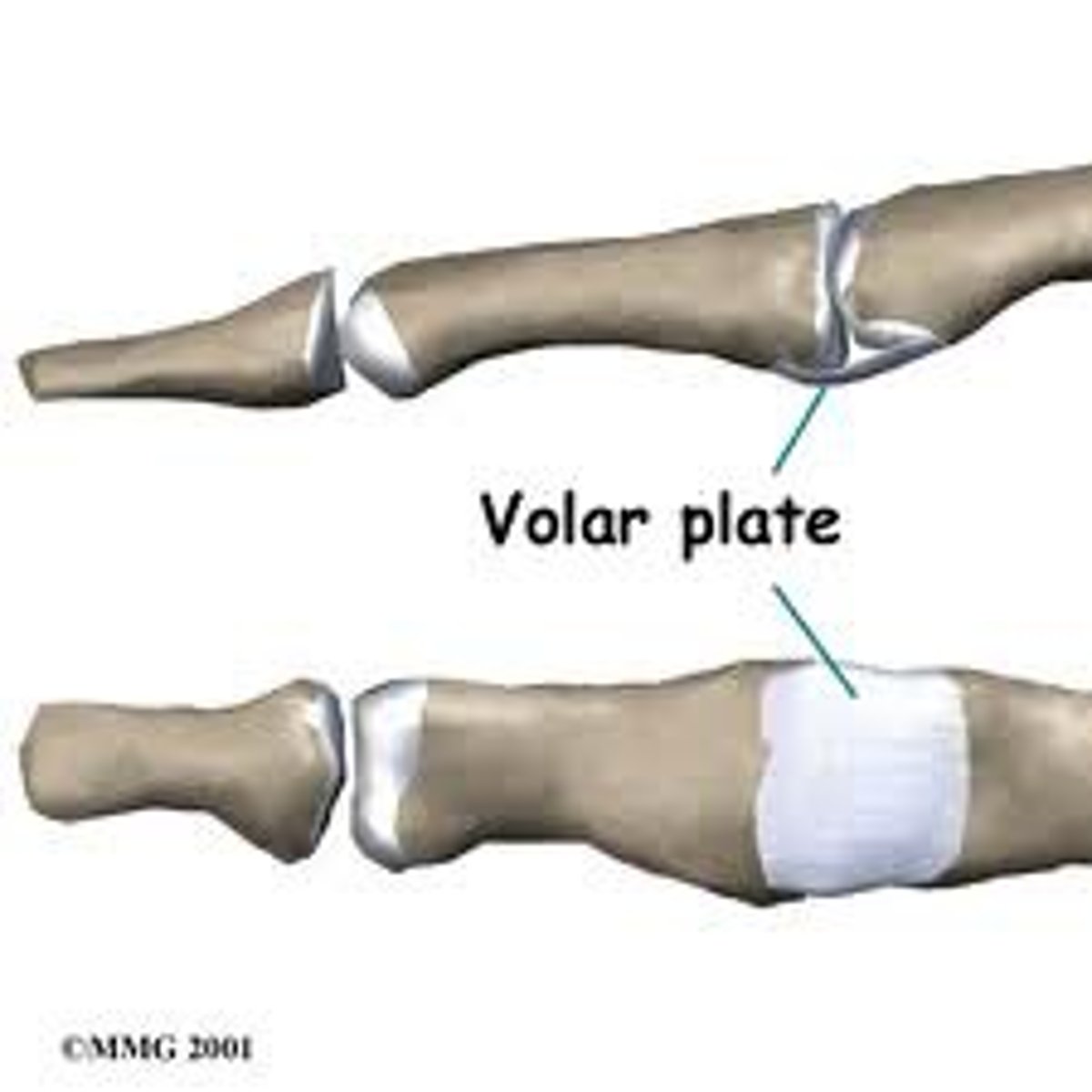
Volar plate
Prevents hyperextension
- Strong fibrocartilage located on volar aspect of the joint spanning across the joint space
- Proximally reinforced by check-rein ligaments on either (lateral) side
- Forms the 'floor' of the finger joints
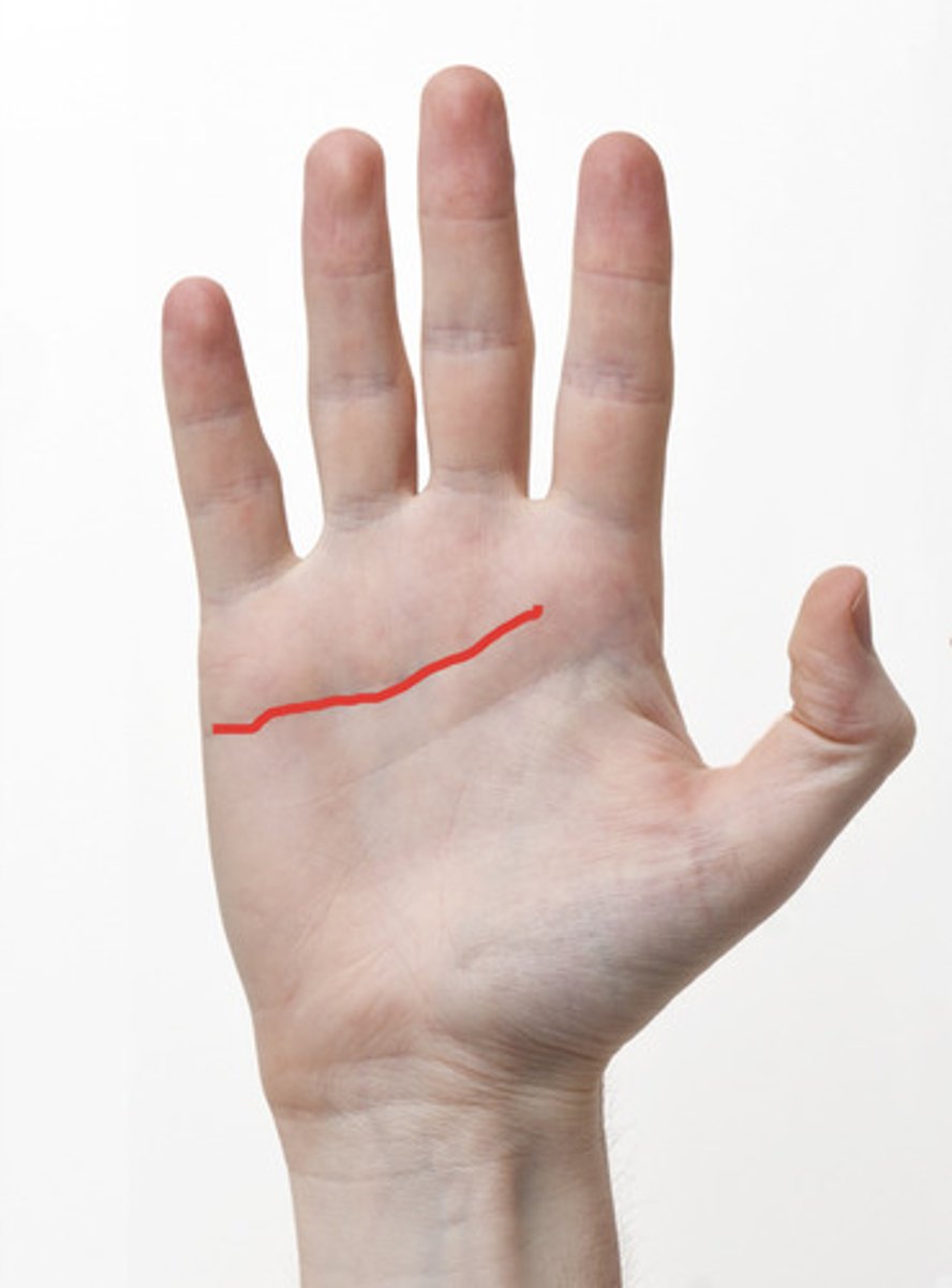
Distal wrist crease
crease that separates the distal and proximal rows of the carpal bones (pisiform to the tubercle of the trapezium)
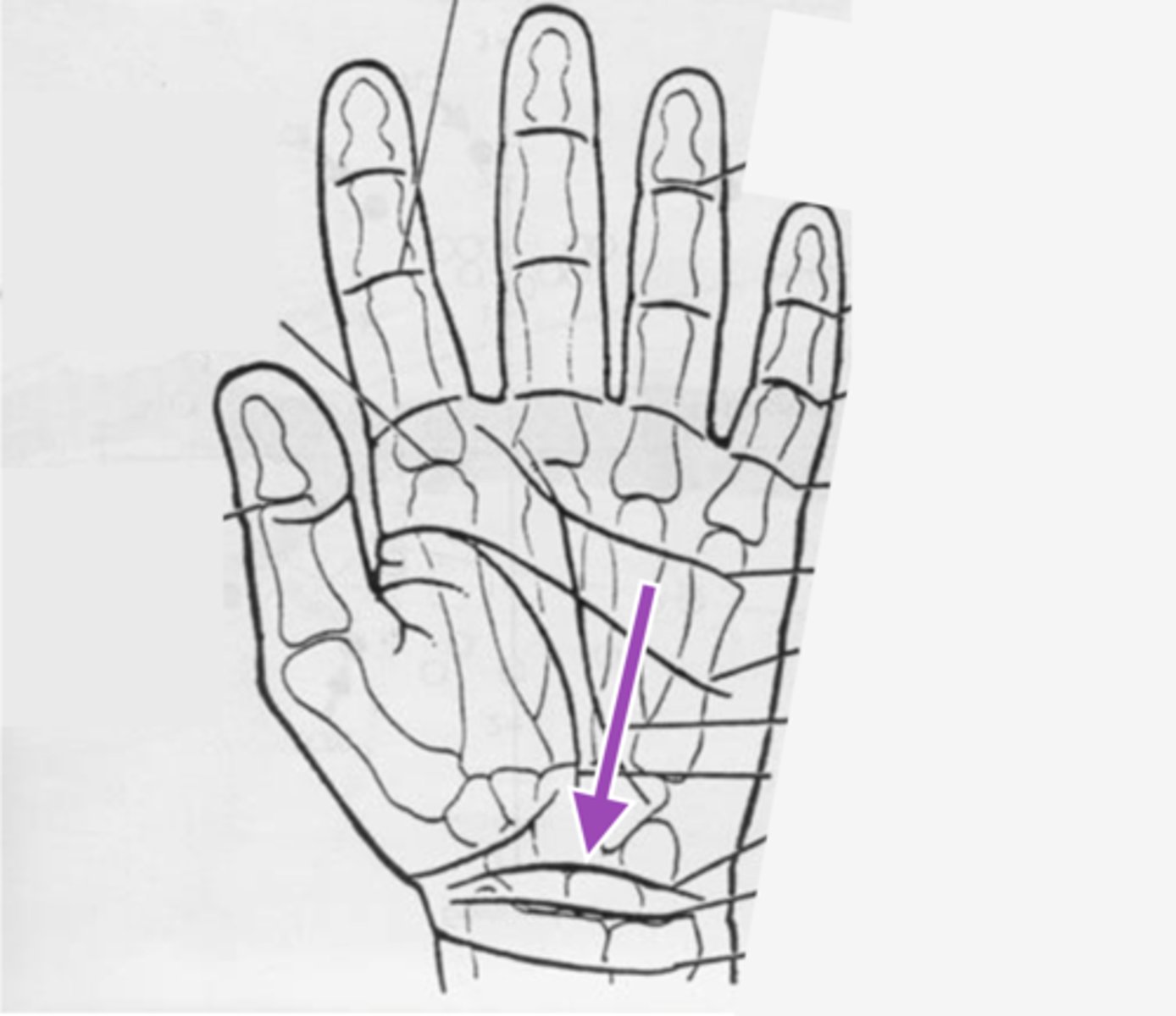
Distal palmar crease
This crease lies at the base of the fingers and marks the location of the proximal pulley:
A. Distal palmar crease
B. Hypothenar crease
C. PIP crease
D. Thenar crease
D. Proximal palmar crease
1st dorsal interosseous muscle
fills posterior side of thumb space (space between 1st and 2nd metacarpal bones)
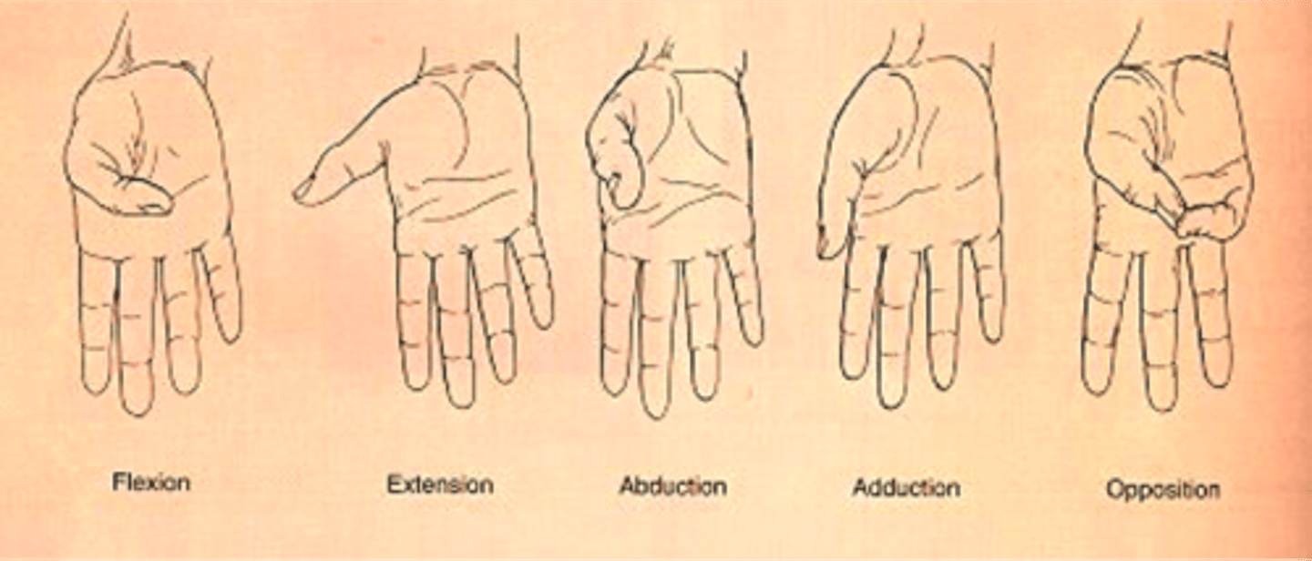
What are the movements of the thumb?
flexion, extension, abduction (radial & palmar), adduction, opposition
Collateral Ligaments (of the hand)
Shared ligamentous structures between MCP, PIP, DIP, IP joints of the fingers and thumb:
Located on radial and ulnar sides of the joint
to provide lateral (side-to-side) stability
2 components:
- Proper collateral ligament: 'cord' like
- Accessory collateral ligament: 'fan' shaped
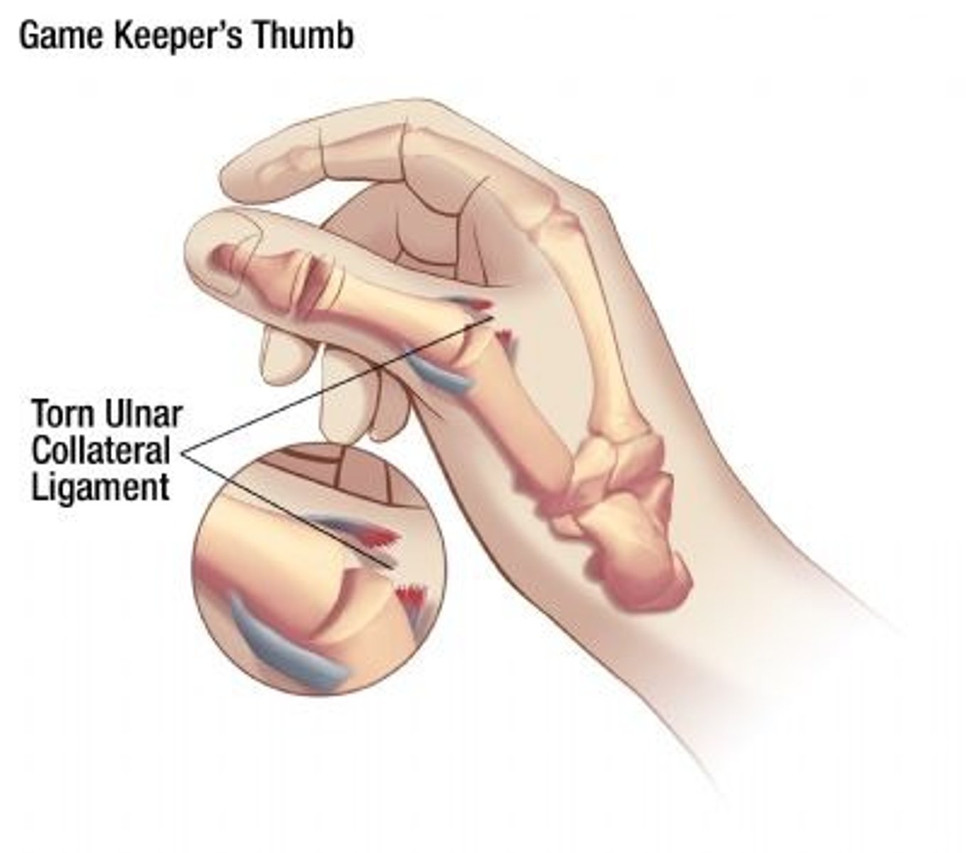
Skier's Thumb
Tear of the ulnar collateral ligament (UCL) of the thumb MCP joint
Mechanism of injury: Fall or direct trauma to the thumb in valgus stress
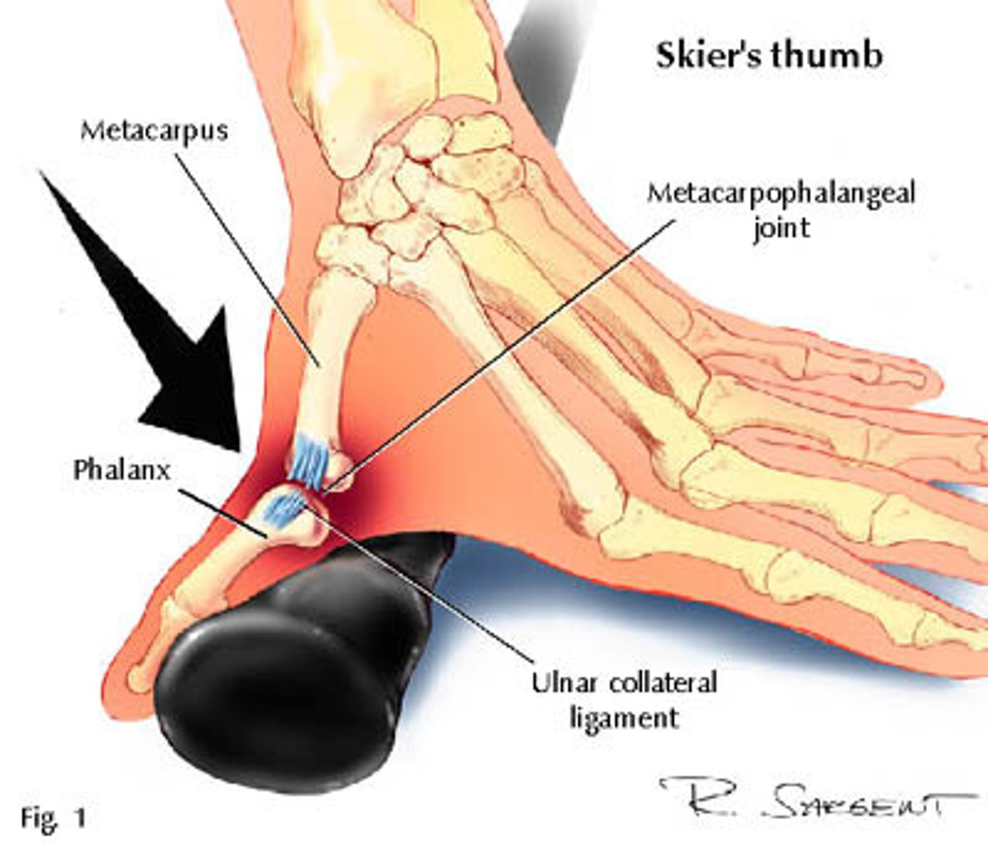
Gamekeeper's thumb
injury to the thumb that results in tearing or stretching of the MP joint or rupture of the ulnar collateral ligament
Chronic tear of the thumb MCP UCL ligament
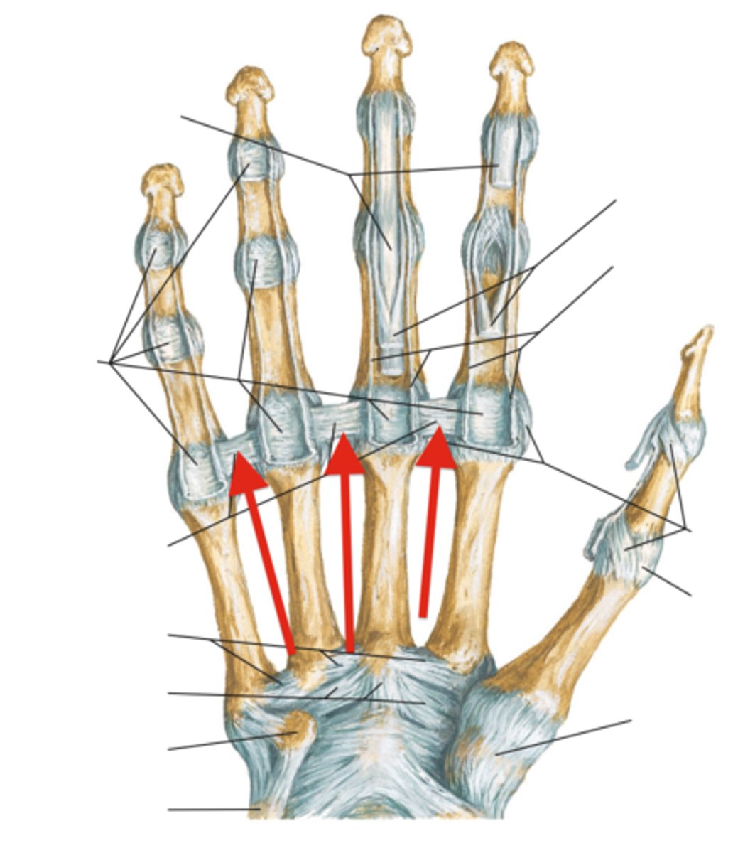
Deep transverse metacarpal ligaments
Strong bands connecting volar plates of adjacent
finger MCP joints
The Proper collateral ligament becomes taut in what motion?
flexion
The Accessory collateral ligament becomes taut in what motion?
extension
Closed pack position (of the finger joints)
Fully congruent joint surfaces held tightly together by maximal tension in joint ligaments.
MP joints: flexion
IP joints: extension
In IP joints, the proper collateral ligaments are taut in all motions. Volar
plates are taut in extension. Making extension what kind of position?
closed pack position
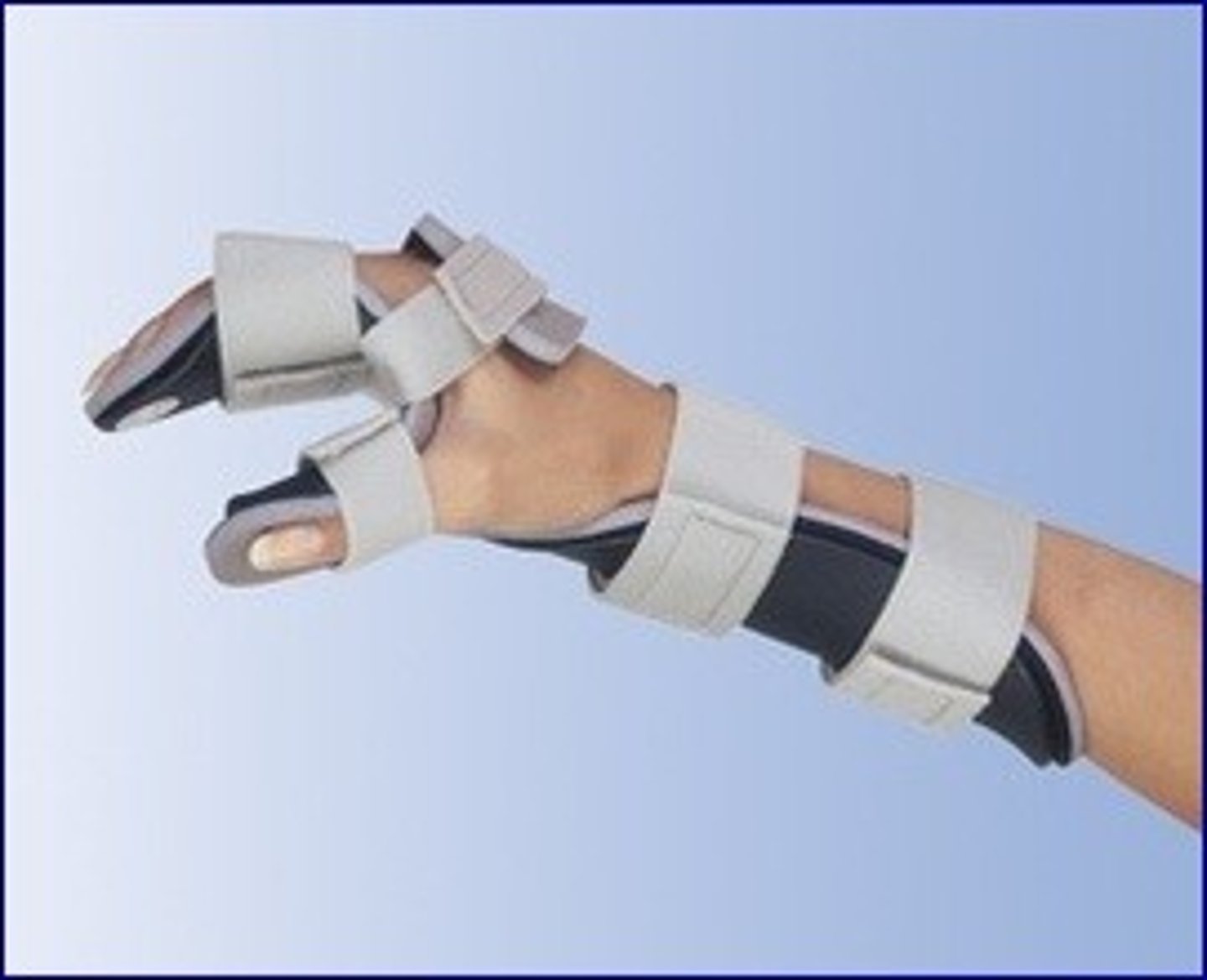
Fracture splinting
To prevent contracture formation, in this intervention:
- MCPs are positioned in 70° flexion
- PIP and DIP joints in full extension
- Also called "intrinsic plus/safe position"
How does joint stability demands in the thumb joints differ from the finger joints?
Thumb is both power and precision whereas the fingers are moreso for power because its more important to be stable
Extrinsic hand muscles
Muscles in the forearm that cause movement in the hands or fingers
What extrinsic hand muscles are the flexors (anterior forearm)?
flexor digitorum profundus (FDP), flexor digitorum superficilis (FDS), and flexor pollicis longus (FPL)
What extrinsic hand muscles are the extensors (posterior forearm)?
Superficial layer:
- Extensor digitorum (ED)
- Extensor digiti minimi (EDM)
Deep layer:
- Abductor pollicis longus (APL)
- Extensor pollicis brevis (EPB)
- Extensor pollicis longus (EPL)
- Extensor indicis (EI)
Flexor retinaculum
Aka transverse carpal ligament
Forms the roof of the carpal tunnel enclosing 9 tendons:
- FPL (1 tendon)
- FDS (4 tendons)
- FDP (4 tendons)
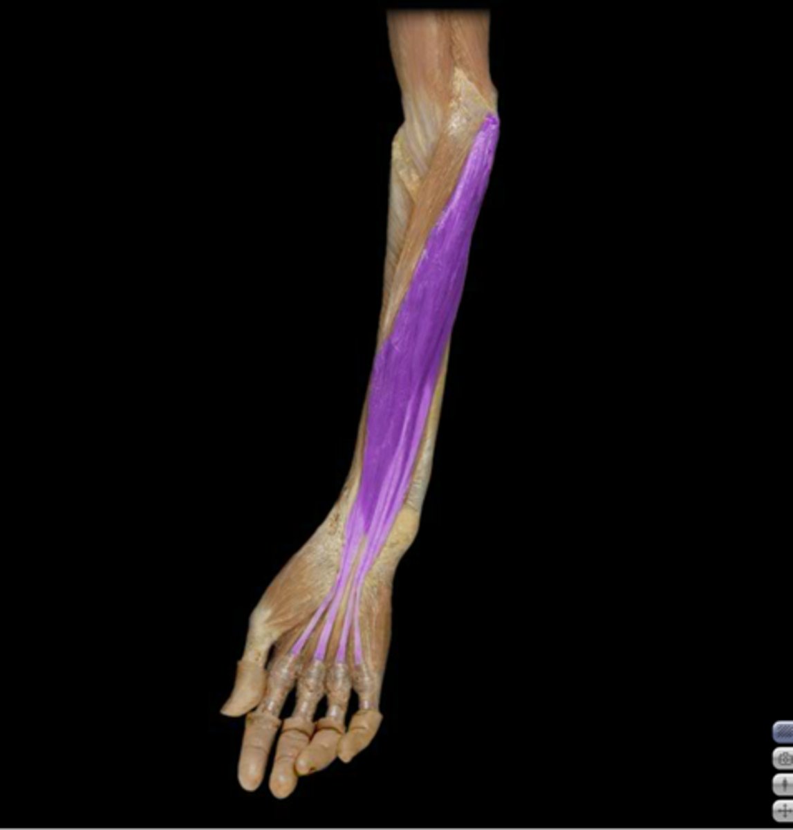
Flexor digitorum superficialis (FDS)
Action: Digit 2-5 flexion at PIP joint
Origin:
- Humeroulnar head: medial epicondyle and
coronoid process of ulna
- Radial head: oblique line of radius
Insertion: Shaft of middle phalanges
Nerve innervation: Median nerve
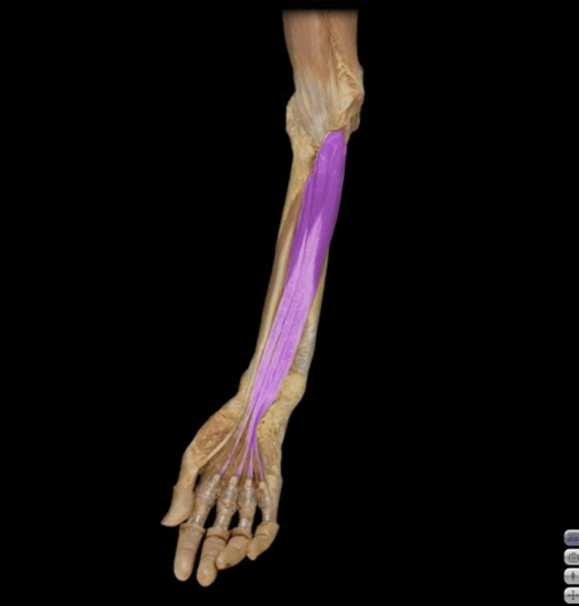
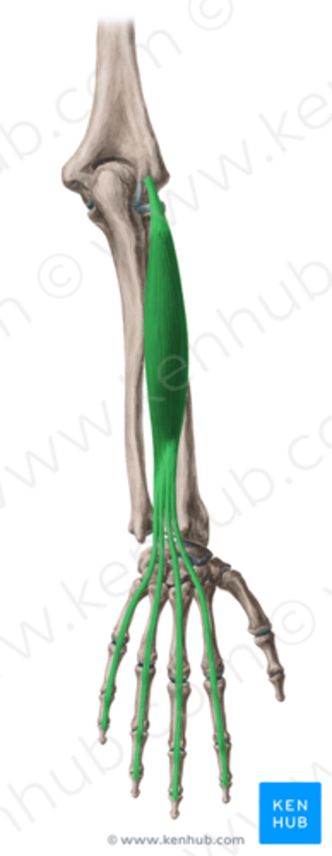
Flexor digitorum profundus (FDP)
Action: Digit 2-5 flexion at DIP joint
Origin:
- Proximal three quarters of anteromedial surface of
ulna and interosseous membrane
Insertion: Base of distal phalanges
Nerve innervation:
Digits 2&3: AIN of median nerve
Digits 4&5: Ulnar nerve
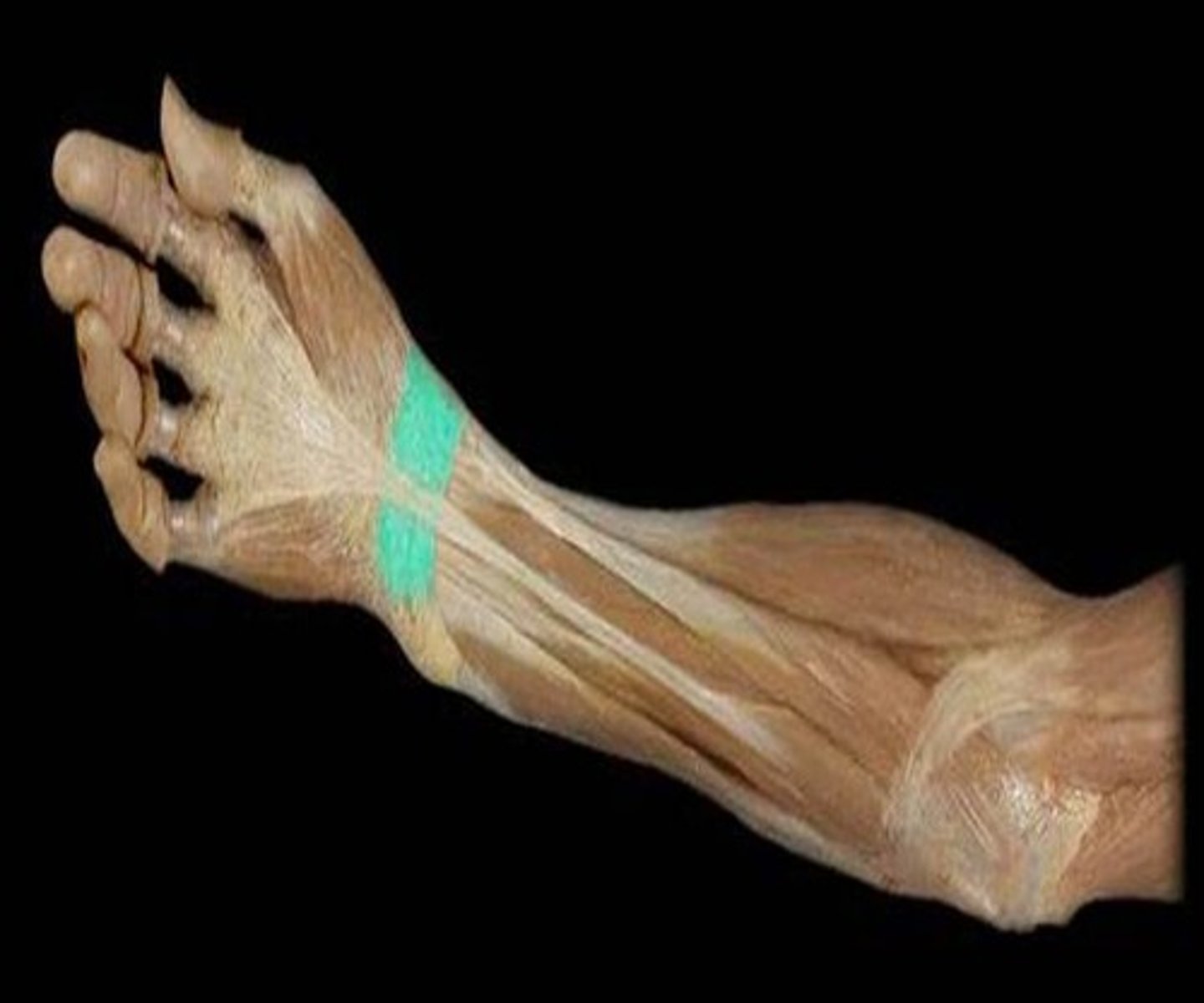
Camper's chiasm
where the FDS runs superficial to the FDP: at its insertion, the FDS splits into two tendon slips wrapping itself around the FDP to allow the FDP to pass through
What type of joint flexion does the FDS handle?
PIP flexion
What type of joint flexion does the FDP handle?
DIP flexion
The _____ does not divide and continues to insert into the distal phalanx
FDP
How would you isolate the action of the FDS?
Flex the targeted digit while holding the rest of the
digits in extension
How would you isolate the action of the FDP?
Hold the targeted digit just proximal to the DIP joint
- Only allow the DIP to flex while keeping the PIP in extension
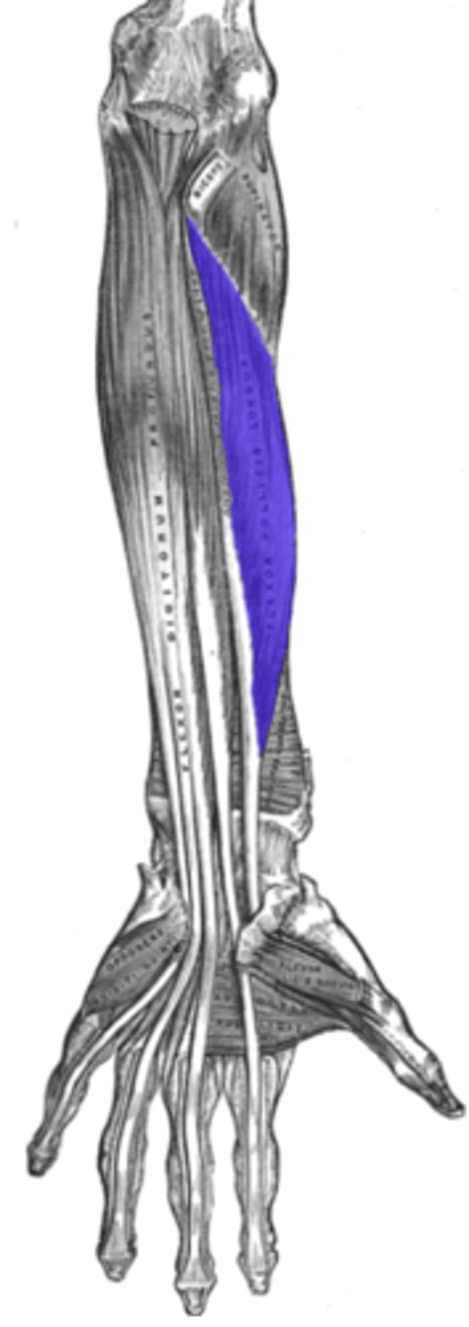
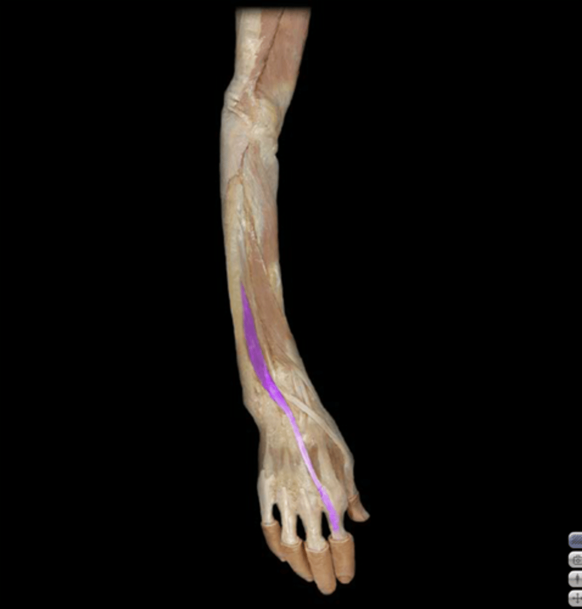
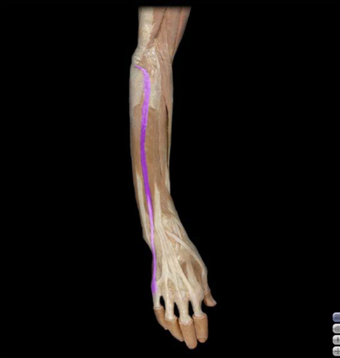
Flexor pollicis longus (FPL)
- Part of the deep layer of anterior forearm muscles
Thumb flexor
Action: thumb flexion at IP and MCP joints
Origin:
- Anterior surface of radius and adjust interosseous membrane
- Interosseous membrane
Insertion: Base of distal phalanx of the thumb
Nerve innervation: AIN of the median nerve
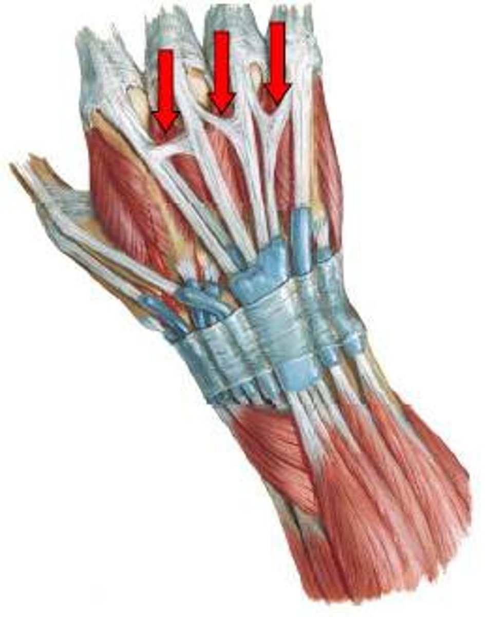
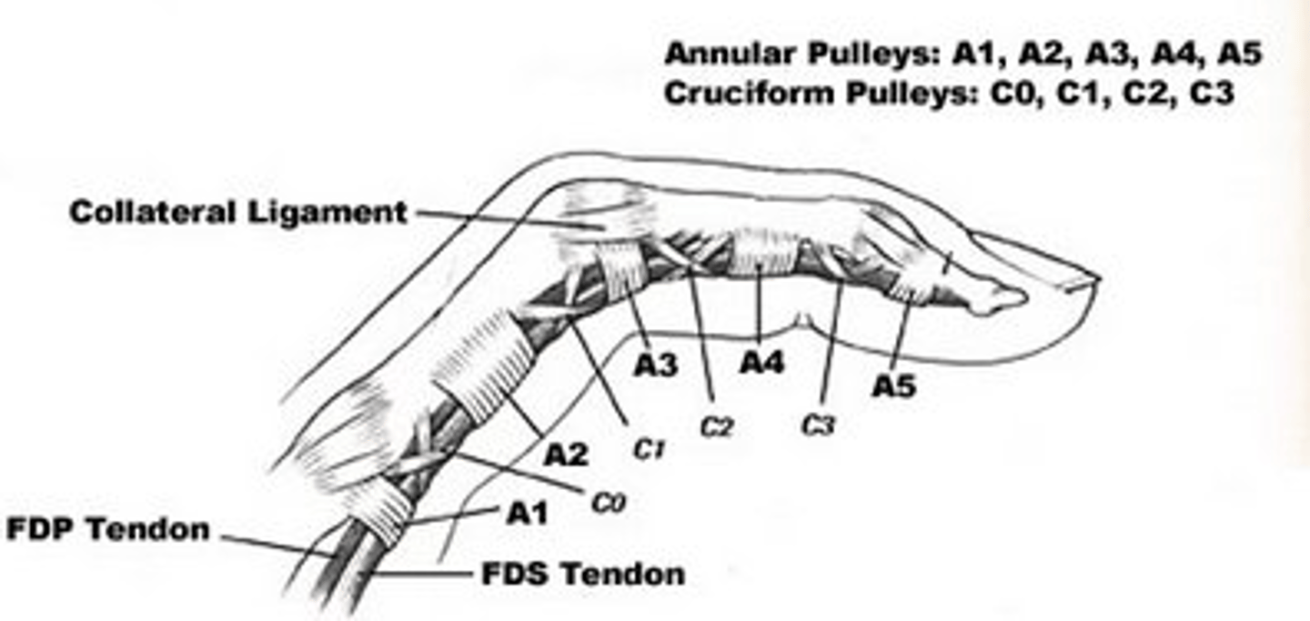
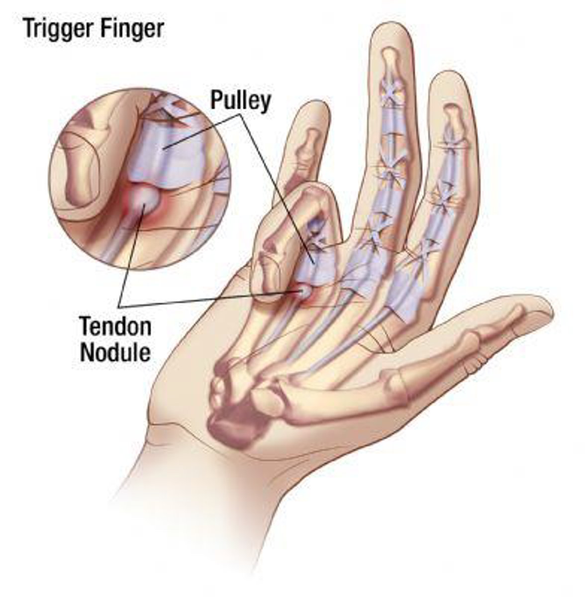
Flexor tendon pulleys
found on the flexor side to prevent bowstringing and consist of A1, A2, A3, and A4 and C1, C2, C3, and C4.
Fingers: 5 annular (A1-A5) and 3 cruciate
- Annular are much stronger than cruciate
- A2 pulley is the strongest
Thumb: 2 annular (A1, A2) and 1 oblique
- A1 located at MCP joint
- Oblique located in the middle of the proximal phalanx: most critical against bowstringing
Trigger finger/ thumb
Pathophysiology: Inflamed nodule forms on the
thickened flexor tendon sheath. Nodule becomes
trapped proximal to the A1 pulley structure when the
digit is trying to extend from full flexion.
Mechanism of injury: overuse/repetitive gripping
What are the finger extensors?
extensor digitorum, extensor indicis, extensor digiti minimi
Extensor Compartments
Thick fibrous band securing the extensor tendons
6 fibro-osseous tunnels form compartments:
- 1st compartment: APL and EBP
- 2nd compartment: ECRB/L
- 3rd compartment: EPL
- 4th compartment: EDC and EDI
- 5th compartment: EDM
- 6th compartment: ECU
Extensor indicis (EI)
Action: extension of wrist and digit 2
Origin: Distal third of ulna and interosseous membrane
Insertion: Digit 2 extensor expansion
Innervation: Posterior interosseous nerve (PIN): continuation of deep branch of the radial nerve
Dorsal extensor compartment: 4th compartment
Extensor digitorum (EDC)
Action: Digit 2-5 extension
Origin: Lateral epicondyle
Insertion: Digit 2-5 extensor expansion
Nerve innervation: Posterior interosseous nerve (PIN): continuation of deep branch of the radial nerve
Dorsal extensor compartment: 4th compartment
Unique feature: Tendons are linked to each other in dorsal hand with juncturae tendinae: connective tissue band
Extensor Digiti Minimi (EDM)
Action: Digit 5 extension
Origin: Lateral epicondyle
Insertion: Digit 5 extensor expansion
Nerve innervation: Posterior interosseous nerve (PIN): continuation of deep branch of the radial nerve
Dorsal extensor compartment: 5th compartment
Juncturae Tendinae (JT)
Connective tissue band linking adjacent
tendons of the EDC proximal to the
MCP joints
- Highly variable structure
- Assist in centralizing the extensor tendon