imaging exam 2 photos
1/80
There's no tags or description
Looks like no tags are added yet.
Name | Mastery | Learn | Test | Matching | Spaced | Call with Kai |
|---|
No analytics yet
Send a link to your students to track their progress
81 Terms
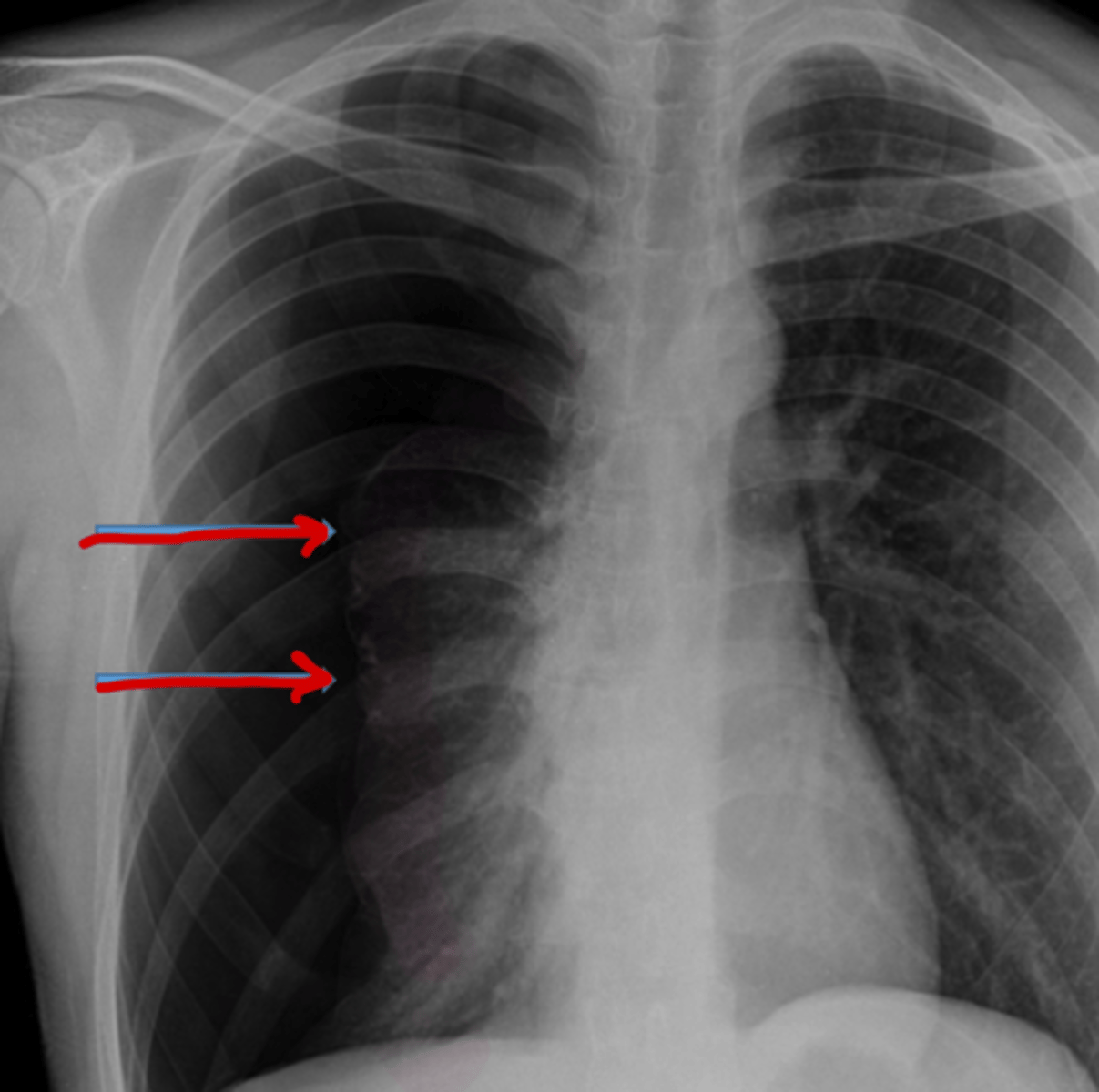
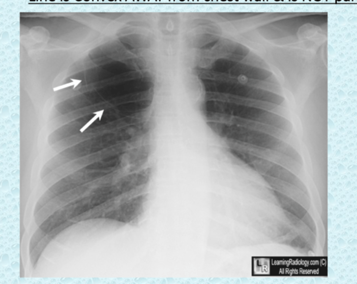
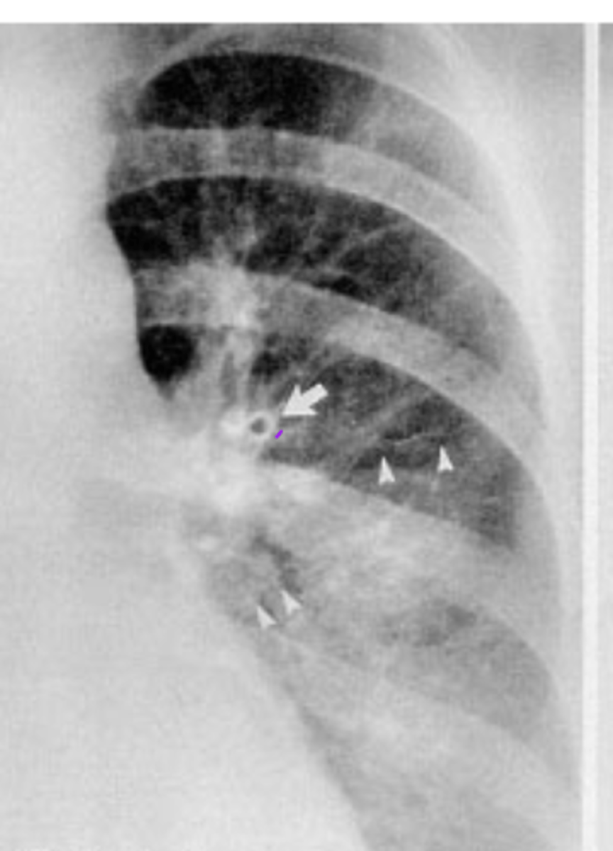
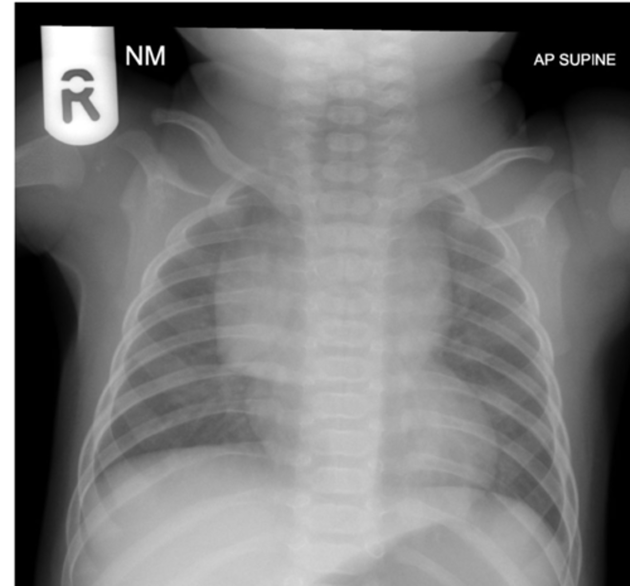
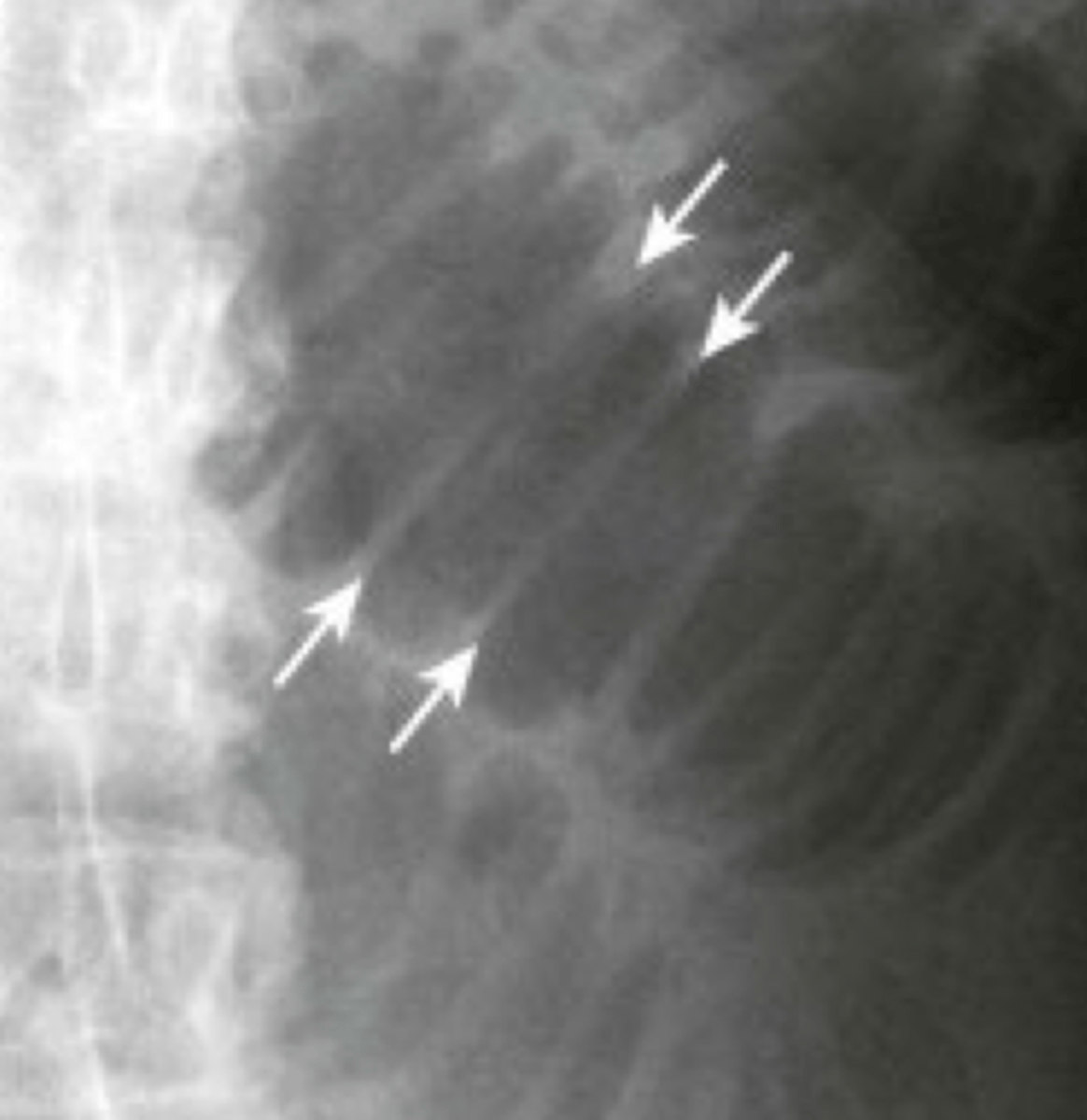
Visceral pleural line, simple pneumothorax
what are these arrows pointing to and what disease process is it associated with?
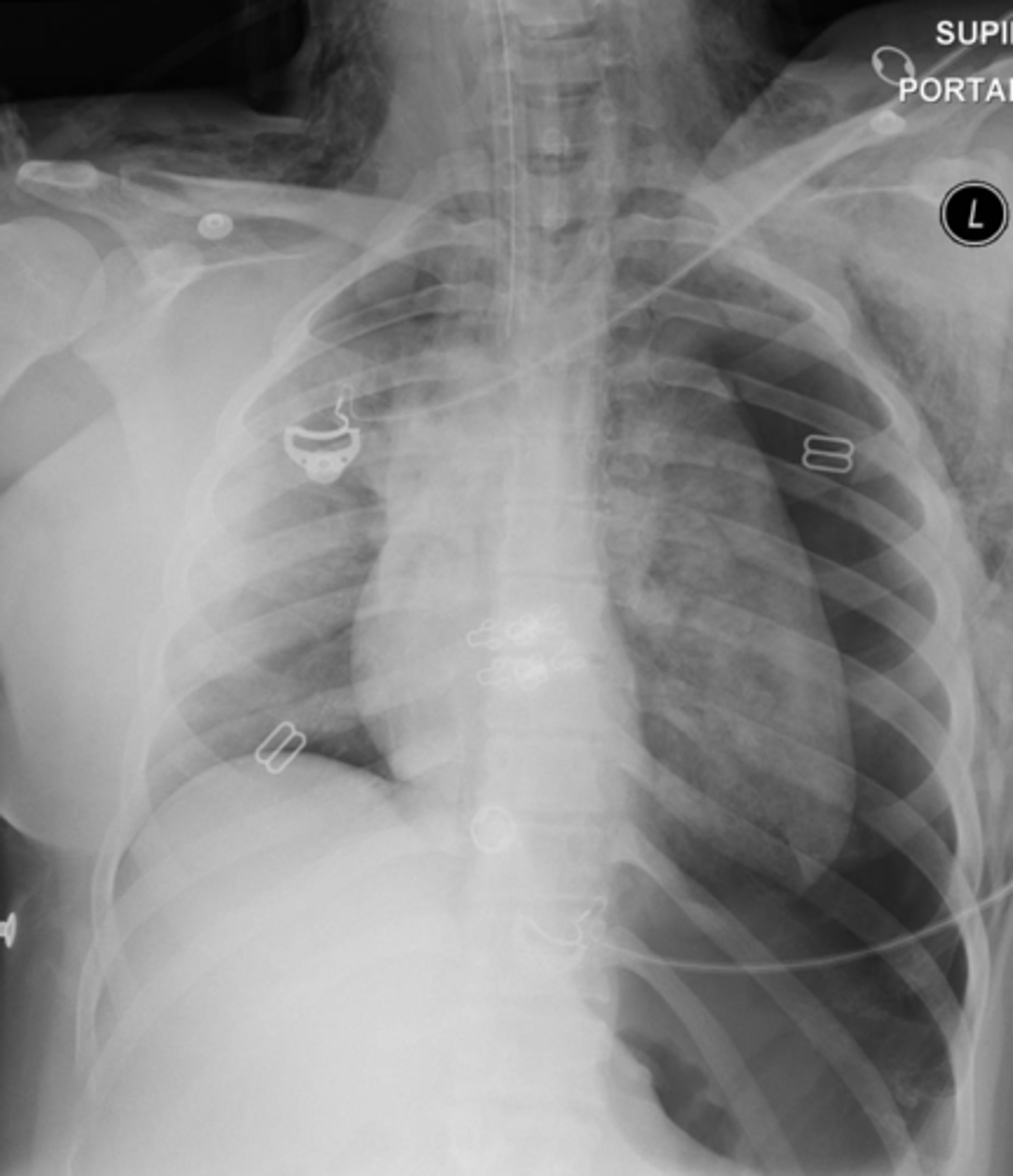
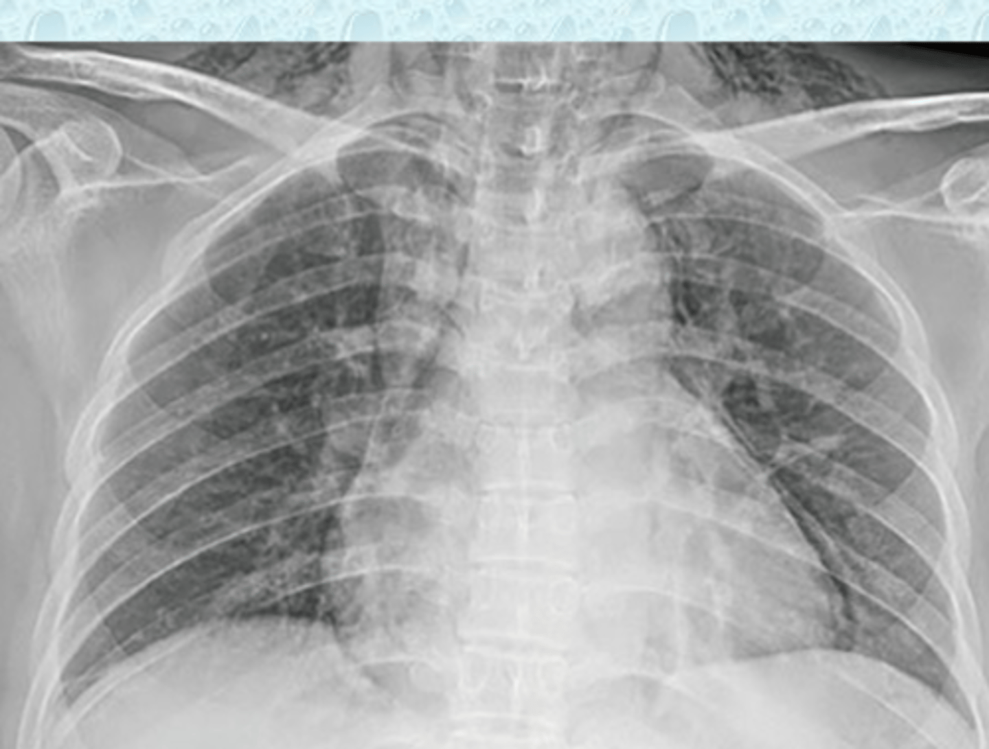
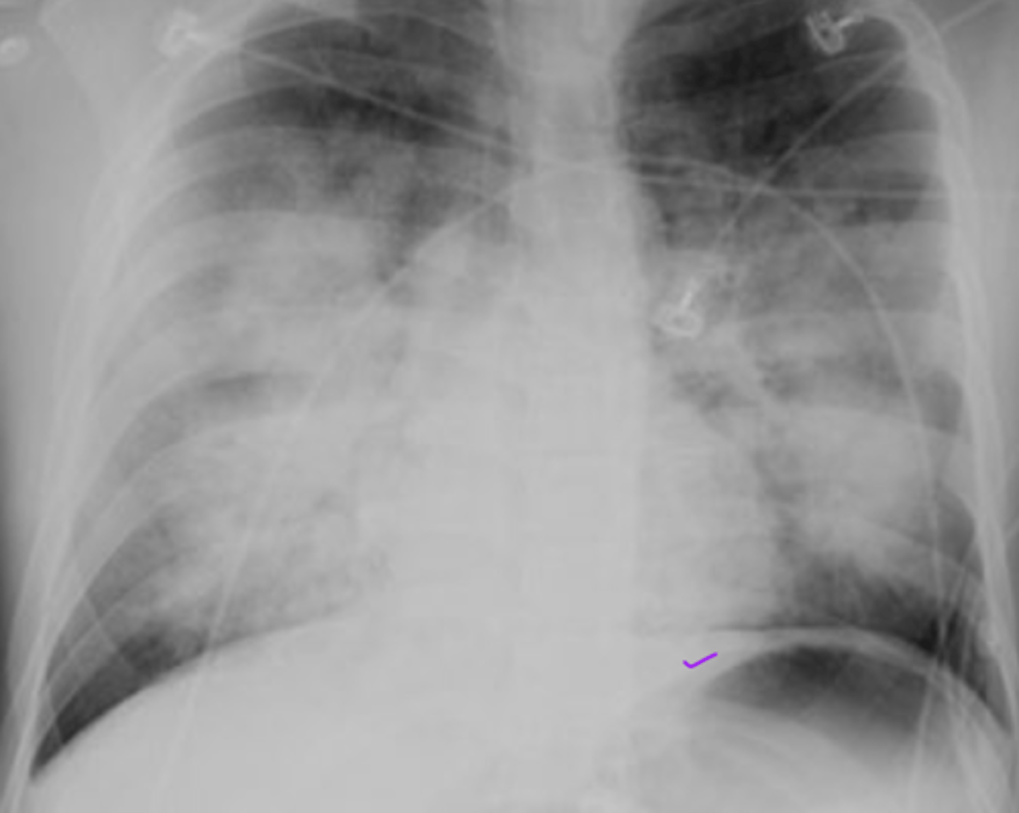
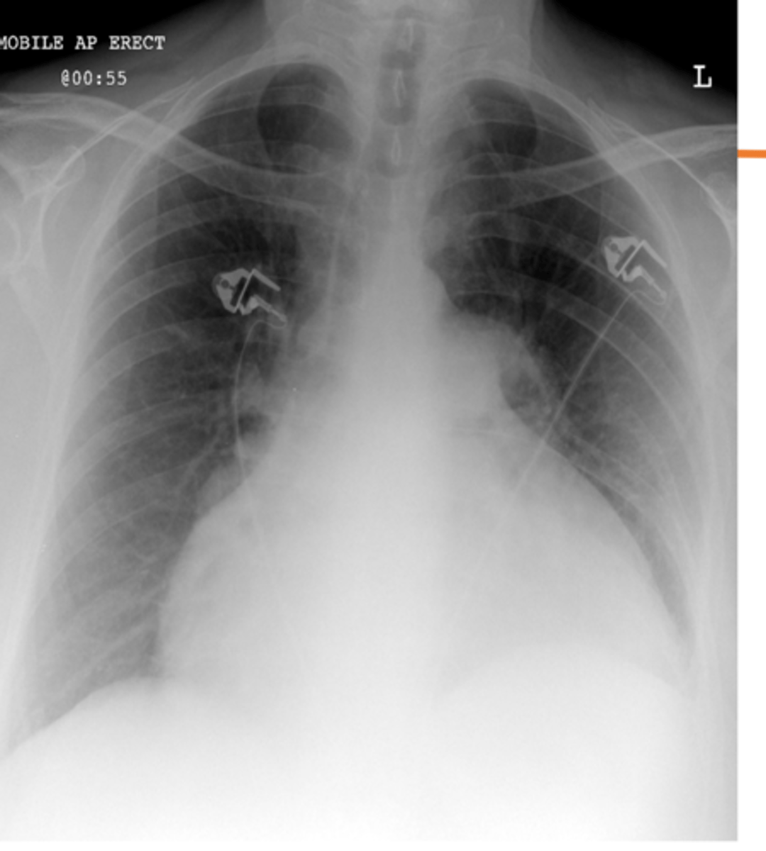
a tension pneumothorax
what does this image show?
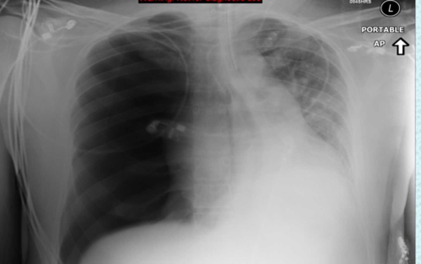
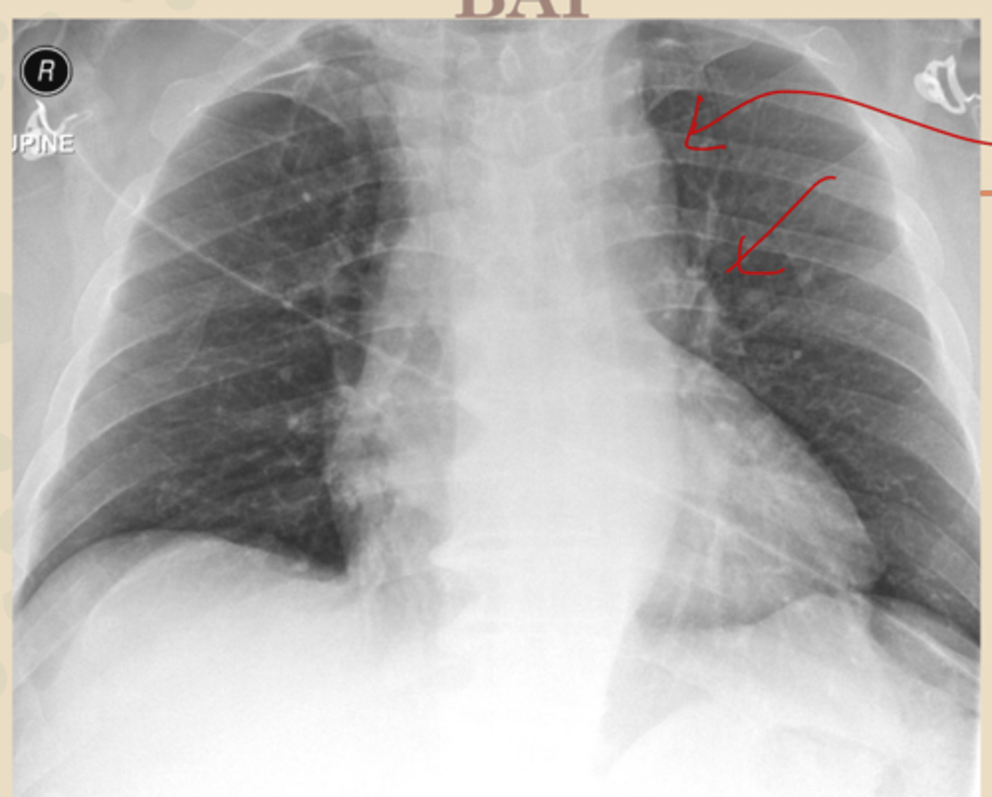
tension PTX- has shifting of mediastinum, flattening of diaphragm, etc
what is this and how do you know
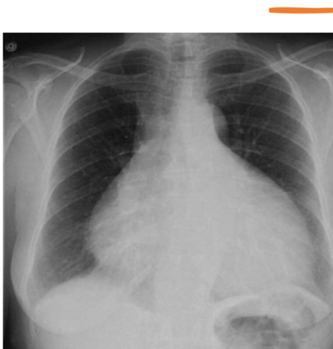
cyst or bleb
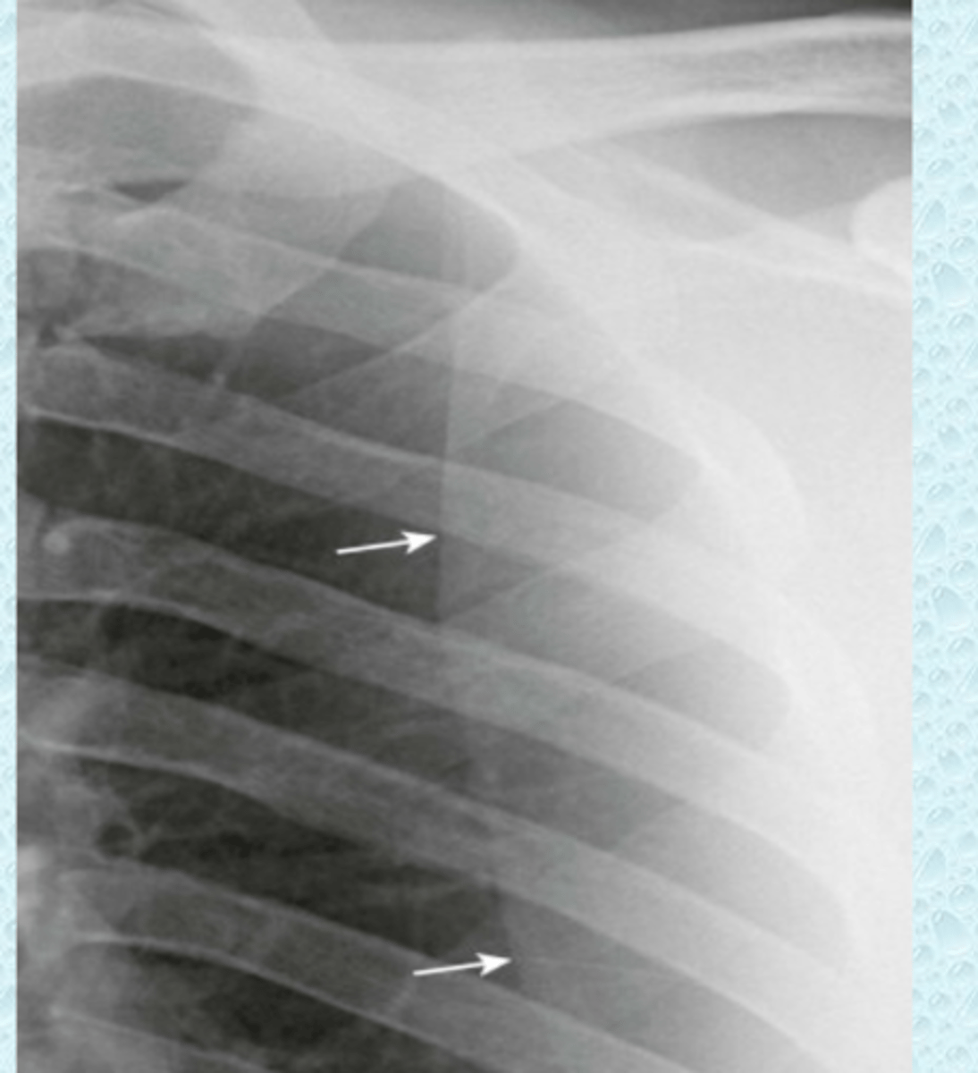
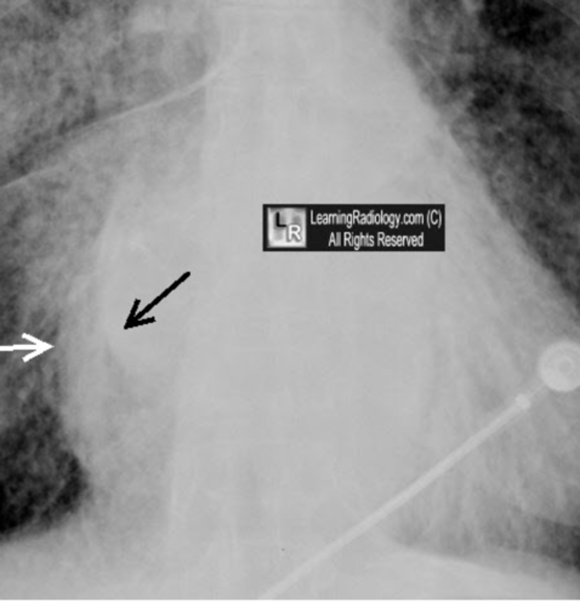
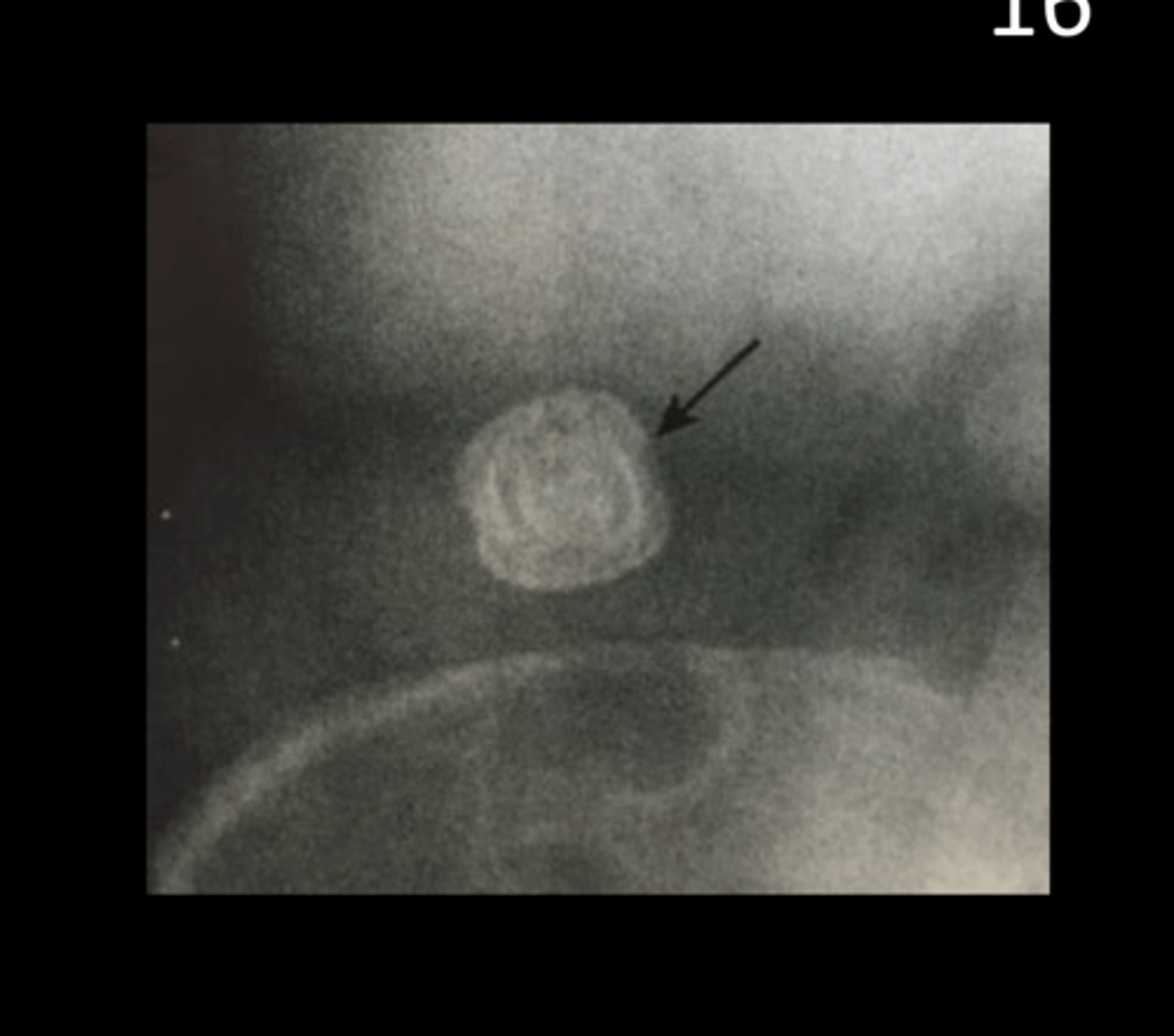
what are the white arrows most likely pointing to
the border of the scapula
what is this
Pulmonary interstitial emphysema
what is this showing
pneumomediastinum- see streaking around the heart and great vessels, continuous diaphragm
what is this showing, how do you know?
pneumopericardium, take a CT scan
what is this, what could you do to confirm?
subcutaneous emphysema
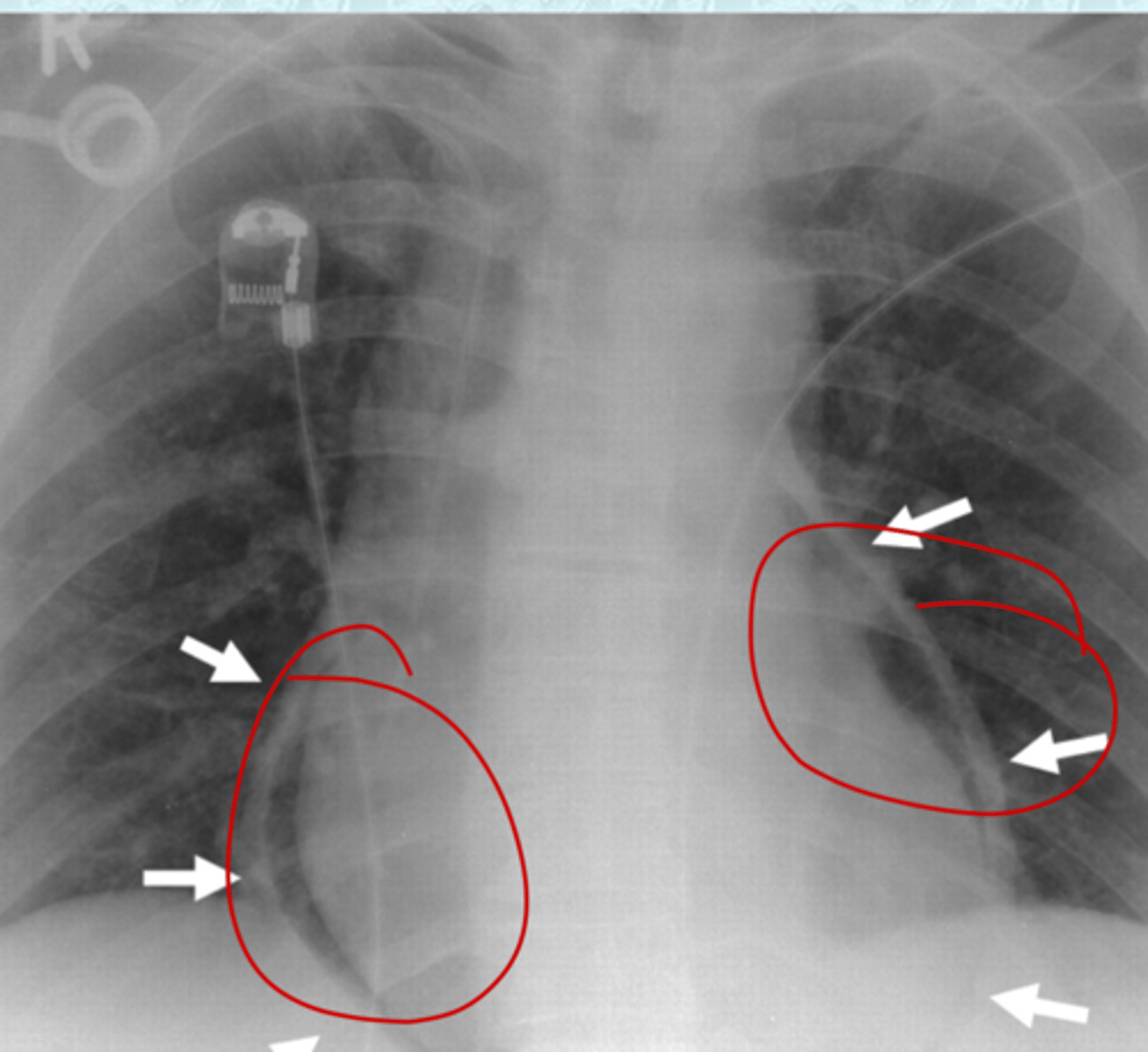
what are these red circles of?
LV heart failure
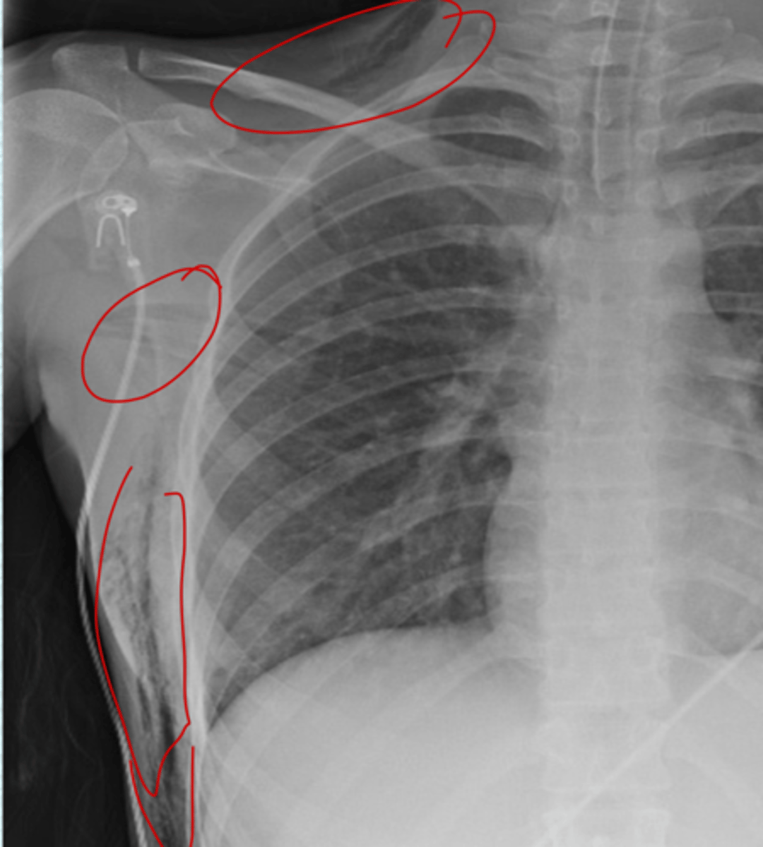
what are all of these indicative of?
Kerley B lines from interstitial edema
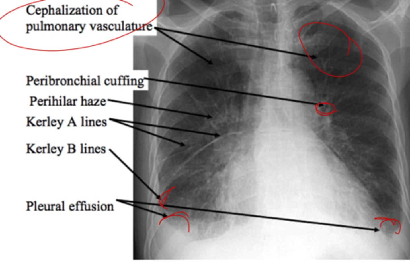
what is this pointing to and what caused it?
peribronchial cuffing, caused by fluid in the peribronchial interstitum
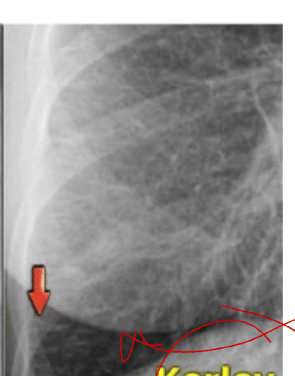
what is this called and what causes it
pulmonary alveolar edema
what is this
blunt aortic injury
what is this
Enlarged cardiac silhouette
what is happening here?
the thymus
this patient, age 2, is showing what?
pericardial effusion, water bottle sign
what is this?
the ascending and descending aorta and the aortic knob
this CXR shows abnormalities in which areas
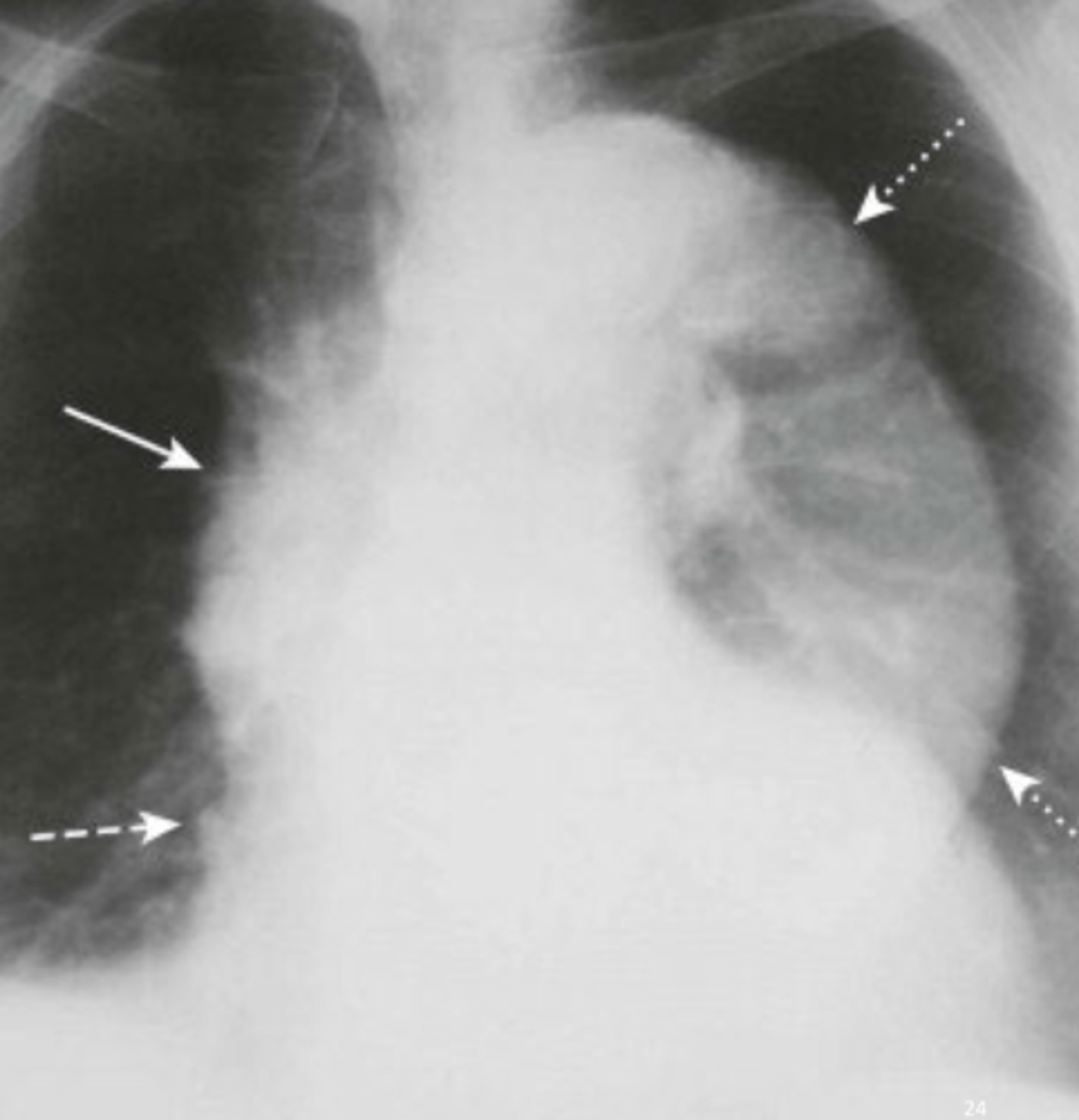
double density sign, indicative of Left atrium enlargement
what is this showing and what is it indicative of
small bowel
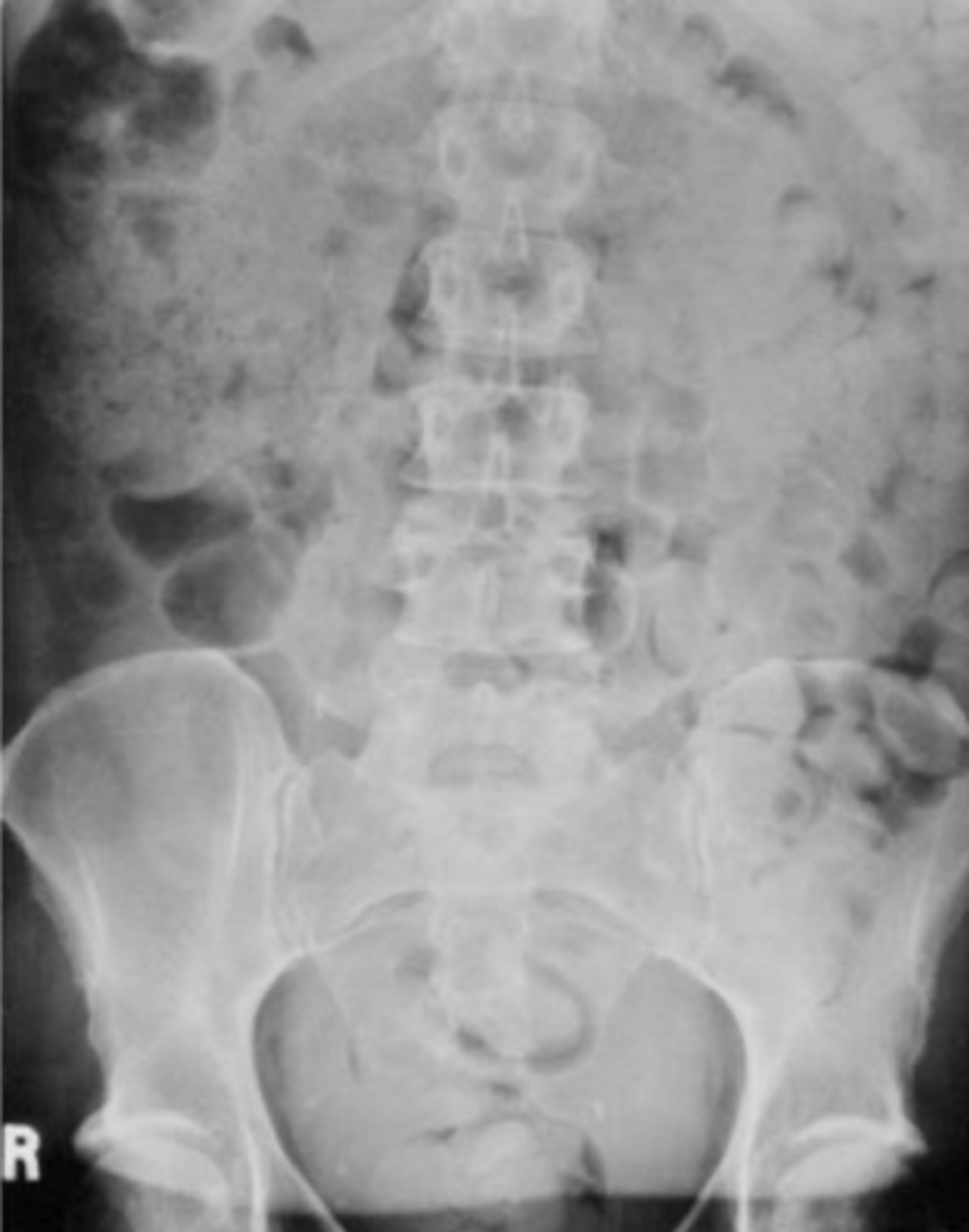
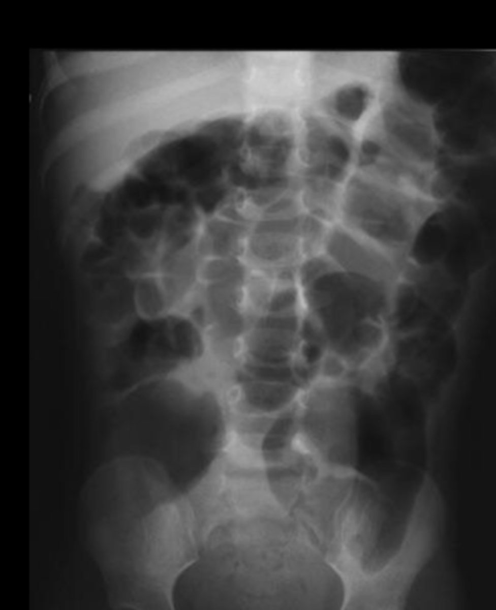
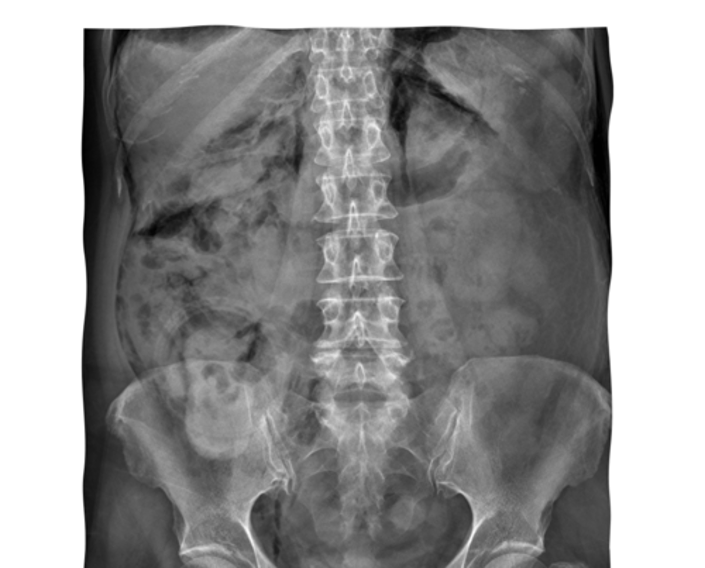
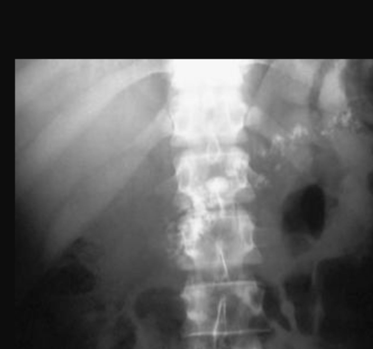
this is a cross section of what
normal gas patterns
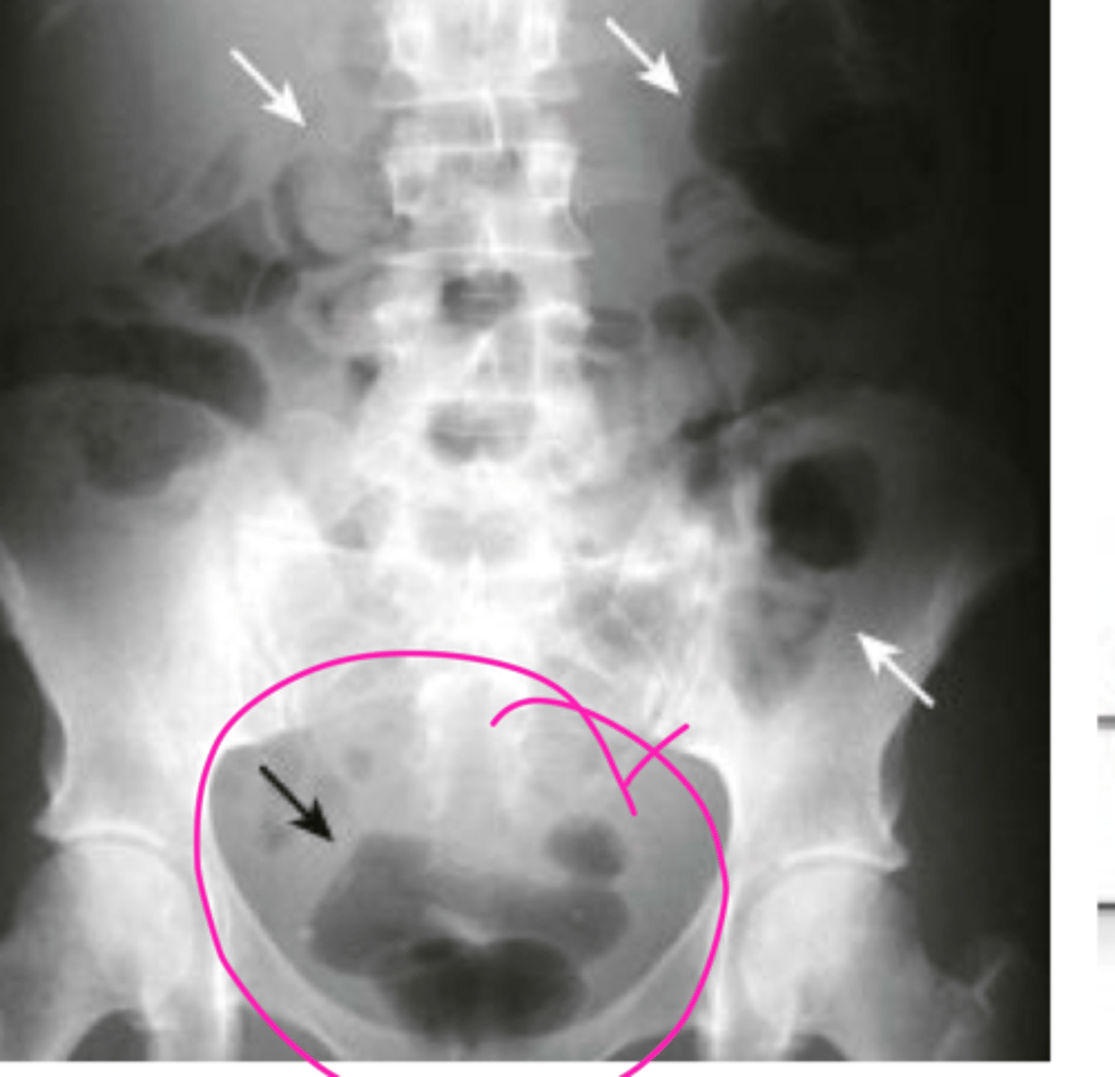
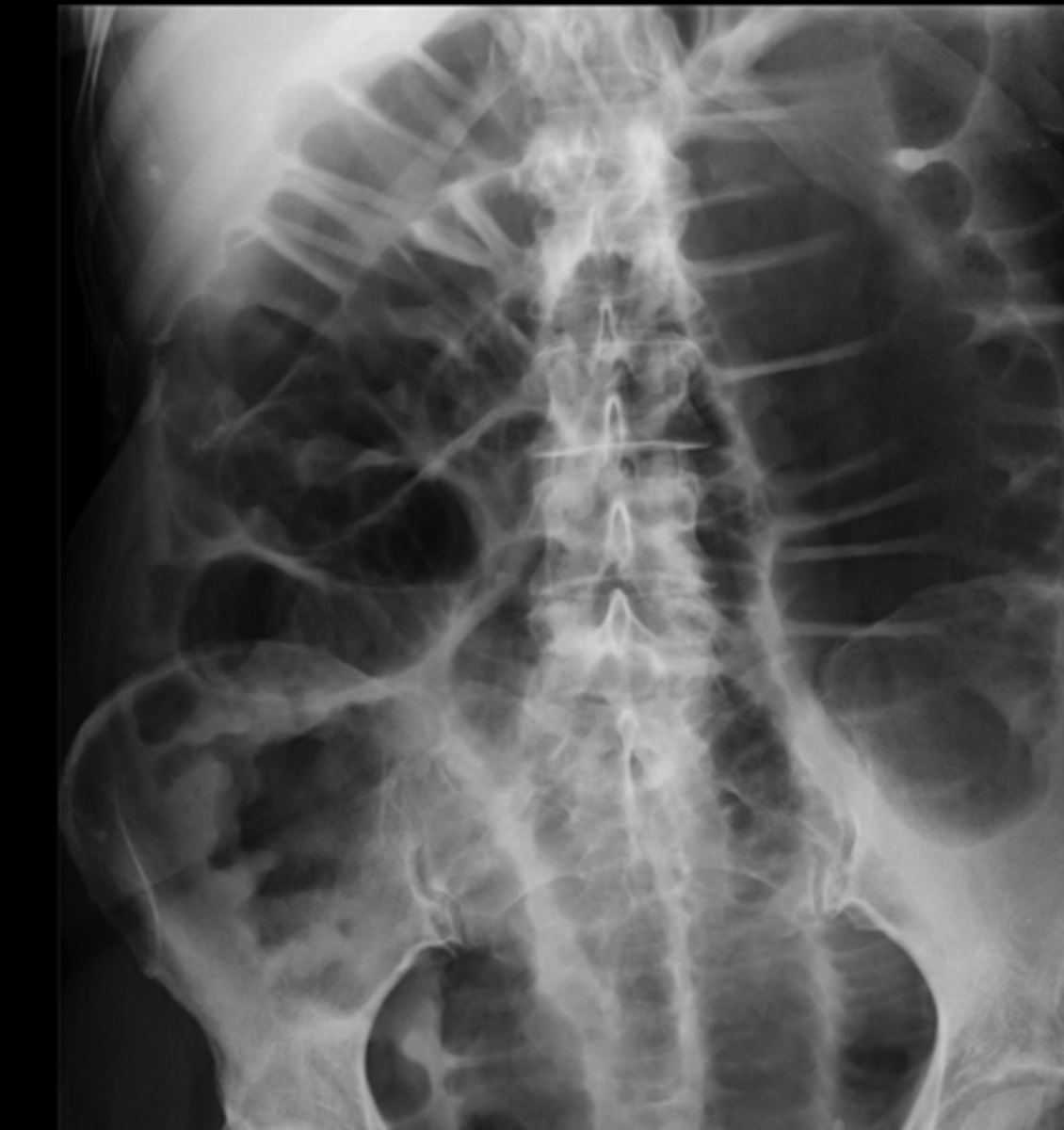
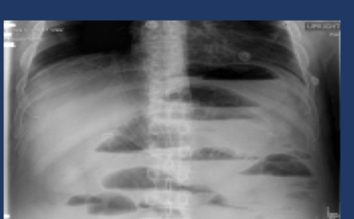
what is this showing
major constipation
what is this showing
kidney calcifications
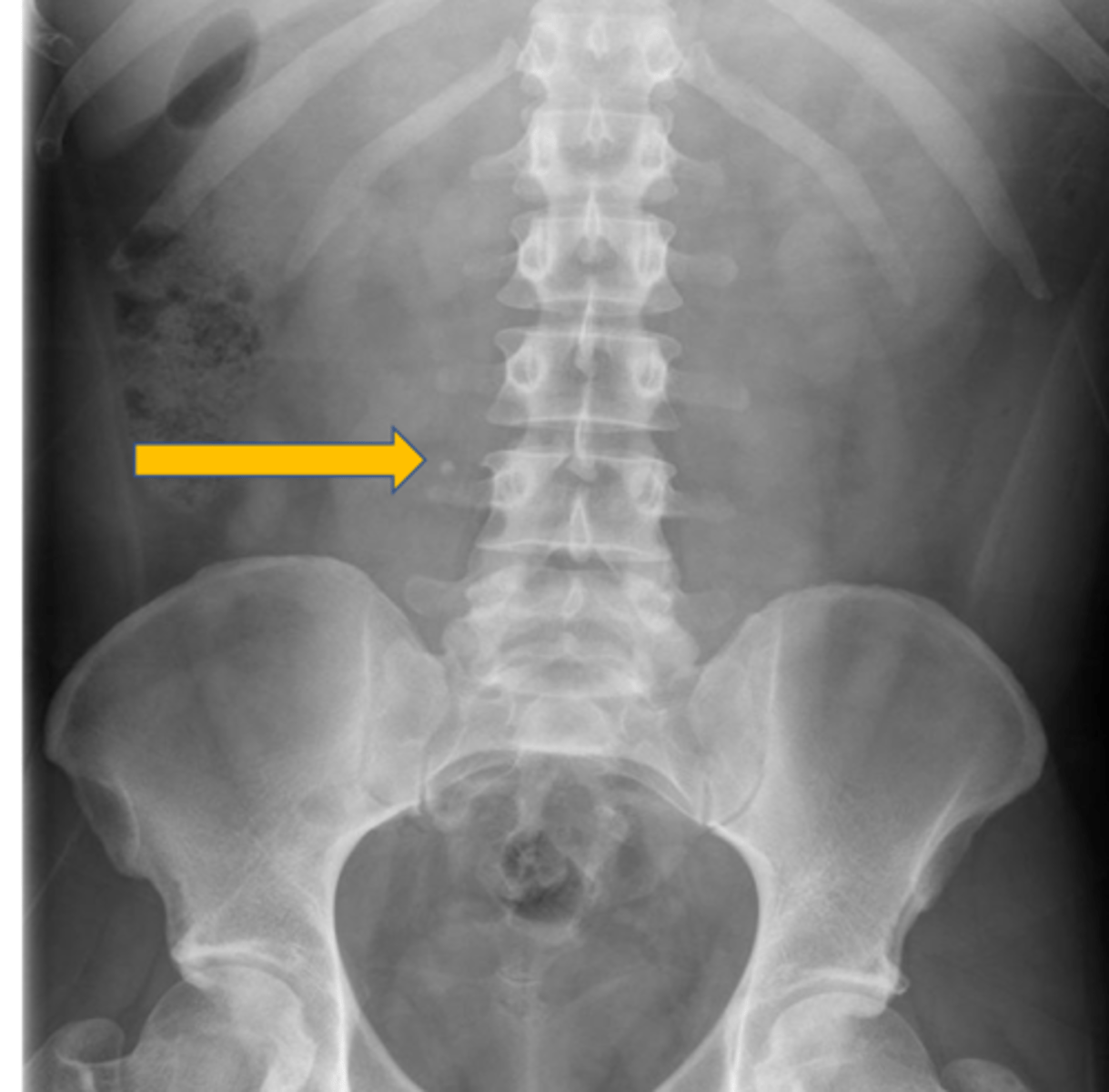
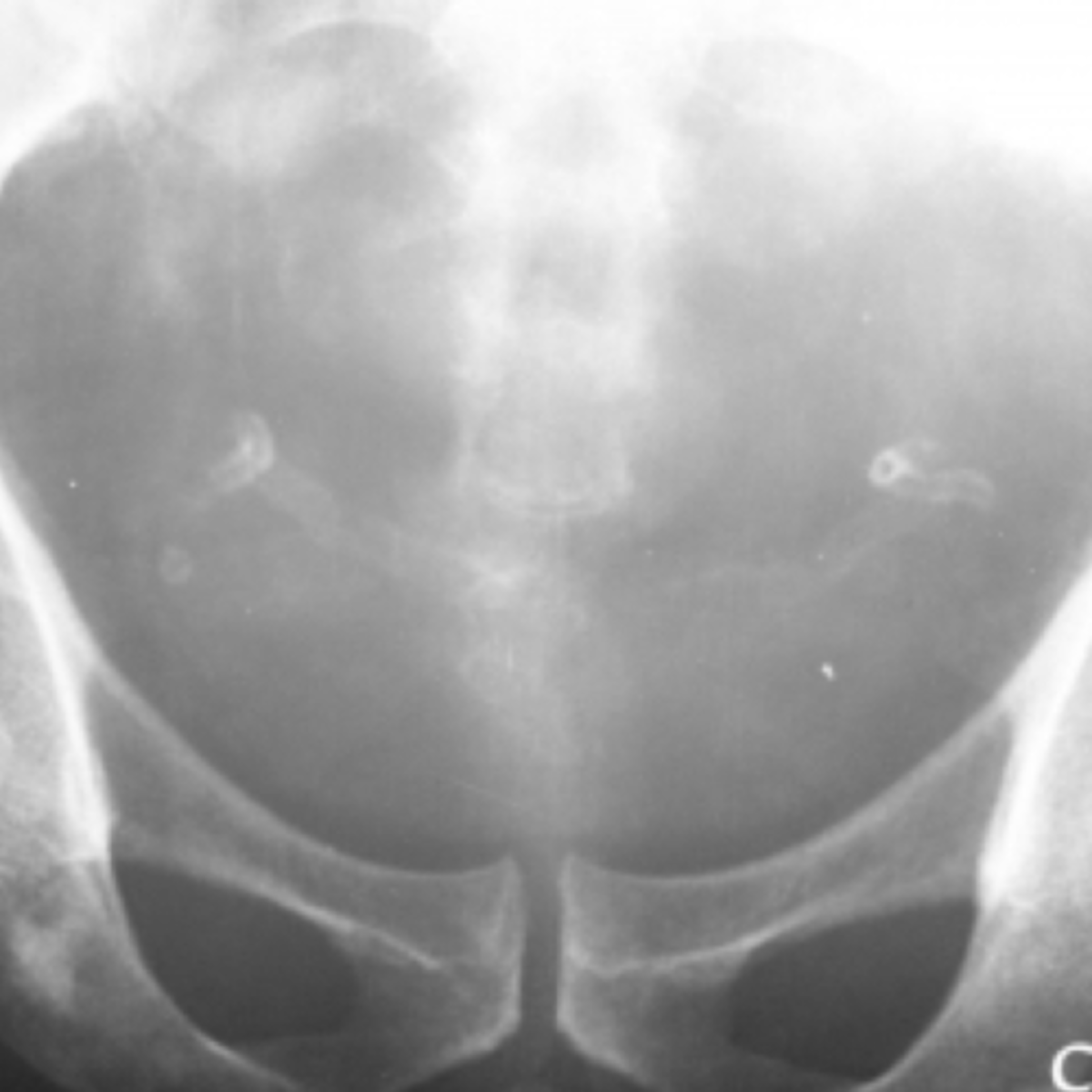
the pink circles are showing
phlebolith
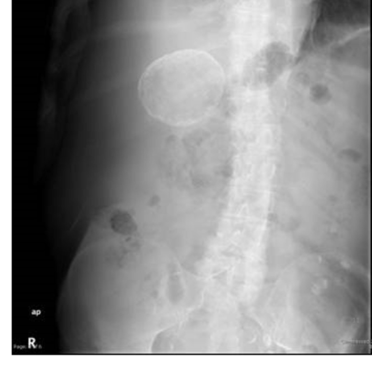
what is this
rib cartilage, could overly the gall bladder and kidney
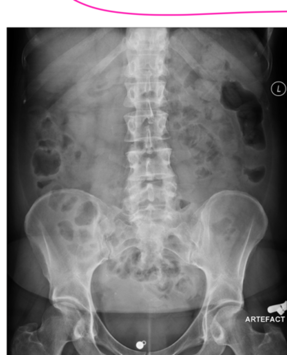
what is this
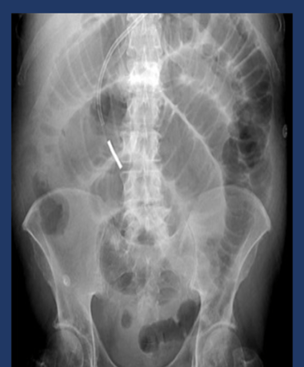
rectal gas, prone view
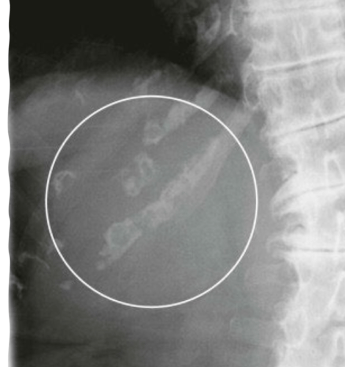
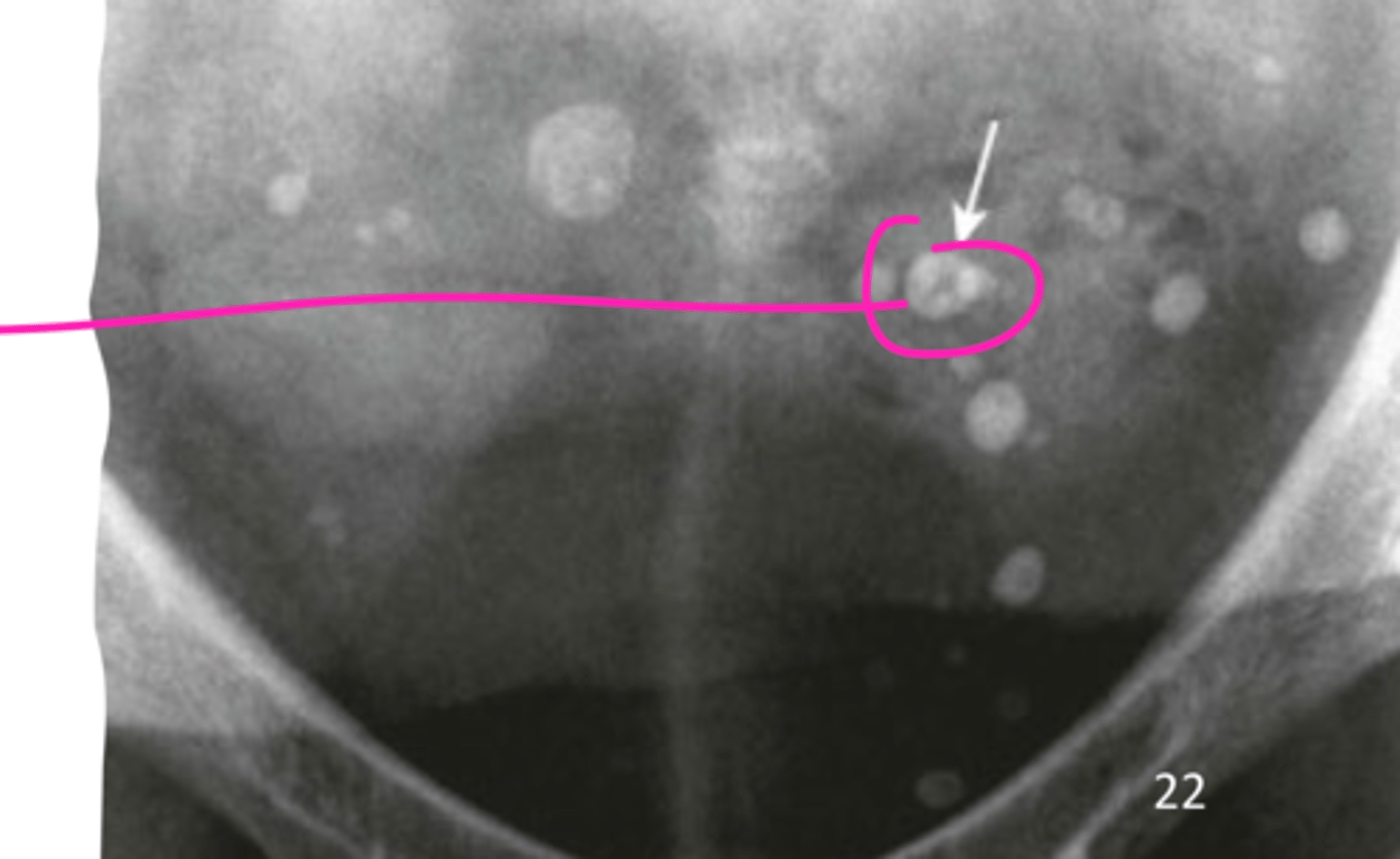
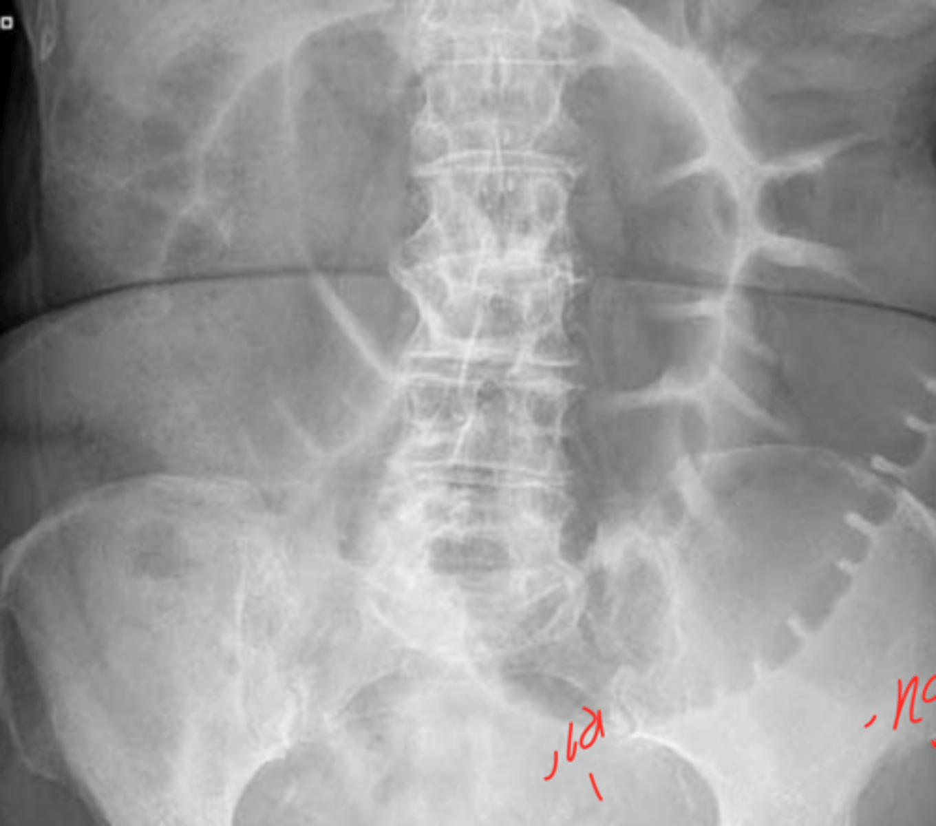
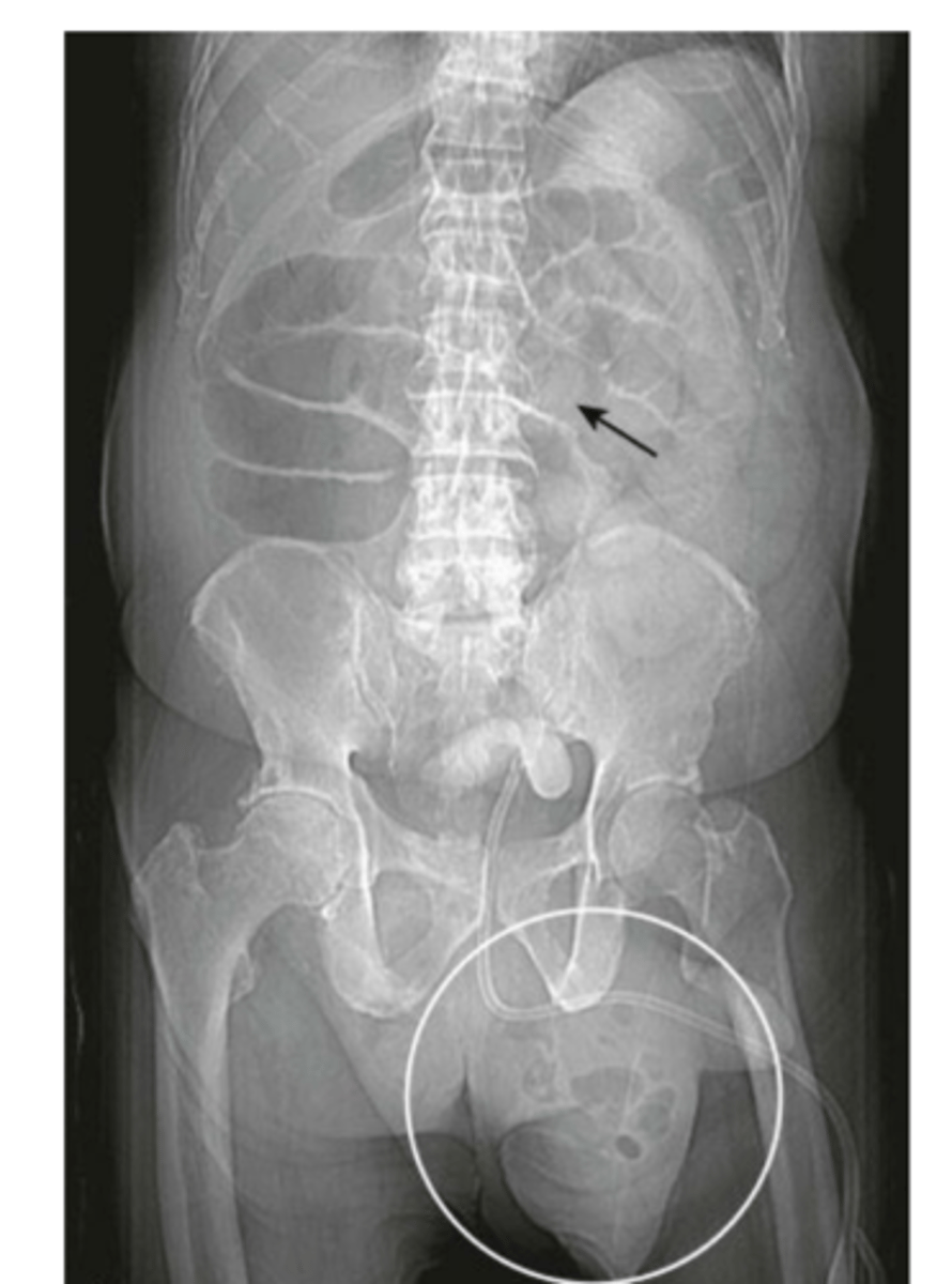
the pink circle is indicating what, what view was this likely taken at
localized ileus
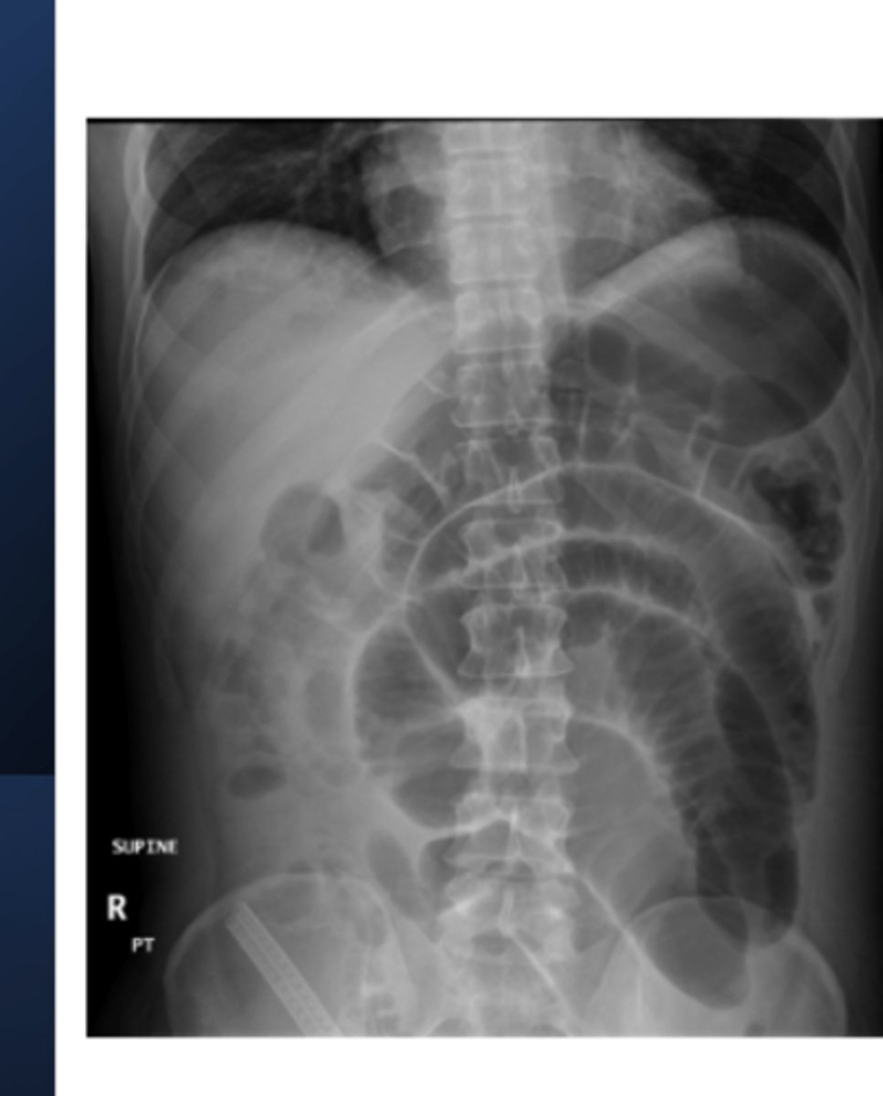
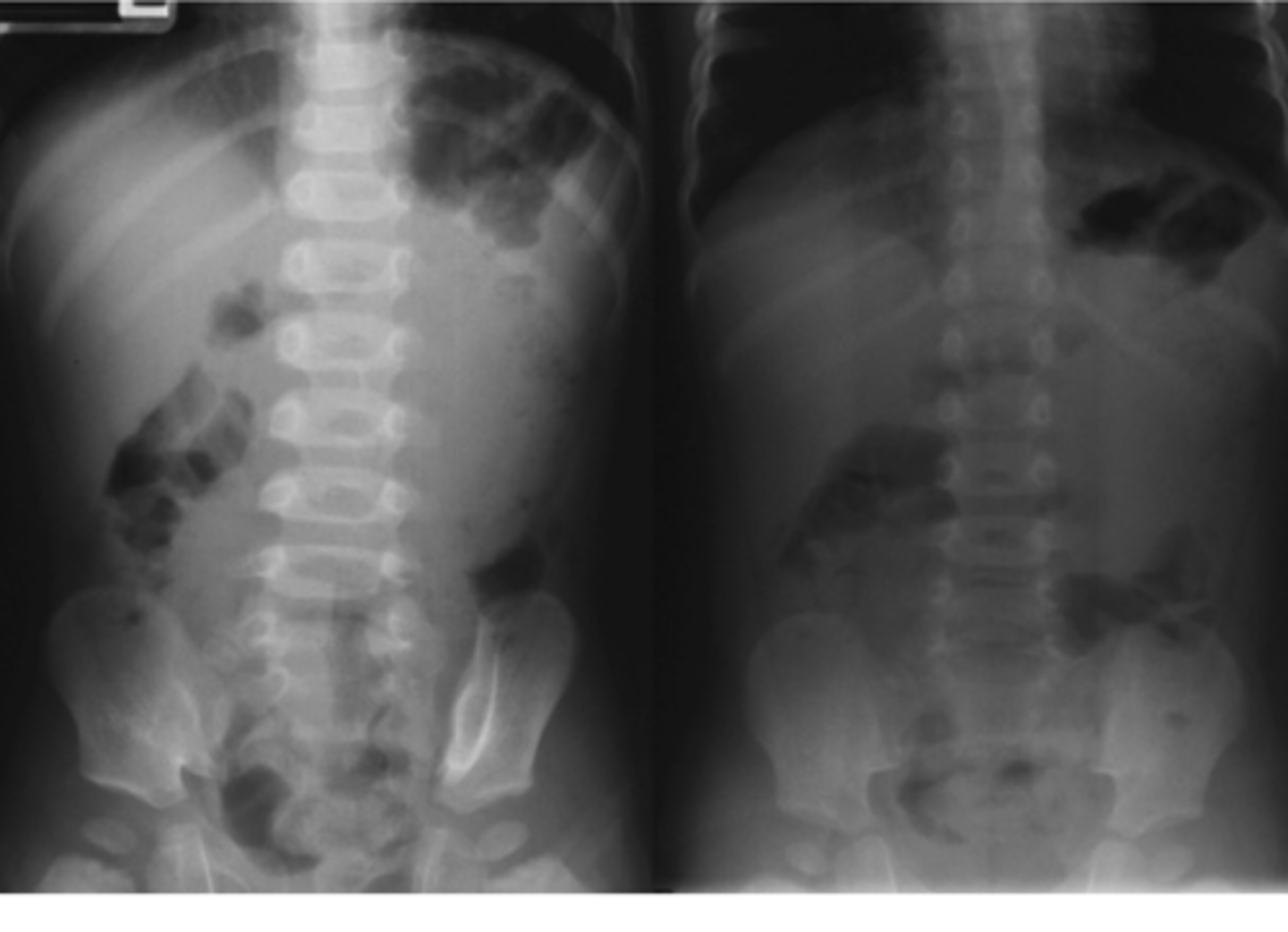
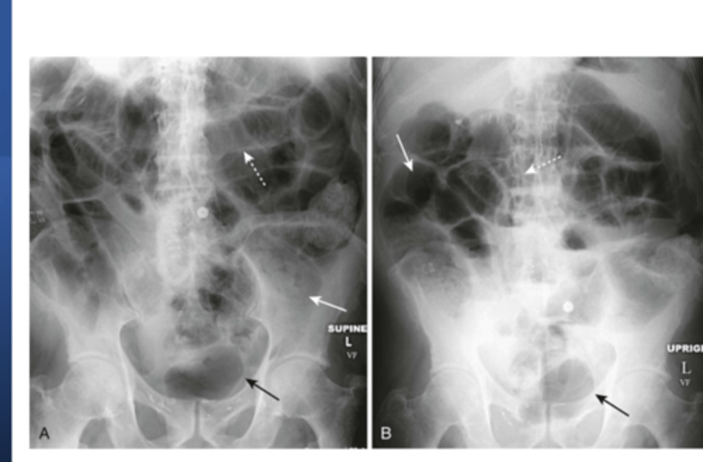
what is this photo showing (two views here)
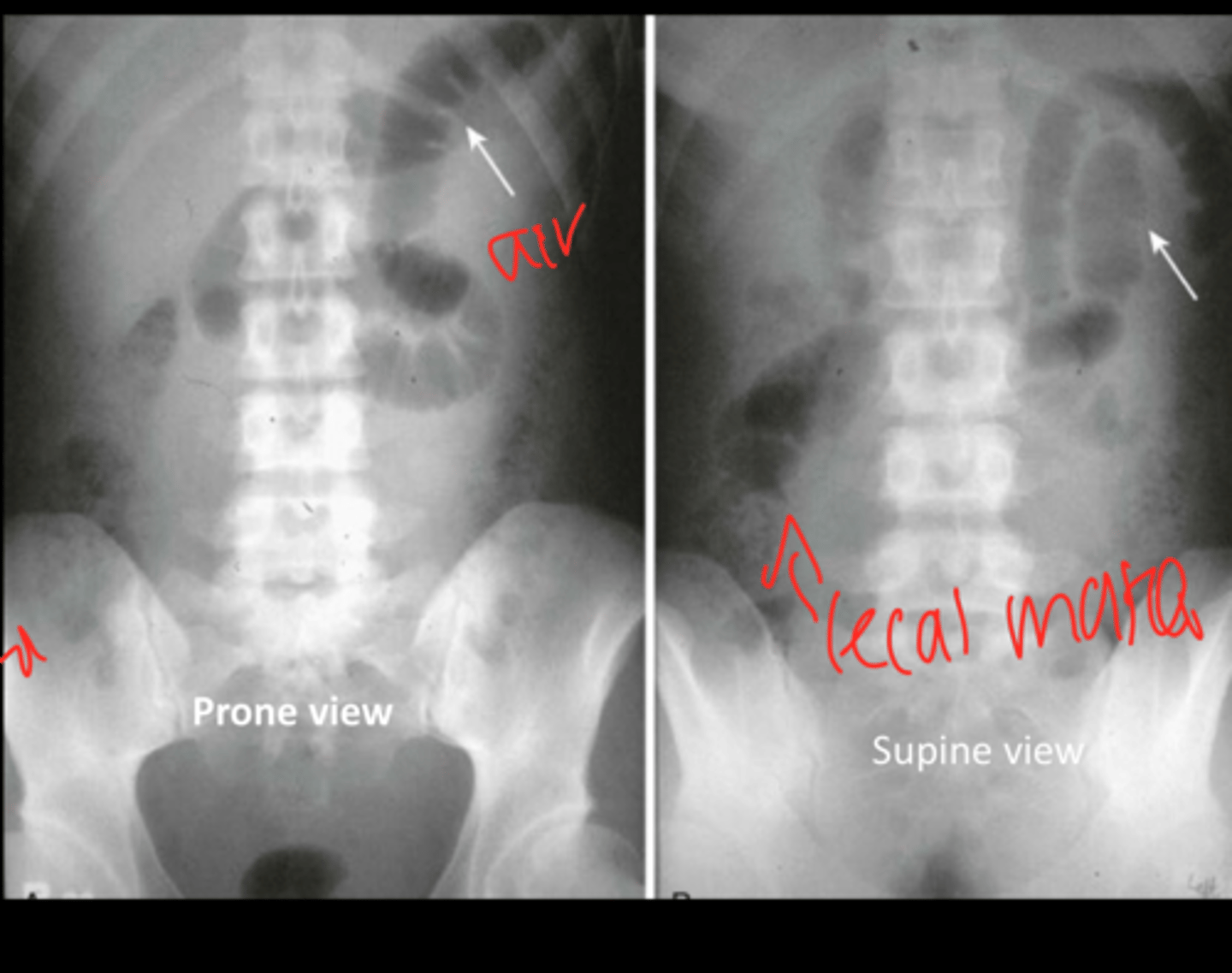
generalized ileus
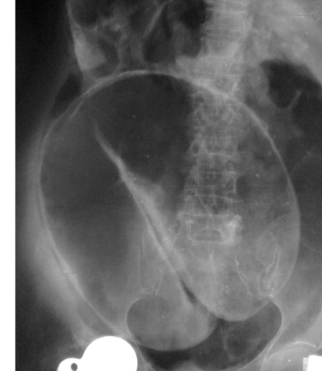
what is being shown here
generalized ileus
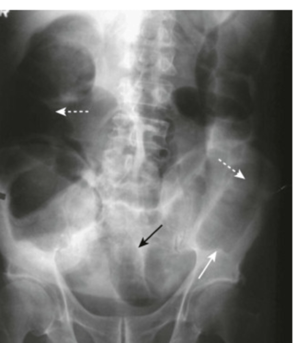
what is this
MLBO
what is this
MLBO
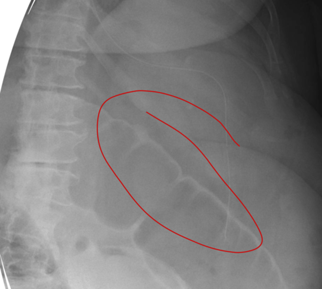
what is this
volvulus
what is this
MLBO
identify what this is
MSBO
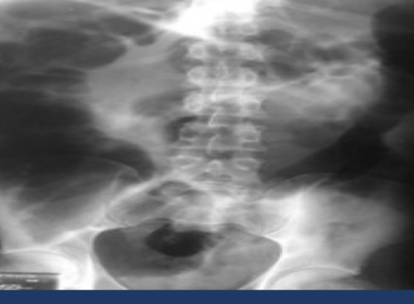
identify what this is
localized ileus
identify what this is
MLBO
identify what this is
MSBO
identify what this is
generalized ileus
identify what this is
MSBO
identify what this is
pneumoperitoneum- crescent sign
identify what this is
riglers sign in pneumoperitoneum
identify what this is
pneump-retroperitoneum
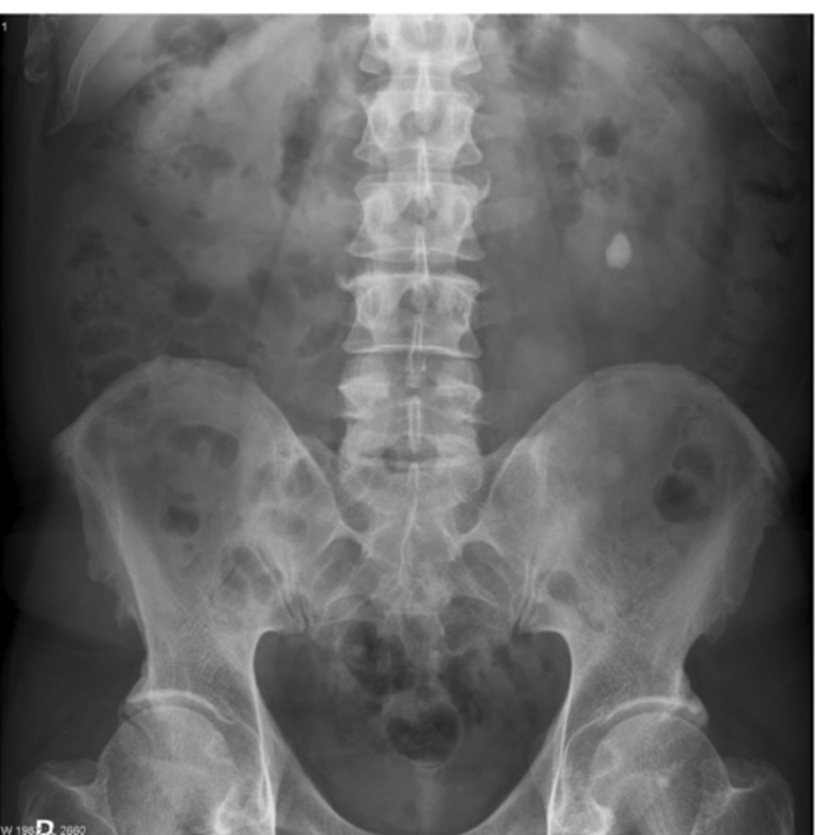
identify what this is
calcification in the ureter
what is this and where is it located
calcification in the kidney
what is this and where is it located
rim like calcification of the gall bladder
What is this
linear calcification of the fallopian tubes
what is this
a lamellar calcification
what is this
amorphous calcification
what is this
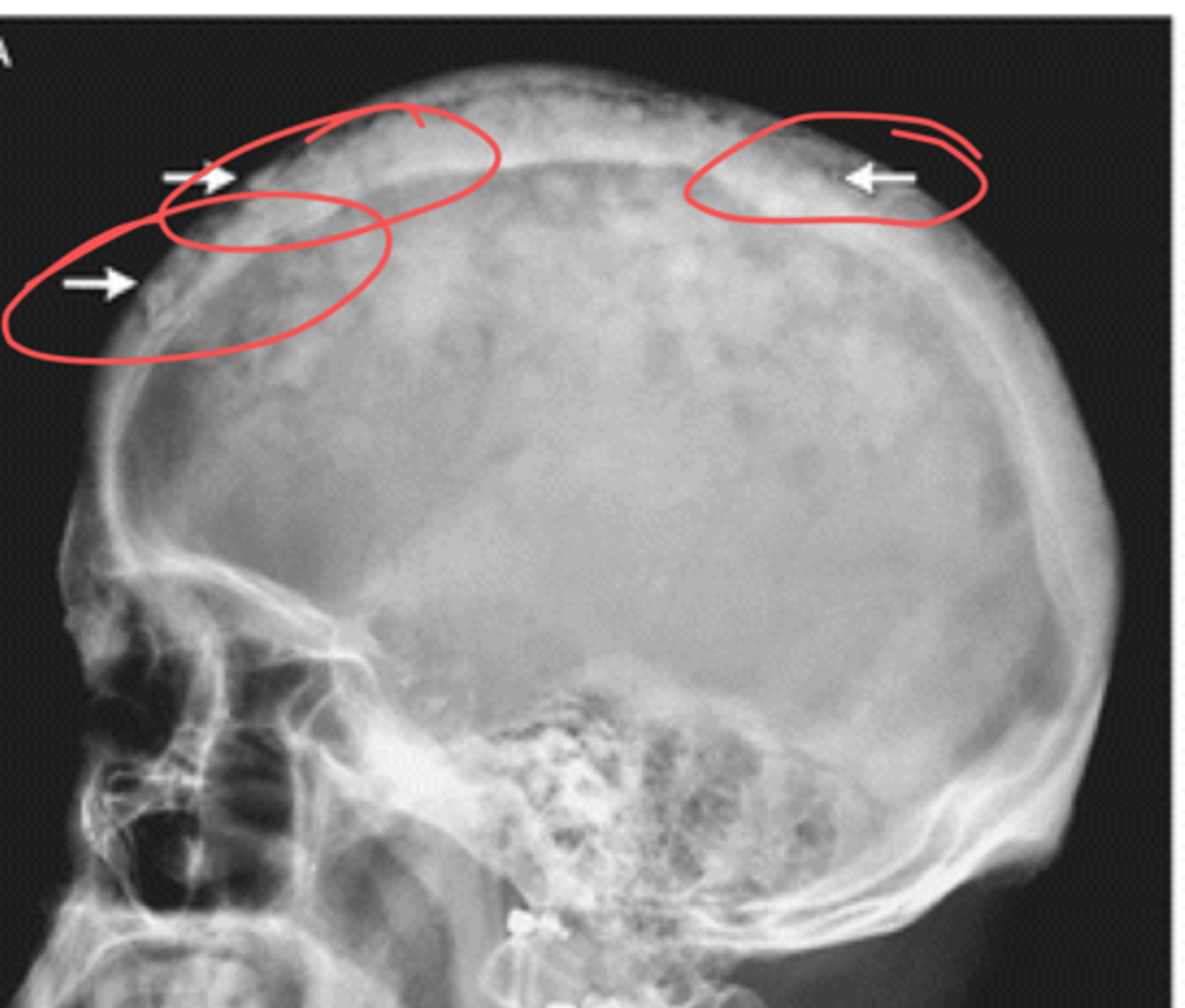
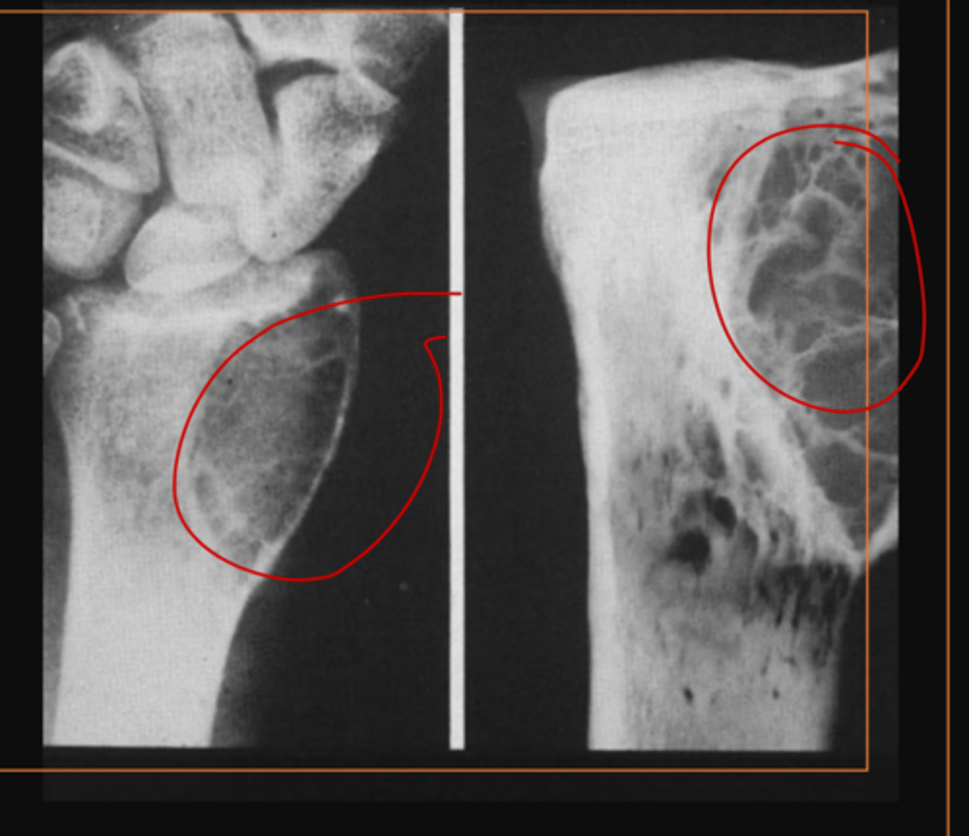
pagets disease
what disease process is this showing
cotton wool spots, caused by pagets disease
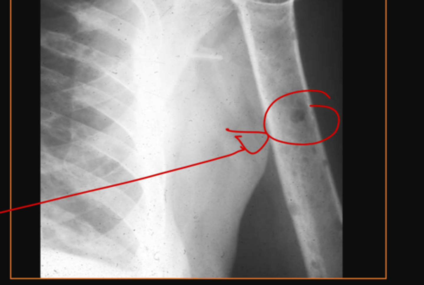
what is this and what causes it
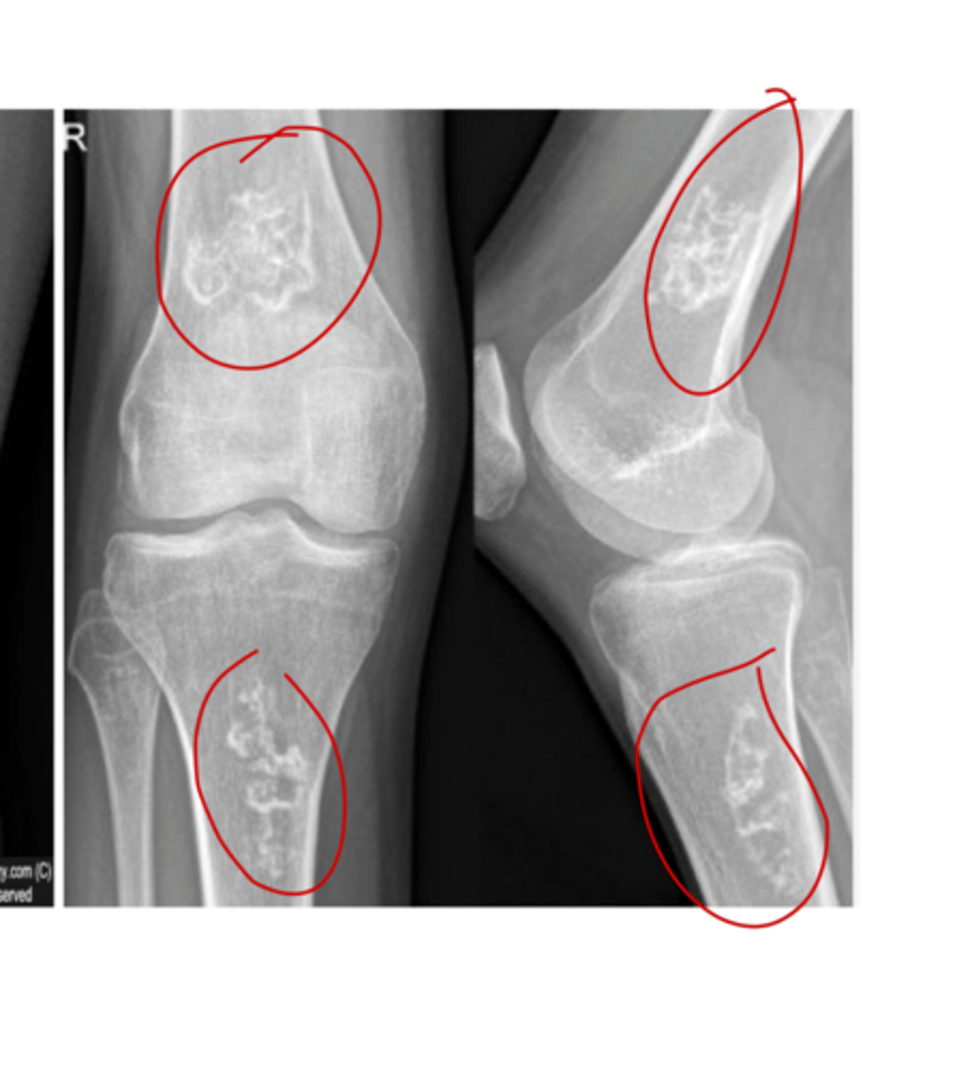
medullary bone infarcts
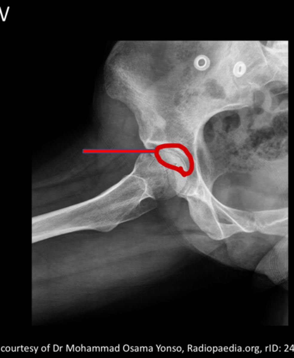
what are these?
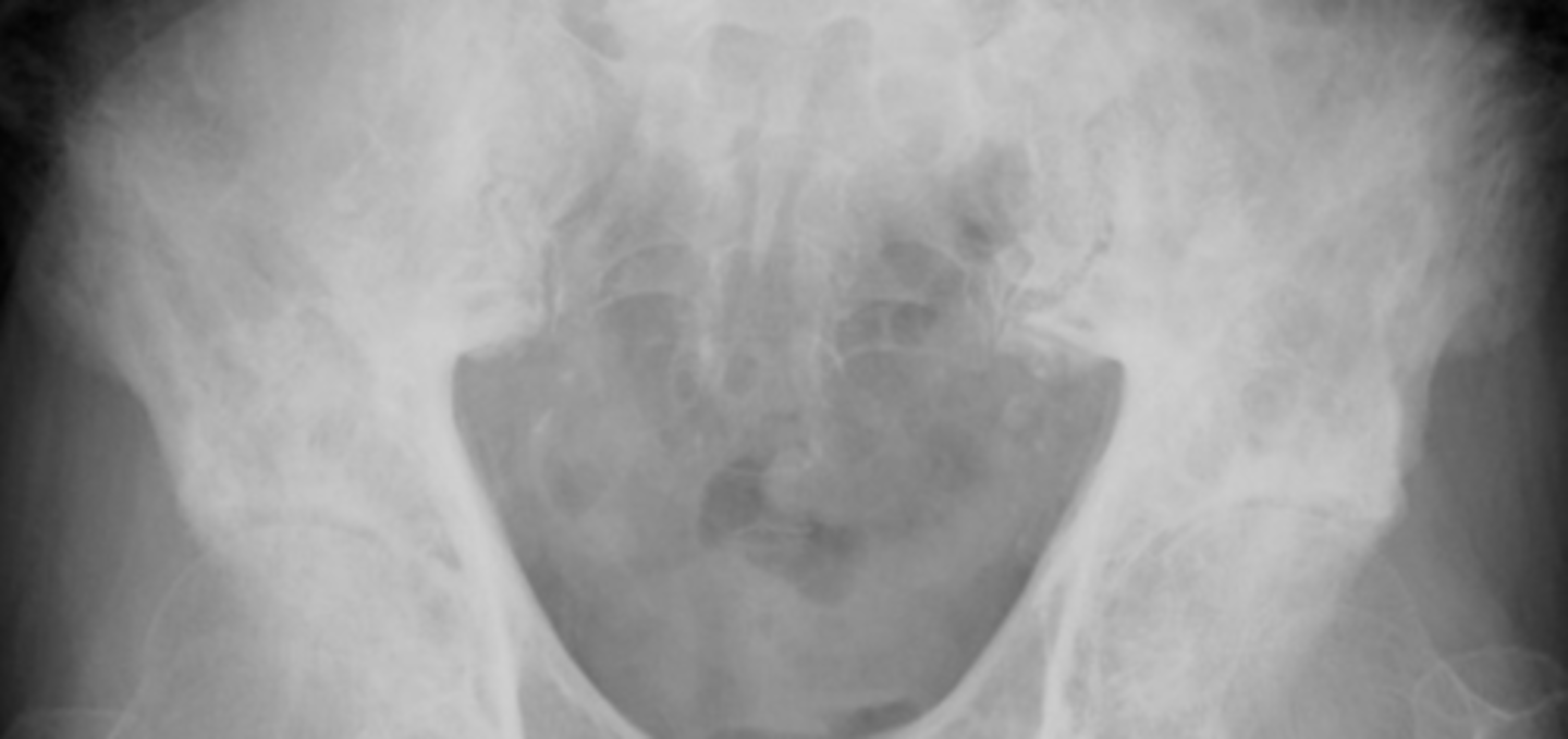
Legg-Calve-Perthes disease
what is this depicting
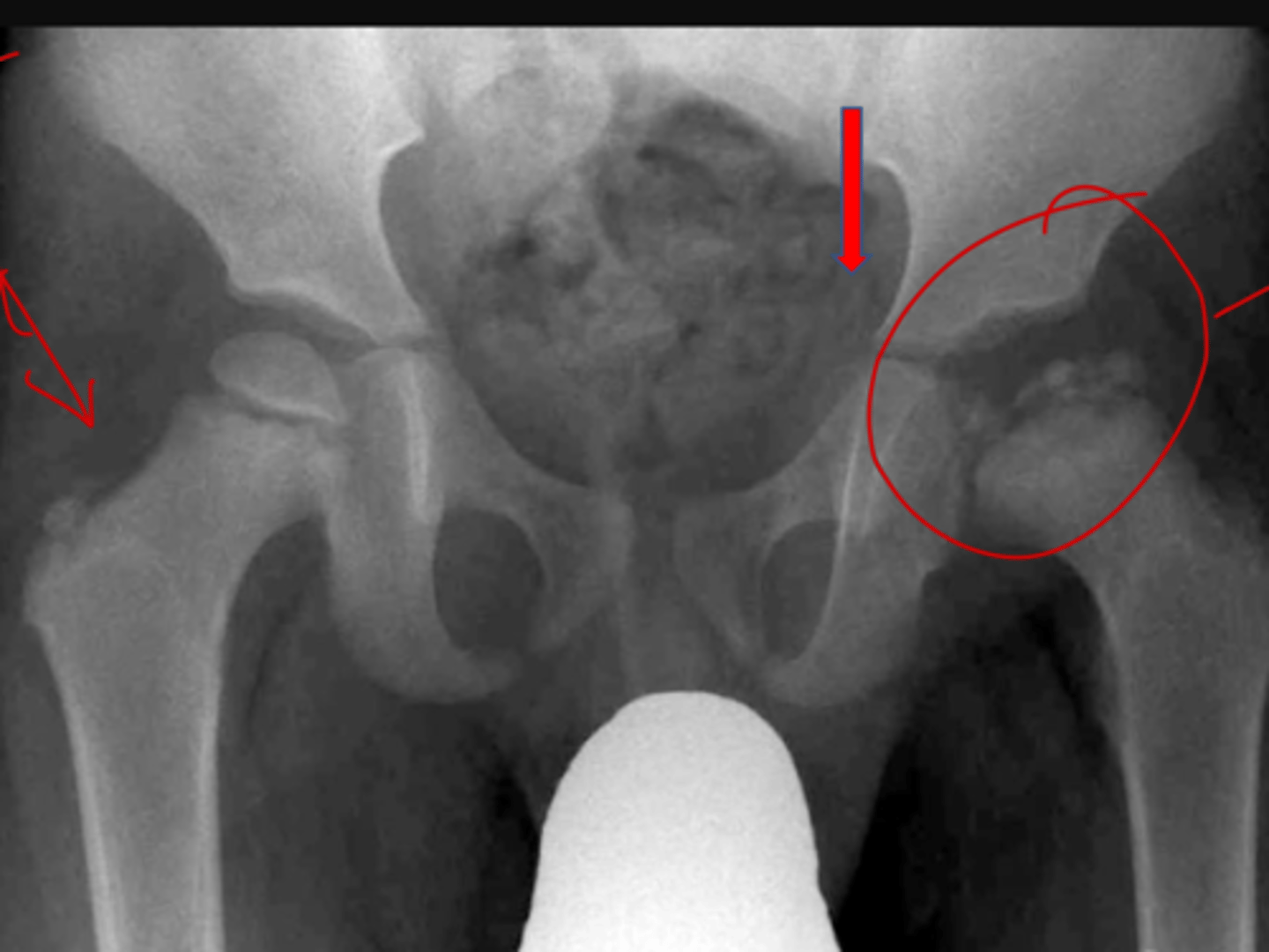
Crescent sign in Legg-Calve-Perthes
what is this showing, what is it indicative of?
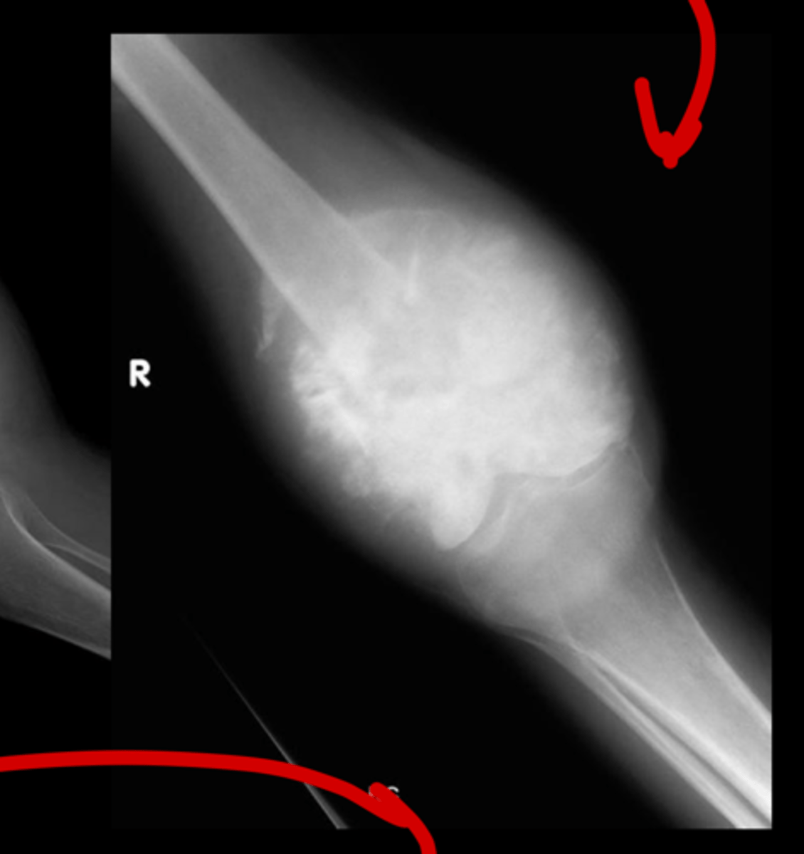
sunburst
what kind of periosteal reaction is this
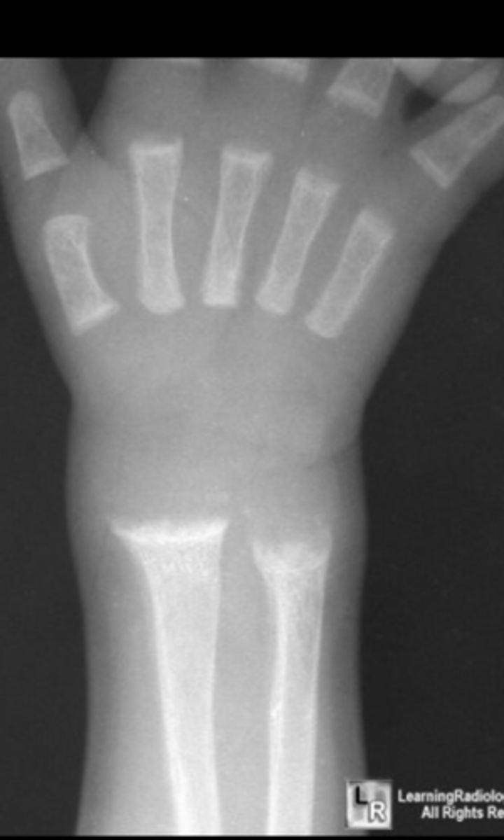
osteoprosis of the foot
what is this
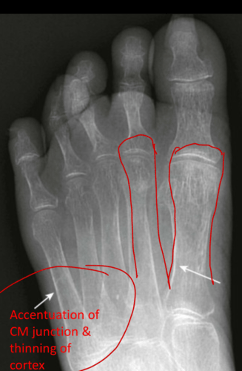
Fraying of the metaphyses in rickets
what is this condition and what feature is this image highlighting
soap bubbles in plasmacytoma
what is this and what disease process
lytic lesions in multiple myeloma
what is this
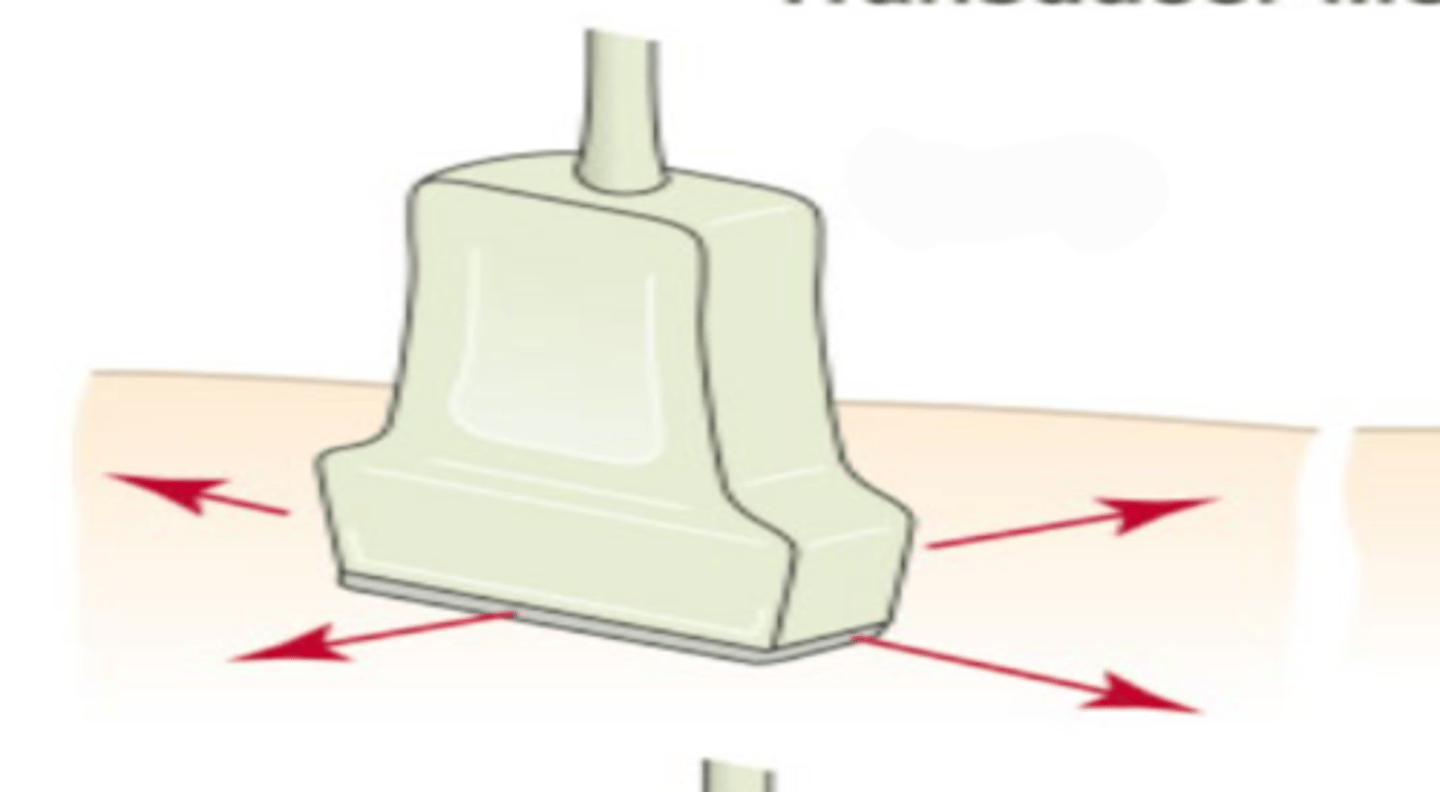
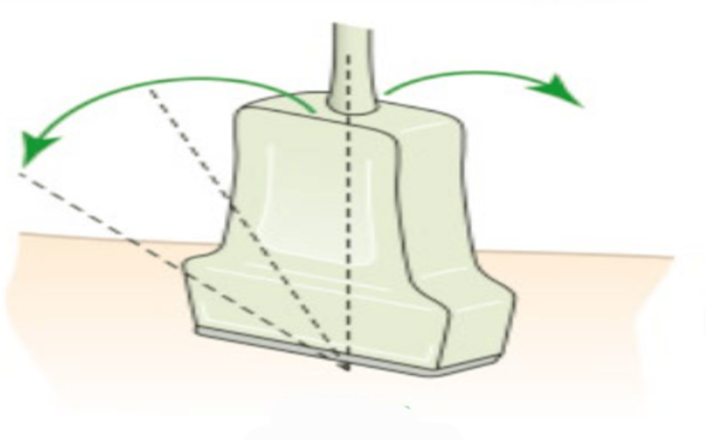
sliding
what is this transducer movement called
rotating
what is this transducer movement called
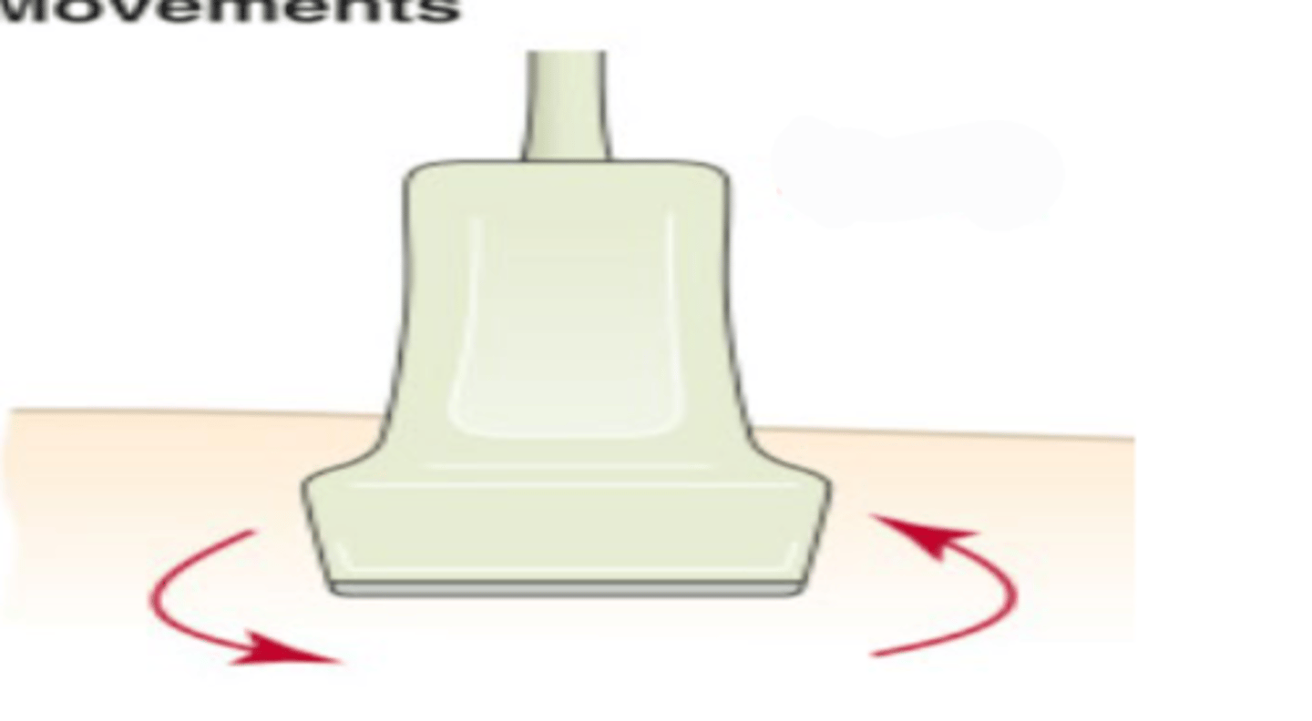
rocking
what is this transducer movement called
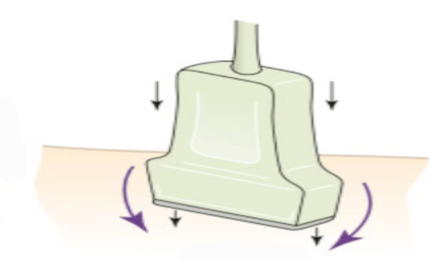
tilting
what is this transducer movement called
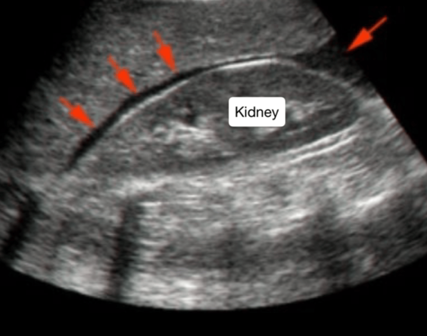
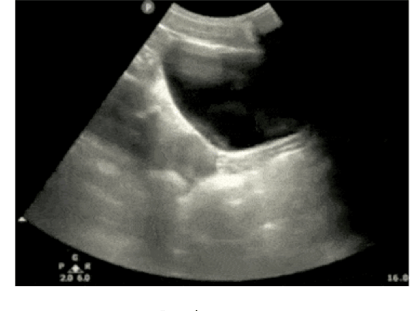
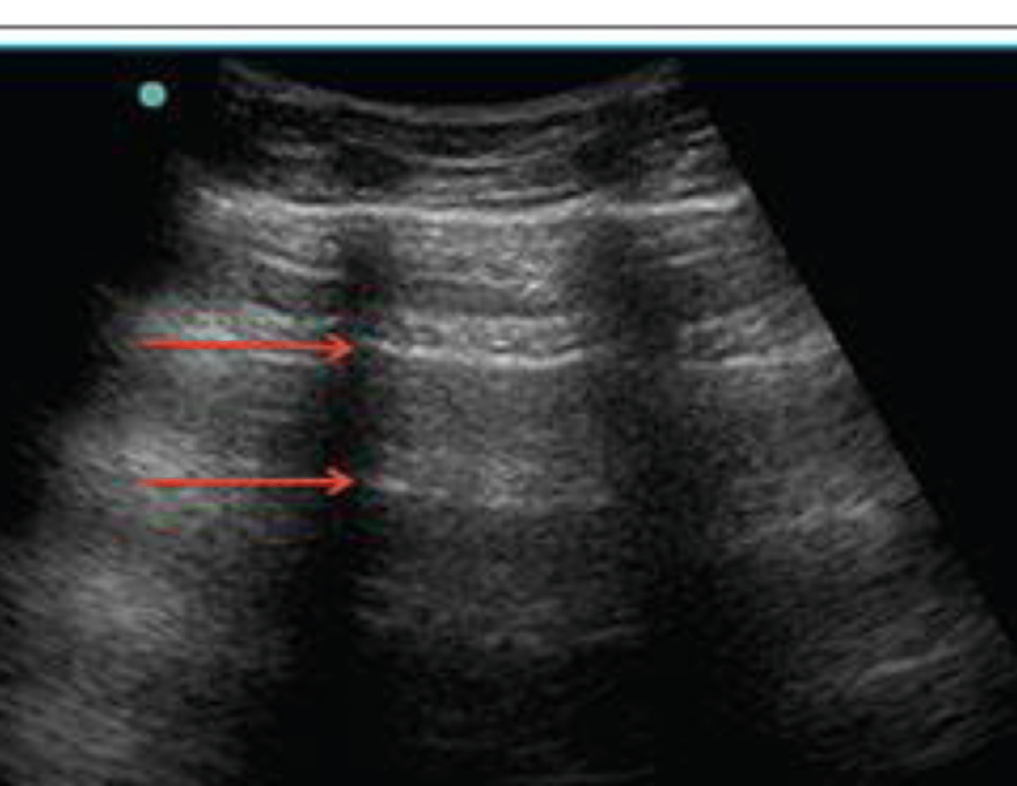
fluid between the kidney and liver
this RUQ view during the EFAST reveals what
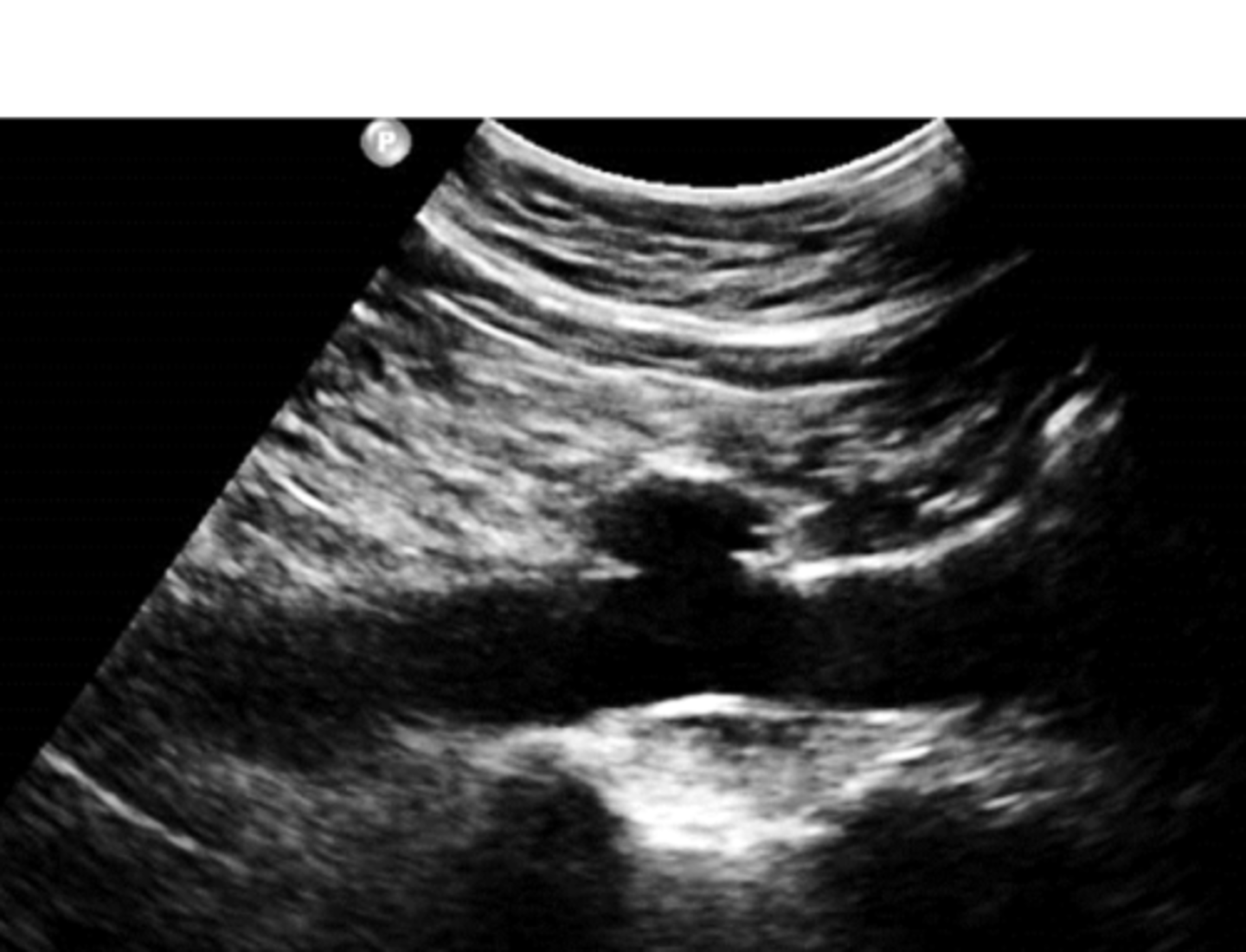
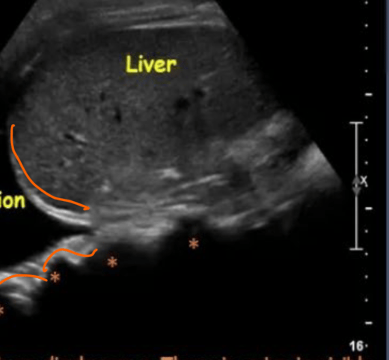
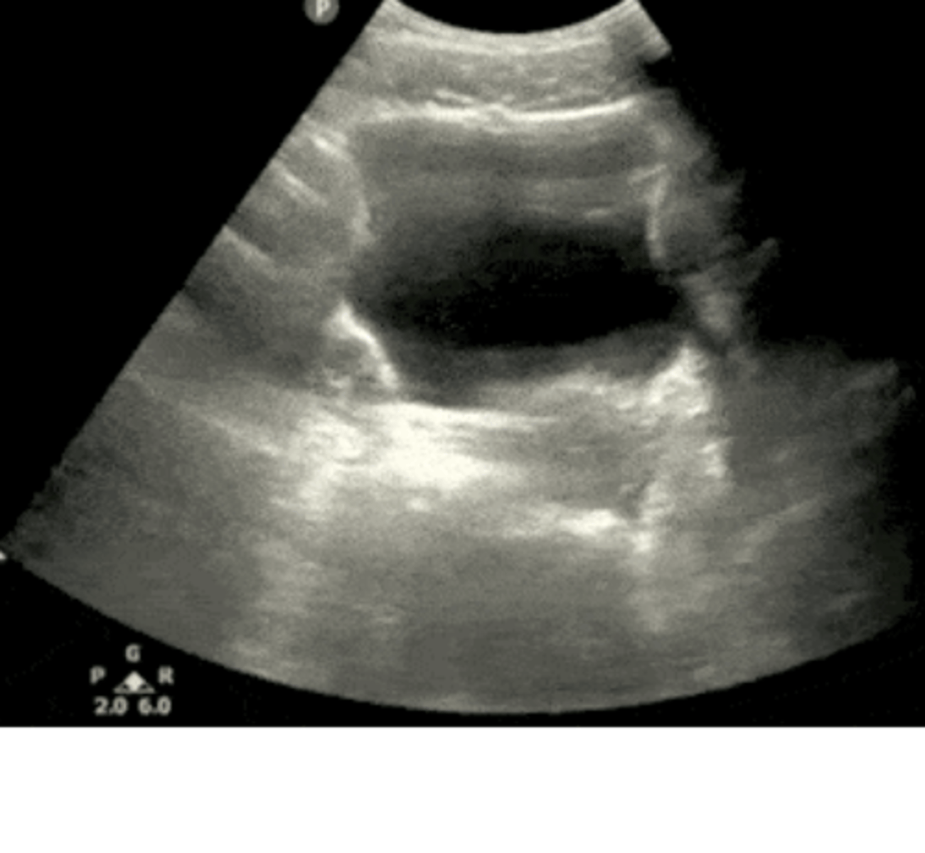
spine sign, pleural effusion
this is a RUQ view, what is it showing and what is it indicative of
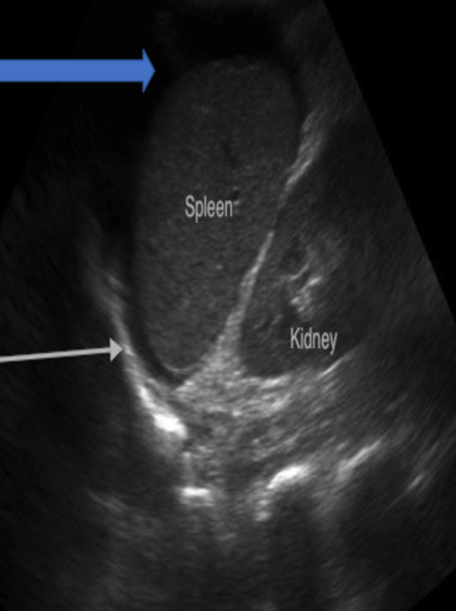
+LUQ
what is this
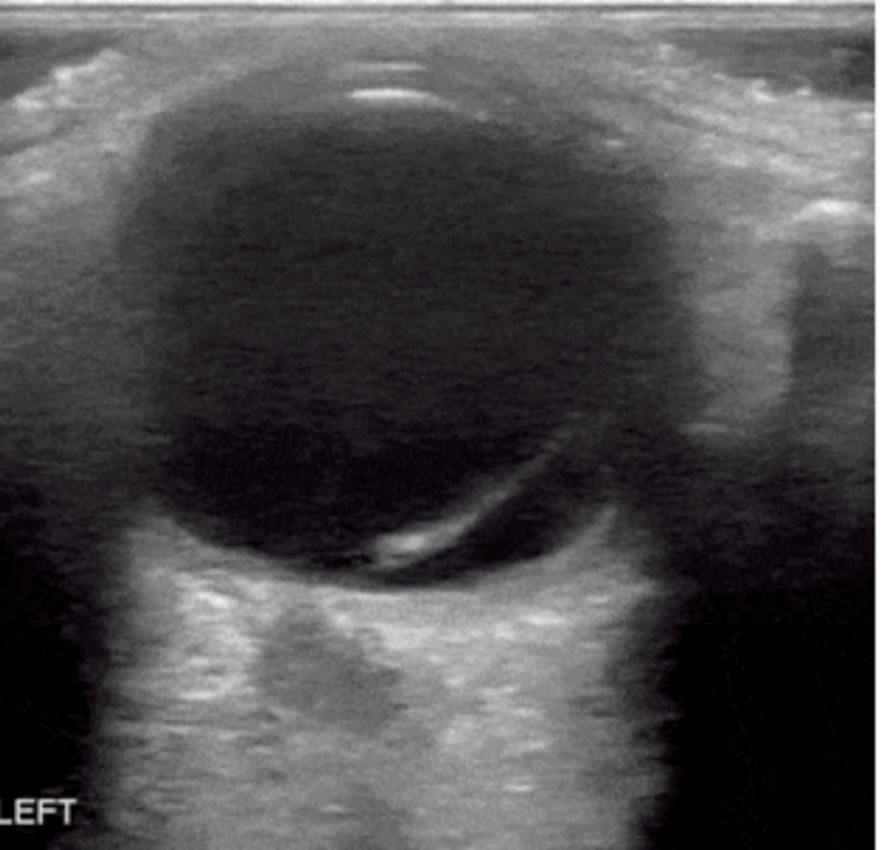
female pelvis, longitudinal view
what view is this US of (be specific)
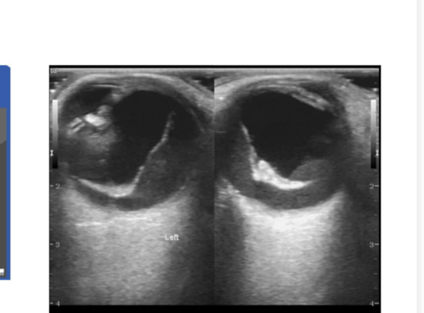
transverse pelvis
what is this view of
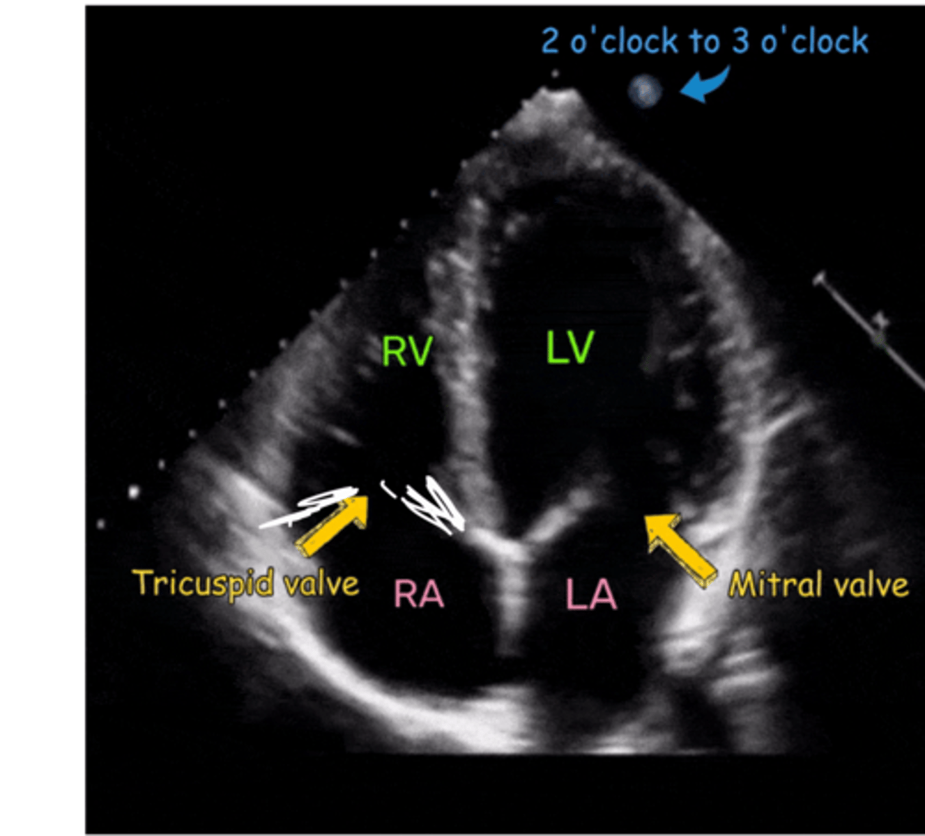
left atrium
identify the green circle area
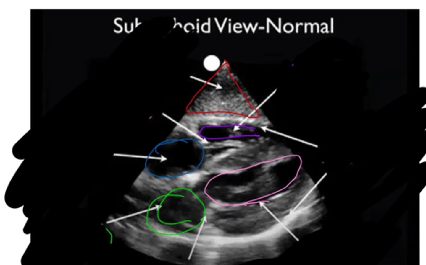
right atrium
identify the blue circle area

left ventricle
identify the pink circle area

right ventricle
identify the purple circle area

Liver
identify the red triangle area

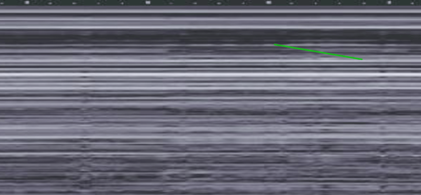
M-mode ultrasound, normal seashore sign
what is this and how was it taken
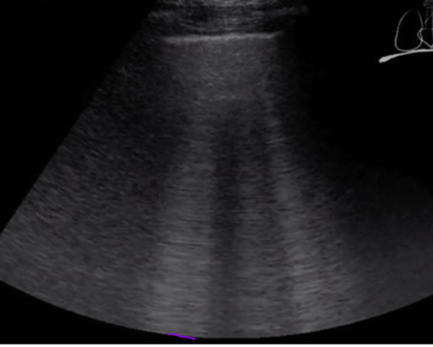
barcode/stratosphere sign, pneumothoraz=x
what is this and what is it indicative of
A-line artifacts
what is the arrow pointing to
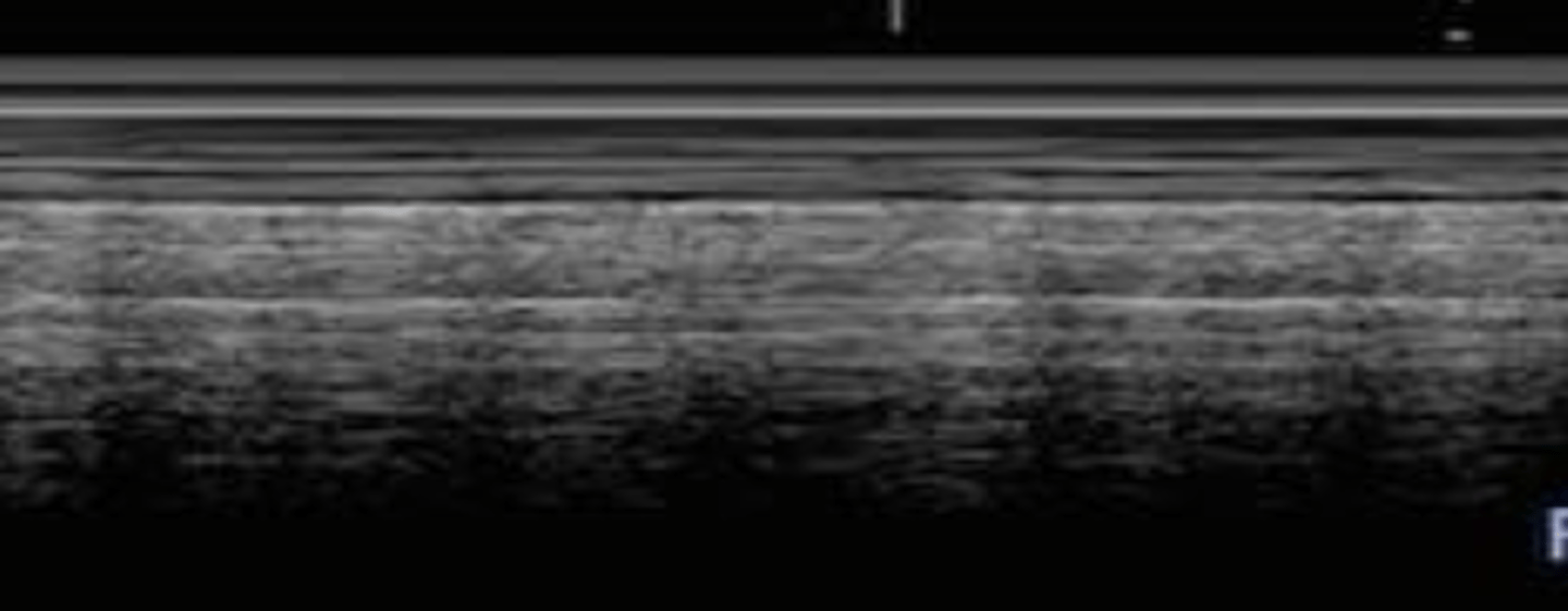
B lines
what are these?
retinal detachment
what is happening here
posterior vitreous detachment
what is this
apical 4 chamber US view
what view was this taken from?
SMA view, mid aorta
what view is this
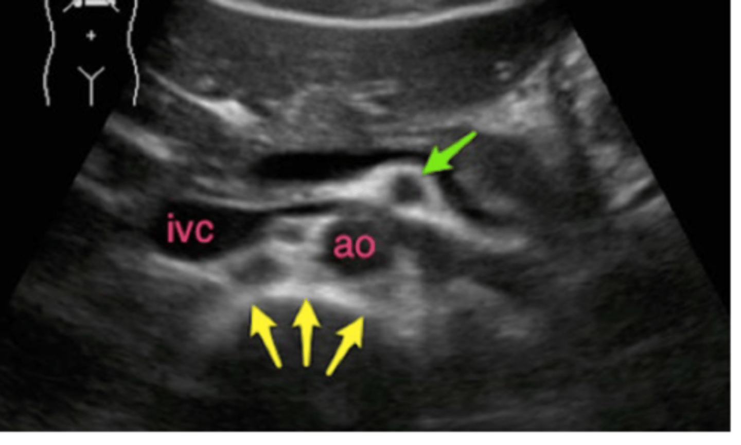
fusiform aortic aneurysm
what is this