FINALS BIO FLASHCARDS
1/170
Earn XP
Description and Tags
save my soul (cards 1-65: DNA, RNA, protein synthesis | cards 66-
Name | Mastery | Learn | Test | Matching | Spaced | Call with Kai |
|---|
No analytics yet
Send a link to your students to track their progress
171 Terms
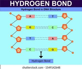
DNA structure
complementary base pairing rule
covalent bonds everywhere except between 2 bases, which is hydrogen bond
double stranded, double helix
one strand in the 3’-5’ direction, one in the 5’-3’ direction
dioxyribose
large and less mobile than RNA
bases - ATCG
RNA structure
complementary base pairing rule (slightly different)
single stranded, single helix
synthesized in the 5’-3’ direction
bases - AUCG
ribose
smaller than DNA, more mobile
nucleic acids
purpose - stores genetic material
monomer - nucleotide
polymer - nucleic acids
sugars - dioxyribose (DNA) or ribose (RNA)
bases - acg and t (DNA) or u (RNA)
shape - helix
antiparallel
antiparallel
parallel, but running in opposite directions
DNA is antiparallel - one strand is in the 3’-5’ direction, one is in the 5’-3’ direction
gene
the information regarding how to produce a specific protein
nucleotide structure
3 parts - phosphate, sugar (dioxyribose or ribose), and base (ATCGU)
deoxyribose
the sugar found in DNA - missing an oxygen
ribose
the sugar found in RNA - has an additional oxygen compared to deoxyribose
Erwin Chargaff’s rules
discovered that the number of adenine (A) bases is equal to the number of thymine (T) bases in DNA and that the number of cytosine (C) bases is equal to the number of guanine (G) bases
Franklin and Wilkins
used x-ray crystallography to photograph a DNA molecule
Watson and Crick
Interpreted the DNA image taken by Franklin and Wilkins and used Chargaff’s rules to determine that the shape of DNA is a double helix
Components of DNA double helix
shaped like a ladder
rail of the ladder - sugar and phosphates
ring of ladder - 2 complementary bases
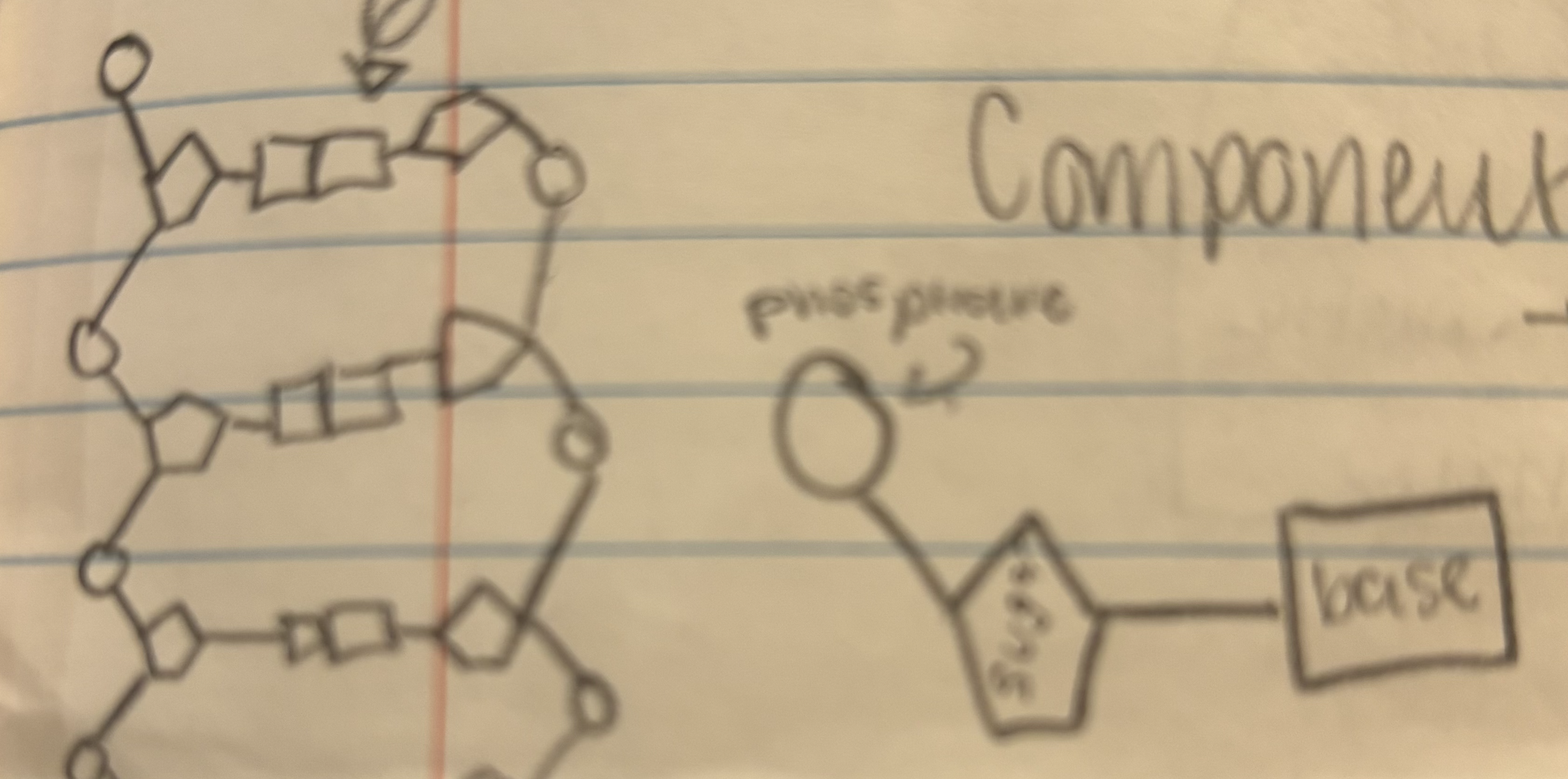
bonds in DNA
covalent bonds everywhere (shared electrons) with 2 hydrogen bonds between A and T and 3 hydrogen bonds between C and G
Direction of DNA
1 strand is in the 3’-5’ direction, the other is opposite, going in the 5’-3’ direction
antiparallel
complementary base pairing rule
in DNA: A bonds with T, C bonds with G
in RNA: A bonds with U, C bonds with G
2 categories of bases
purines and pyrimidines
purine
a molecule with 2 ring structures (A & G)
smaller word, bigger structure
pyrimidine
a molecule with 1 ring structure (C & T)
bases for DNA
ATCG
bases for RNA
AUCG
DNA replication
during the S phase of interphase, DNA replicates to ensure there is enough for the daughter cells
3 steps: unzip the DNA, complementary nucleotides bind, binding continues until strand is unzipped
step 1 - DNA replication
unzip the DNA
unzip the DNA - DNA replication
replication occurs at different places on the DNA at the same time
helicase unravels the helix to begin a new origin of replication
as it unravels, supercoiling occurs downstream
topoisomerase prevents this
step 2 - DNA replication
complementary nucleotides bind
complementary nucleotides bind - DNA replication
DNA polymerase adds nucleotides to each strand one at a time
can only work in the 3’-5’ direction
the leading strand (5’-3’) is fine because of this, it doesn’t stop, and there are no breaks in the addition of nucleotides
the lagging strand’s (3’-5’) nucleotides have to be added in chunks (called okazaki fragments)
At the end, DNA ligase seals the gaps created from there being multiple origins of replication and the okazaki fragments
okazaki fragments
the fragments created because DNA polymerase can only add nucleotides from the 3’-5’ direction
found on the lagging strand
sealed together by DNA ligase
helicase
an enzyme that unravels the DNA during DNA replication
origin of replication
the space where helicase begins to unravel the DNA
there can be multiple on the same strand
topoisomerase
the enzyme that prevents supercoiling by cutting and resealing the DNA twist
DNA polymerase
adds new nucleotides to the DNA strand
can only work in the 3’-5’ direction
leading strand
the strand of DNA whose nucleotides are added continuously
always in the 5’-3’ direction
lagging strand
the strand of DNA whose nucleotides cannot be added continuously (due to the fact that DNA polymerase can only work in the 3’-5’ direction), so its nucleotides are added in chunks called okazaki fragments
always in the 3’-5’ direction
step 3 - DNA replication
complete
complete - DNA replication
semi-conservative replication
semi conservative replication
DNA replication with part of the molecule being recycled from a previous strand
the parent strand serves as a template for new bases to be added onto to form a complementary strand
each new DNA molecule is made up of a “new” strand and an “old” strand
DNA’s instructions
contains instructions to make proteins
ribosomes
location of DNA synthesis
located in cytoplasm
why is RNA used to make proteins
DNA is too large to leave the nucleus, so RNA is used as an intermediate to carry the instructions to make the proteins to the ribosomes
central dogma of genetics
DNA is used to make RNA, RNA is used to make proteins
transcription
production of RNA from DNA
3 parts - initiation, elongation, termination
location - nucleus
translation
production of proteins from RNA
location - ribosomes (cytoplasm)
3 parts - initiation, elongation, termination
initiation (TRANSCRIPTION)
an enzyme called RNA polymerase II (POL II) adds RNA nucleotides* one at a time
*(the same as DNA but uses U instead of T)
transcription factors bind to the promoter region because they recognise an area of repeating T and A nucleotides (called the TATA box)
transcription factors encourage POL II to bind to that region
when POL II binds, the DNA double helix unwinds
promoter region
the area that transcription bind to during initiation of transcription
elongation (TRANSCRIPTION)
POL II moves down the strand, unwinding the DNA
as it unwinds, it adds complementary RNA nucleotides
as it moves down, DNA reattaches further back
multiple POL II enzymes can attach to the same DNA molecule and work on synthesizing the same DNA strand
rate of transcription
60 nucleotides/second
termination (TRANSCRIPTION)
transcription continues until POL II reaches a segment of the DNA called the terminator, at which point the strand of mRNA is cut free
result - a strand of pre-mRNA that needs modification to be functional
modifications
a 5’ cap is added to the 5’ end of the mRNA strand
the cap - a modified guanine nucleotide
function - to protect the strand and to be a location for the ribosome to attach to
a tail is added to the 3’ end of the mRNA strand
tail = a series of 30-200 adenine nucleotides called a poly-a tail
protects the strand and helps with movement from the nucleus to the cytoplasm
RNA splicing
there are sections of DNA that do not code for anything in eukaryotes
these strands (introns) are removed by the spliceosome
leaves the exons which are combined to form a complete strand of RNA
introns
the sections of RNA that do not code for any protein
exons
the sections of RNA that code for protein
spliceosome
the protein that does the removing of the introns
protein review
monomer - amino acids
polymer - polypeptide (chain of amino acids held together by peptide bonds)
types of RNA
mRNA - messenger RNA - carries the code of DNA to ribosomes to make protein (made in nucleus, travels to cytoplasm)
rRNA - ribosomal RNA - combines with protein to form the ribosome
tRNA - transfer RNA - brings amino acids to the ribosome
translation summary
purpose - makes protein from the message carried in RNA and the message DNA holds
location - cytoplasm (ribosomes)
starts w - RNA
ends w - proteins
3 steps - initiation, elongation, termination
multiple ribosomes can attach to the same mRNA strand
transfer RNA
pairs of 3 nucleotides form a codon (located in mRNA)
tRNA brings amino acids - has the corresponding anticodon which binds to the codon and brings the amino acid to the ribosomes
genetic code
instructions for making the specific amino acids
43 (64) possible codons
some codons code for the same amino acids (21 possible amino acids)
ribosomal RNA (rRNA)
makes up most of a ribosome
constructed in 2 subunits called the large subunit and the small subunit
when the ribosome isn’t making protein, these 2 pieces are seperate in the cytoplasm
ribosome structure
3 locations
A site: a new t-RNA comes and binds here, carrying an amino acid
P site: has a tRNA with the growing amino acid chain
E site: the tRNA exits, having lost its amino acid
initiation (TRANSLATION)
the mRNA and the 1st tRNA carrying the 1st amino acid bind to the small subunit of the ribosome, signaling for the large subunit to bind as well
all of these pieces combined are called the initiation complex
the ribosome starts translating when it reads AUG (the start codon which codes for methionine)
the mRNA is going to be pulled through the ribosome
elongation (TRANSLATION)
2 STEPS - codon recognition, peptide bond formation
codon recognition
a tRNA with the complementary anticodon (carrying an amino acid) comes into the A site and bonds with the mRNA
peptide bond formation
the ribosome pulls a section of the mRNA through. This brings the tRNA through so the tRNA that was in the P site is now in the E site and the tRNA in the A site is now in the P site. As this happens, the tRNA in the p—>G transition loses its amino acid, which binds to the tRNA in rhe a—>p transition
elongation (TRANSLATION)
when certain codons (stop codons) are reached, release factors will come and bind to the a site
this signals the complex to separate into the 2 subunits, mRNA, tRNA, and the polypeptide
what is the mRNA and anticodons for the following DNA strand: TACGCTAGTACGATT
mRNA: AUGCGAUCAUGCUAA
anticodons: UACGCUAGUACGAUU
start codon
AUG
diploid
a cell with 2 sets of chromosomes (pairs of homologous chromosomes)
ex - somatic cell
haploid
a cell with one set of chromosomes (1 homologous, not a pair)
ex - gamete (sex) cell
number of chromosomes in humans
46
homologous chromosomes
code for the same trait, although they may code for them differently
1 passed onto offspring in each pair (random selection which one)
gene
a section of DNA which codes for 1 protein
always starts with TAC
humans have 2 versions of each gene
Gregor Mendel
Australian monk - part of an order that followed St. Augustine
being in the order gave him 2 things -
time
land
led to him experimenting with pea plants (see experiment flashcard)
FATHER OF GENETICS
animal reproduction
reproduce through the sperm fertilizing the egg
plant reproduction
use the pollen and the ovule
many can reproduce asexually (self pollination) because they can produce both gametes
Mendel wanted to see what traits offspring inherit from different parents
Mendel’s experiment
goal: to see how an organism gets a train when the parents have different options for that trait
he…
removed stamens from a plant and transferred pollen onto that plant’s pistil
this ensured cross-pollination
used pea plants because they have a short generation time (6 months)
used purebread plants
used either-or traits
generation time
the time it takes to go from the parent generation to a sexually mature offspring
purebread
both alleles are either both dominant or both recessive (not heterozygous)
both homologous chromosomes have the same coding
either-or traits
traits with only 2 possibilities, which are clearly different
ex - detached vs attached earlobes
not an ex - height
alleles
an option of a trait on one homologous chromosome
homozygous
a pair of 2 alleles that code for the same trait the same way
ex - AA, aa
heterozygous
a pair of 2 alleles that code for the same trait differently
ex - Aa (capital letter must go first)
genotype
the genes that are inherited in the chromosomes - shown in allele notation
phenotype
the trait that is expressed in the organism
law of dominance
when 2 differnet alleles are inherited, the dominant trait is shown
dominant allele
the allele that is shown whenever inherited (shown either in heterozygotes - Aa or homozygous dominant - AA)
recessive allele
the allele that is shown in the phenotype only when paired with the same recessive allele (only shown when homozygous recessive - aa)
carrier
an organism that has but also does NOT express a trait
people heterozygous for a trait are carriers of their recessive trait
Allele notation
Capital letter- shows dominant trait, always written first when paired with a lowercase letter
lowercase letter - shows recessive trait
mating notation
P - parent generation
F1 - offspring of parent gen
F2 - offspring of first offspring gen (offspring of F1 gen)
Laws of Meiosis
law of segregation, law of independent assortment
law of segregation
of an organisms 2 homologous chromosomes, it will only pass on 1 to its gamete cells (and thus to its offspring)
law of independent assortment
during metaphase 1, the side that the 2 homologous chromosomes line up on is random (meaning the chromosome that the parent passes onto its offspring is random)
punnett square
shows possible offspring and probability of a certain trait being passed onto an organism
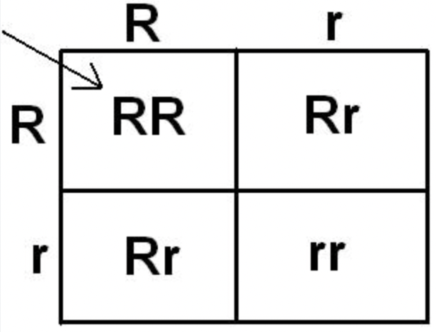
test cross
performed to determine unknown genotype (if unknown, the phenotype = dominant trait)
STEPS:
cross the organism with another organism of the recessive genotype