Cram!!
1/218
There's no tags or description
Looks like no tags are added yet.
Name | Mastery | Learn | Test | Matching | Spaced | Call with Kai |
|---|
No analytics yet
Send a link to your students to track their progress
219 Terms
Appendicular Skeleton
Division of the skeleton responsible primarily for movement; includes upper and lower limbs and their girdles.
Pectoral Girdle
A bony ring that functions as your shoulder, consisting of the clavicle (collar bone) and the scapula (shoulder blade), which are attached by the upper limb of the axial skeleton.
Pectoral Girdle: Function
Structural Support: Attaches the bones of the upper limb to the axial skeleton.
Muscle Attachment: Serves as an anchoring point for the muscle tendons and ligaments.
Mobility: Allows the upper limb to move in various directions without any limitations.
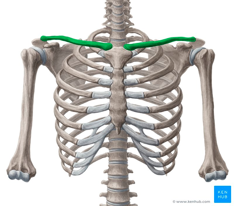
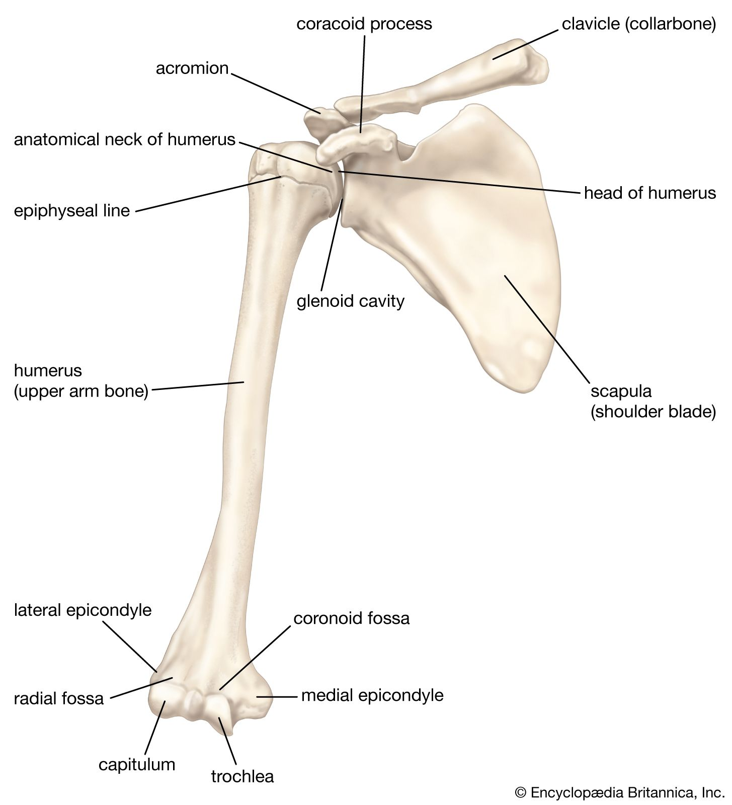
Clavicle
S-shaped bone that connects the upper limb to the sternum, known as the collarbone.
Clavicle: Function
Structural Support: Holds the pectoral girdle away to allow the arm to move freely from the shoulder joints.
Force Transmission: Can safely route mechanical forces and weight from the upper limb to the axial skeleton for movement.
Protection: Shields nerves and blood vessels when traveling between the axial skeleton and the upper limb.
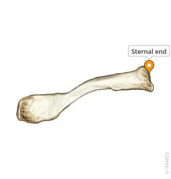
Sternal End
Medial end that articulates with the manubrium of the sternum.
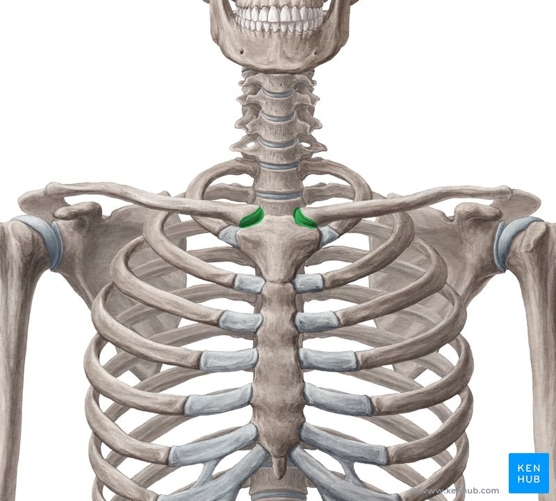
Sternoclavicular Joint
The joint connects the clavicle to the sternum through the sternal end of the clavicle.
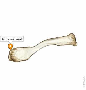
Acromial End
Lateral end that articulates with the acromion of the scapula.
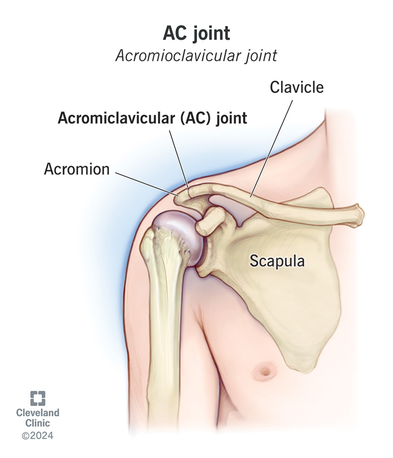
Acromioclavicular Joint
The joint connects the clavicle to the acromion of the scapula through the acromial end of the clavicle.
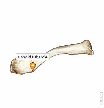
Conoid Tubercle
On the inferior-lateral end that attaches for the conoid ligament on the clavicle, in which attaches the clavicle and scapula
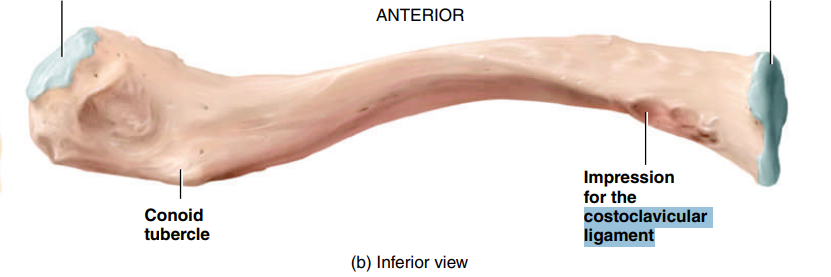
Costoclavicular Ligament
A band of connective tissue that attaches both the clavicle and scapula to the first rib.
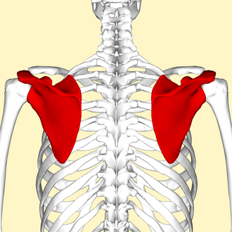
Scapula
A triangular flat bone on the posterior thorax (lower back), known as the shoulder blade
Scapula: Function
Mobility: Allows the shoulder to pivot and point in multiple directions for a full range of motion.
Structural Support: Provides a stable base that allows the humerus (upper arm bone) to move without dislocation.
Muscle Attachment: Anchors different kinds of muscles that provide strength and leverage to move the arm.
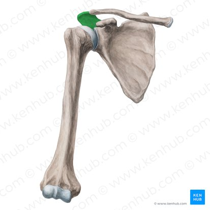
Acromion
Extension of the scapular spine that forms the highest point of the shoulder on the lateral end.
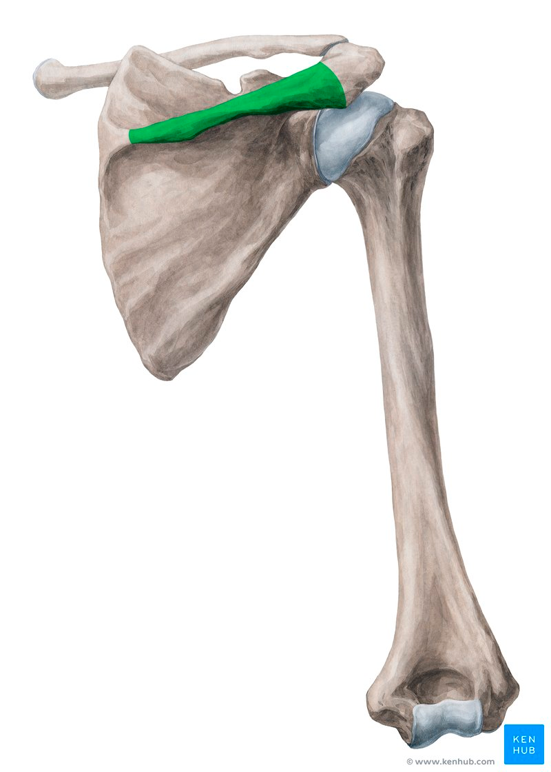
Spine
A long prominent ridge runs diagonally across the posterior surface of the scapula
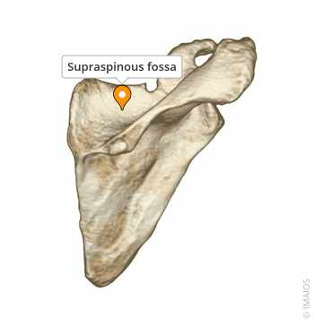
Supraspinous Fossa
Posterior depression superior to the spine of the scapula as the attachment site for supraspinatus muscle.
Infraspinous Fossa
Posterior depression inferior to the spine of the scapula as the attachment site for infraspinatus muscle.
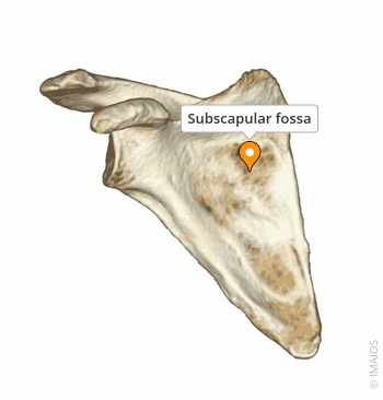
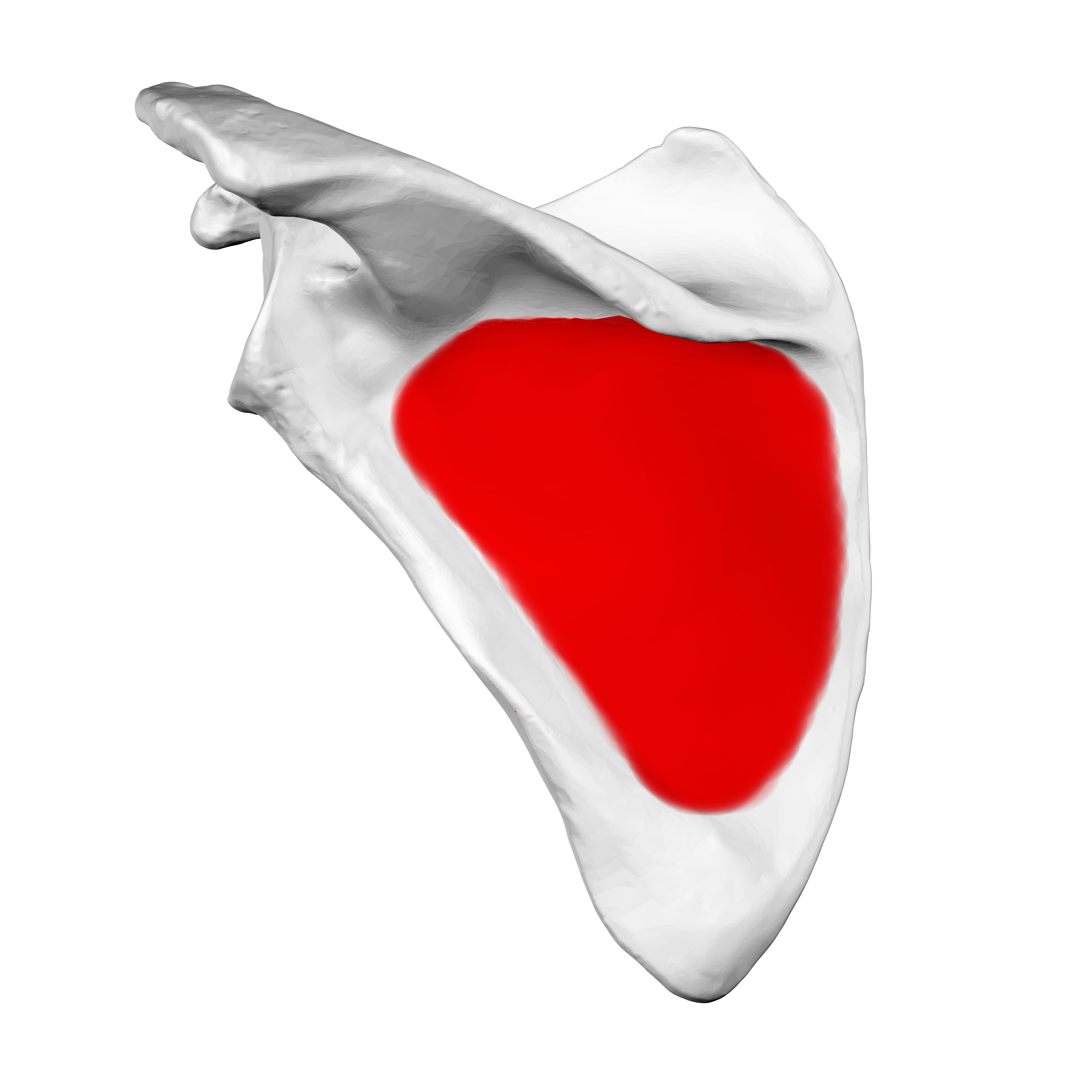
Subscapular Fossa
Anterior depression of the scapula as the attachment site for subscapularis muscle.
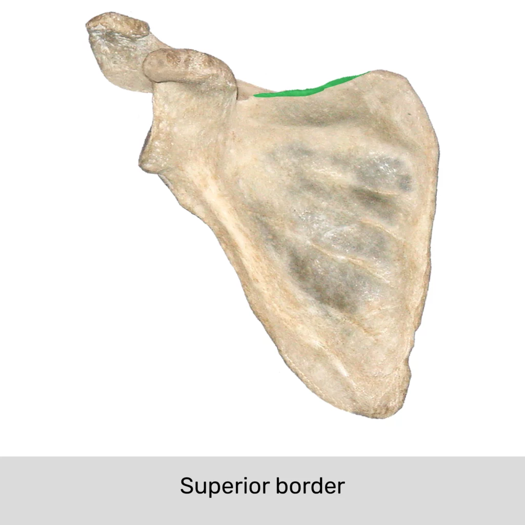
Superior Border
The upper edge of the scapula.
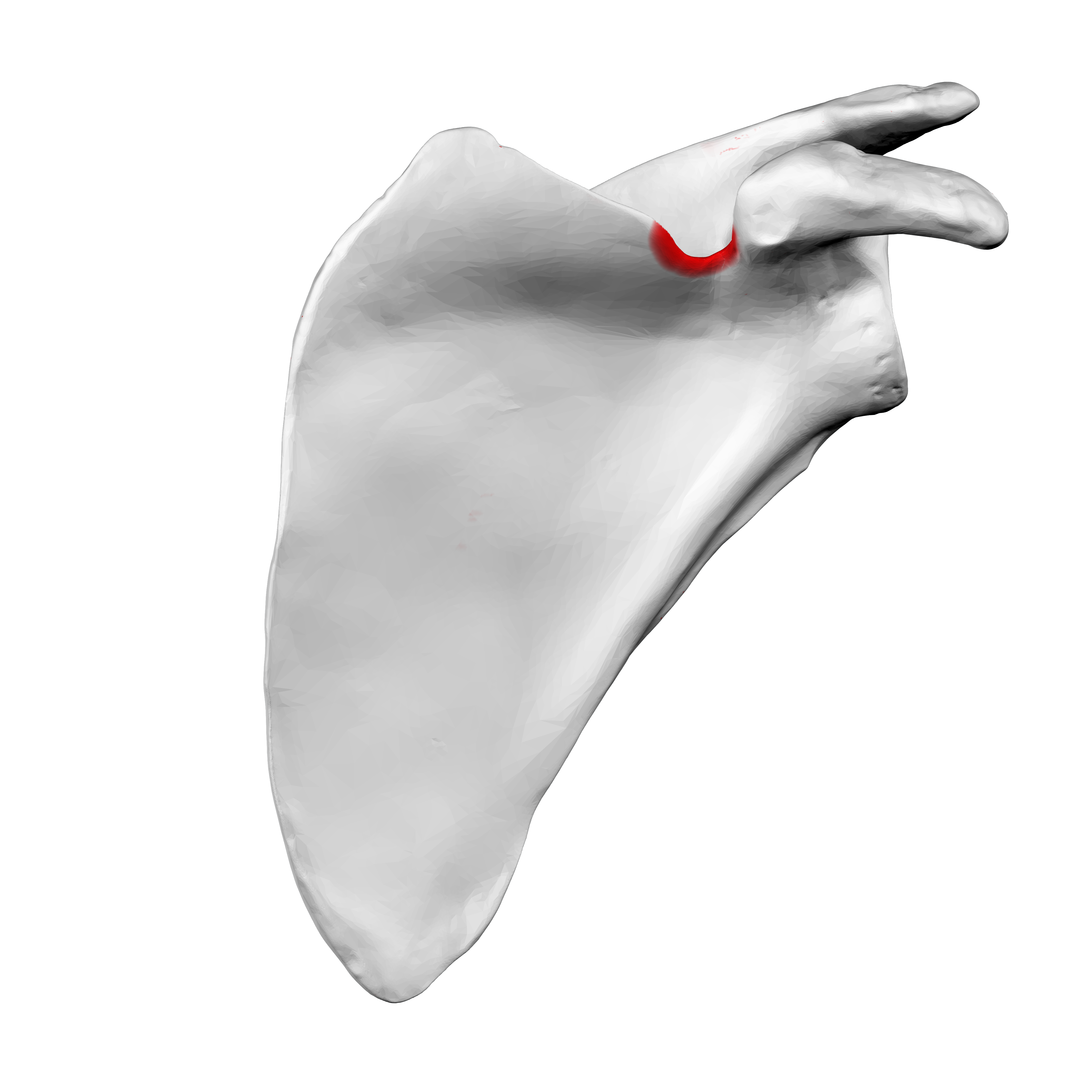
Suprascapular Notch
A prominent indentation on the superior border of the scapula for the passage of suprascapular nerve
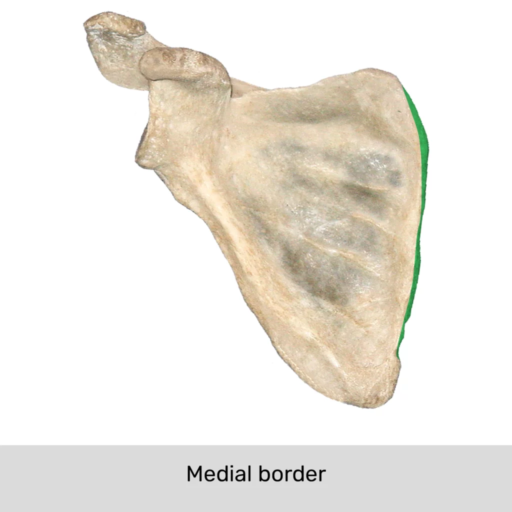
Medial (Vertebral) Border
The thin edge of the scapula closest to the vertebral column.
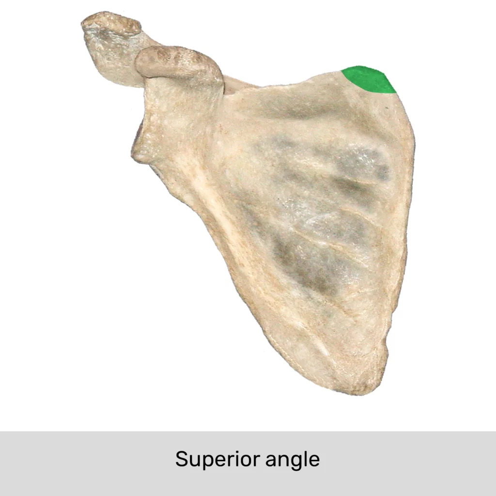
Superior Angle
The point where the superior border and medial border meet.
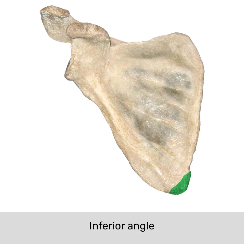
Inferior Angle
The point where the medial border and the lateral border meet.
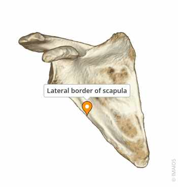
Lateral (Axillary) Border
The thick edge of the scapula closest to the arm.
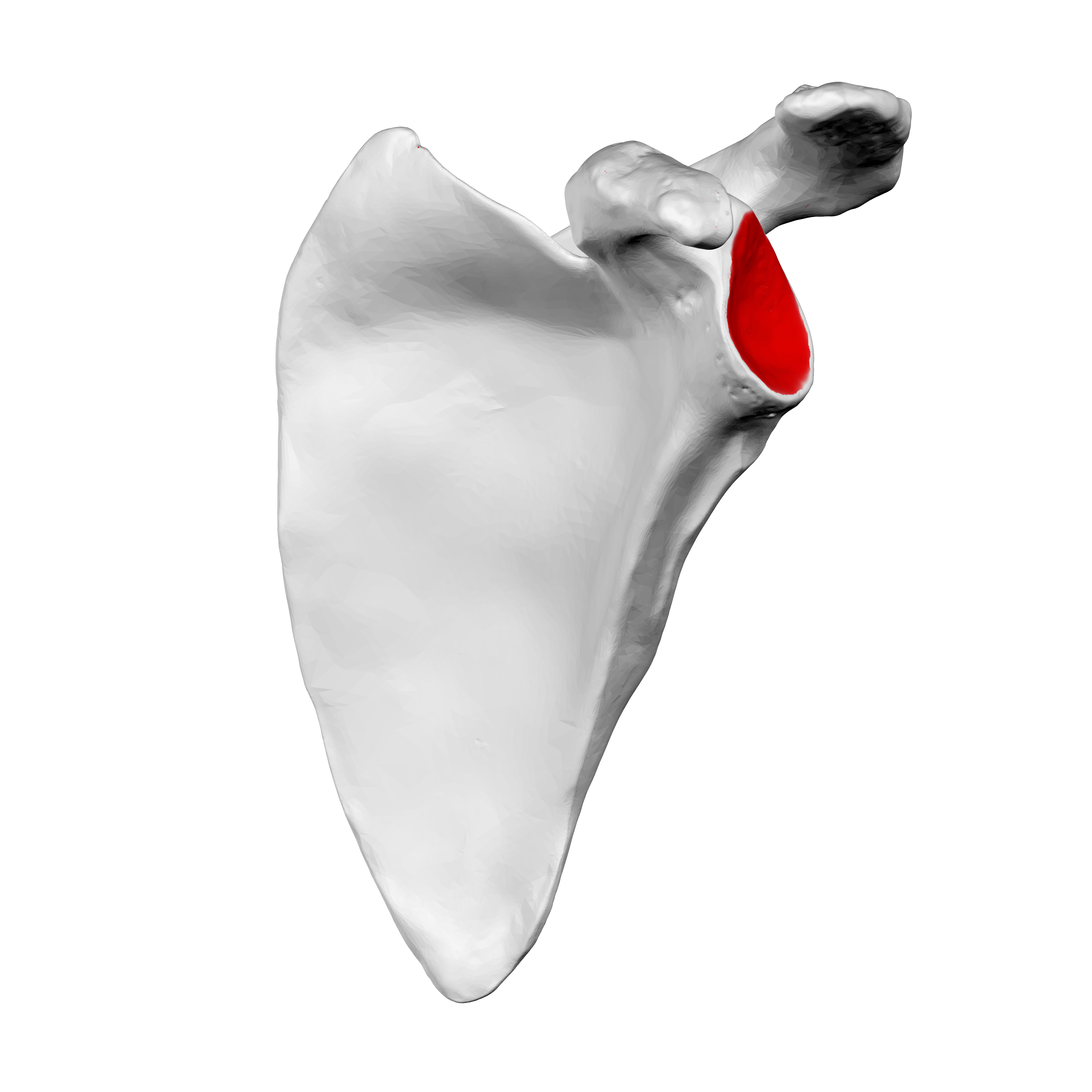
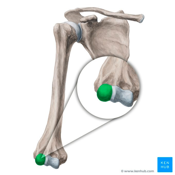
Glenoid Cavity
A shallow depression that serves as an attachment site for the humerus from the glenohumeral (shoulder) joint
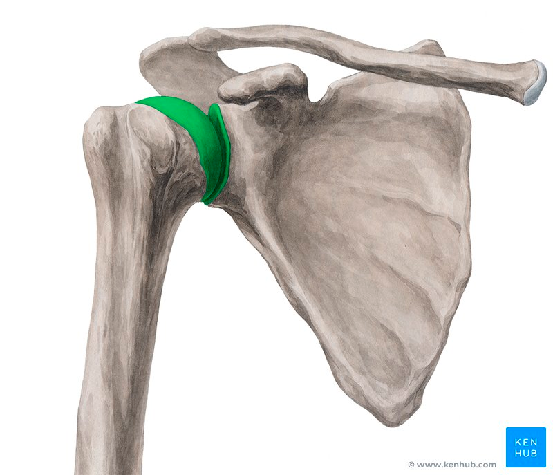
Glenohumeral Joint
A ball-and-socket joint connects the scapulae to the humerus through the glenoid cavity for arm rotation.
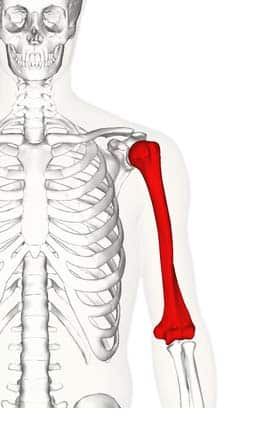
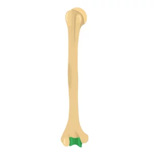
Humerus
Longest and largest bone of the upper limb that articulates proximally with the scapula and distally with the ulna and radius.

Head of Humerus
Proximal rounded portion that articulates with the glenoid cavity to form glenohumeral (shoulder) joint

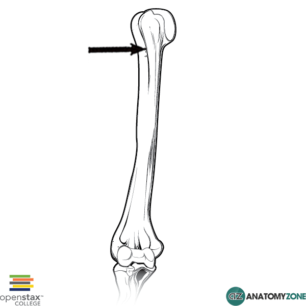
Anatomical Neck
Groove distal to the head as the former growth plate.

Greater Tubercle
Lateral projection distal to the anatomical neck for muscle attachment.

Lesser Tubercle
Anterior projection on the anatomical neck for muscle attachment.
Intertubercular Sulcus
Groove between the greater and lesser tubercles.
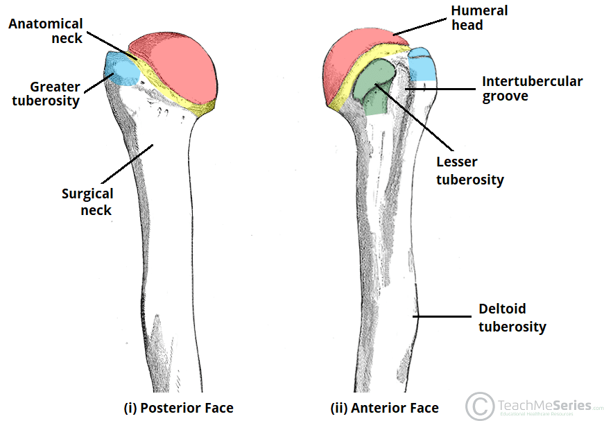
Surgical Neck
A narrow, constricted section located just below the greater and lesser tubercles of the humerus, and are a common fracture site
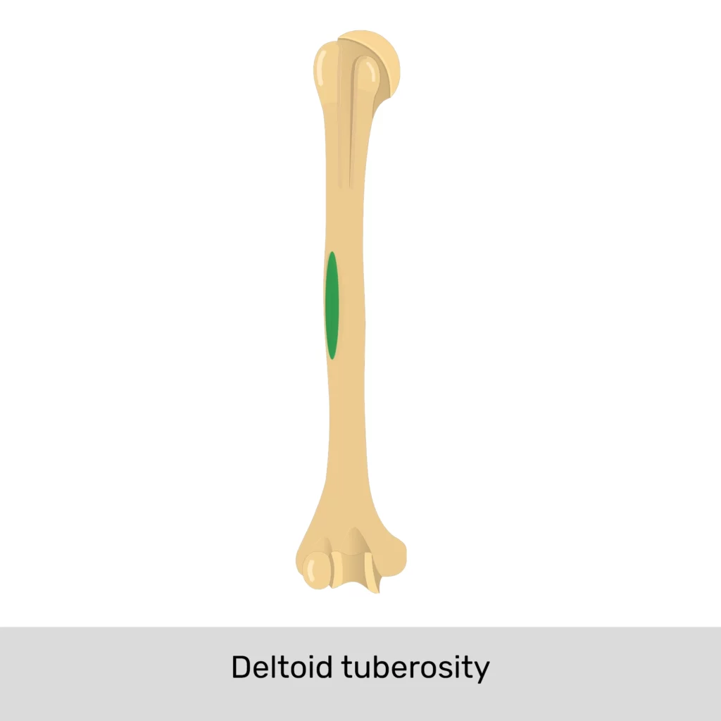
Deltoid Tuberosity
Roughened, V-shaped area for attachment of the deltoid muscle.
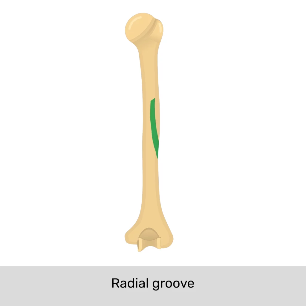
Radial Groove
Groove on the posterior humerus that contains the radial nerve.
Capitulum
Lateral condyle of the humerus that articulates with the radius.
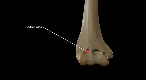
Radial Fossa
An anterior depression above the capitulum that receives the head of the radius during flexion
Trochlea
The medial knob-like condyle of the humerus articulates with the ulna.
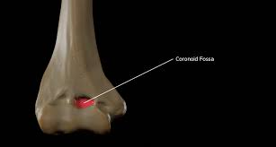
Coronoid Fossa
An anterior depression that receives the coronoid process of the ulna during flexion.
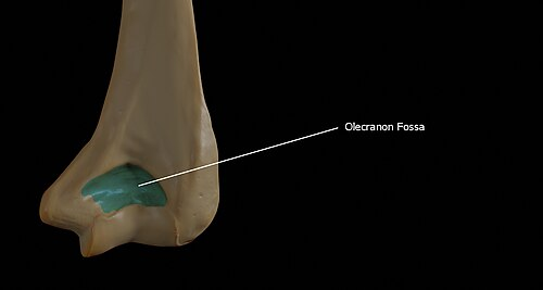
Olecranon Fossa
A large posterior depression that receives the olecranon of the ulna during extension.
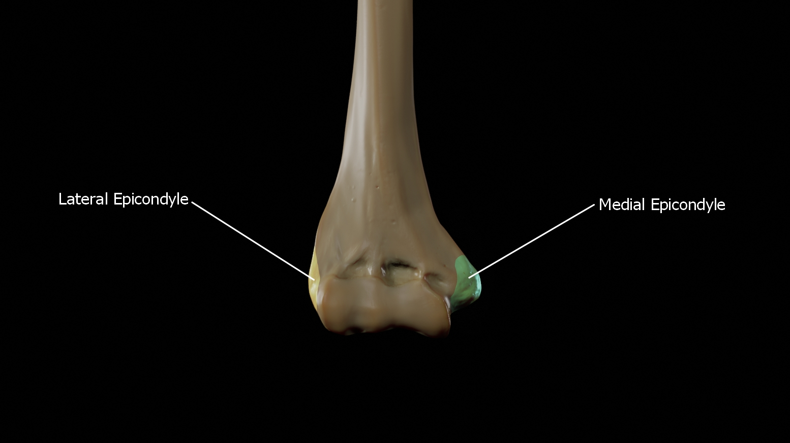
Medial Epicondyle
A rough attachment site for forearm muscles on the medial side.

Lateral Epicondyle
A rough attachment site for forearm muscles on the lateral side.
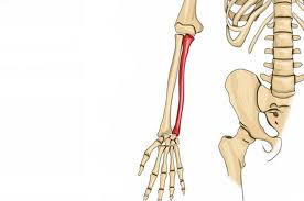
Ulna
Medial forearm bone located on the pinky side.
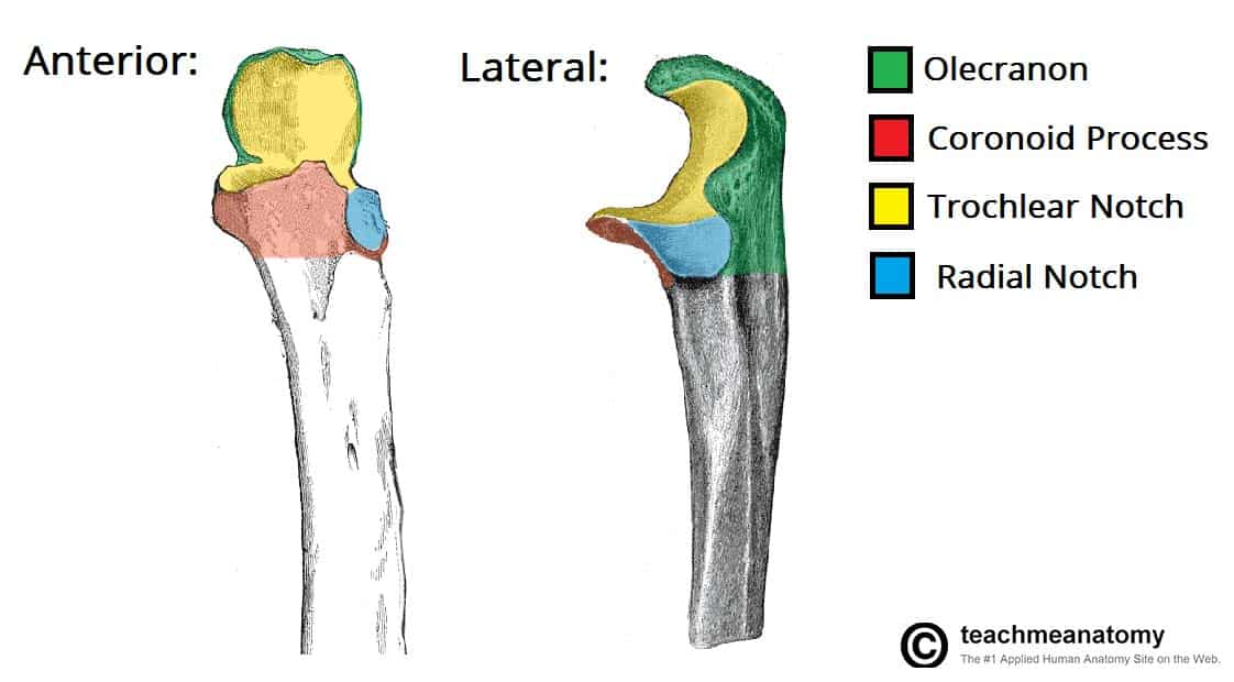
Olecranon
A thick, curved proximal projection forming the point of the elbow, acting as a lever for straightening the arm.

Coronoid Process
A thick, triangular, anterior projection of the ulna that articulates with the trochlea to form trochlear notch, acting as the hinge joint in the elbow.

Trochlear Notch
A large, C-shaped depression located at the proximal end of the ulna, formed by the olecranon and coronoid process.

Radial Notch
A depression on the lateral-inferior to the trochlear notch that attaches to the head of the radius for elbow function and forearm mobility
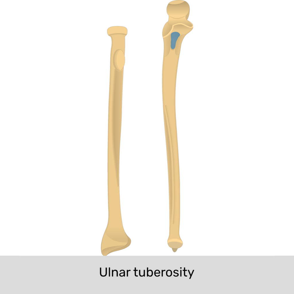
Ulnar Tuberosity
A rough, elevated prominence on the anterior-inferior to the coronoid process that attaches to the brachialis muscle
Styloid Process of Ulna
The posterior projection on the distal ulna end for collateral ligament attachment to the wrist.
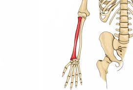
Radius
The lateral forearm bone runs parallel to the ulna on the lateral (thumb) side of the forearm, and it runs narrow at the proximal end and widens at the distal end.
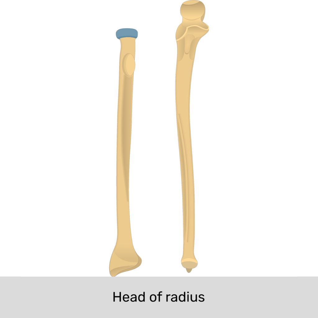
Head of Radius
A disc-shaped proximal end that articulates with the capitulum of the humerus and radial notch of the ulna.
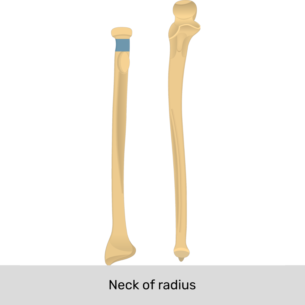
Neck of Radius
The narrowed section of the radius bone located just beneath the disk-shaped radial head, connecting it to the radial shaft and tuberosity
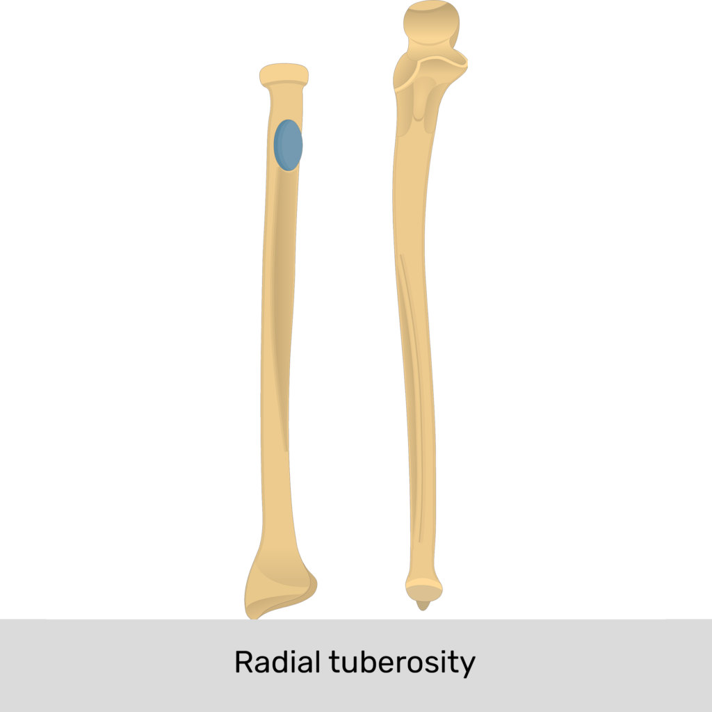
Radial Tuberosity
A roughened area inferior to the neck of the radius that serves an attachment site for tendons of the biceps brachii muscle.
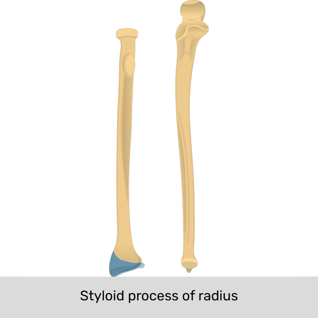
Styloid Process of Radius
The distal lateral projection under the thumb that attaches to the brachioradialis muscle and the radial collateral ligament to the wrist, or supports the lateral side of the wrist joint.
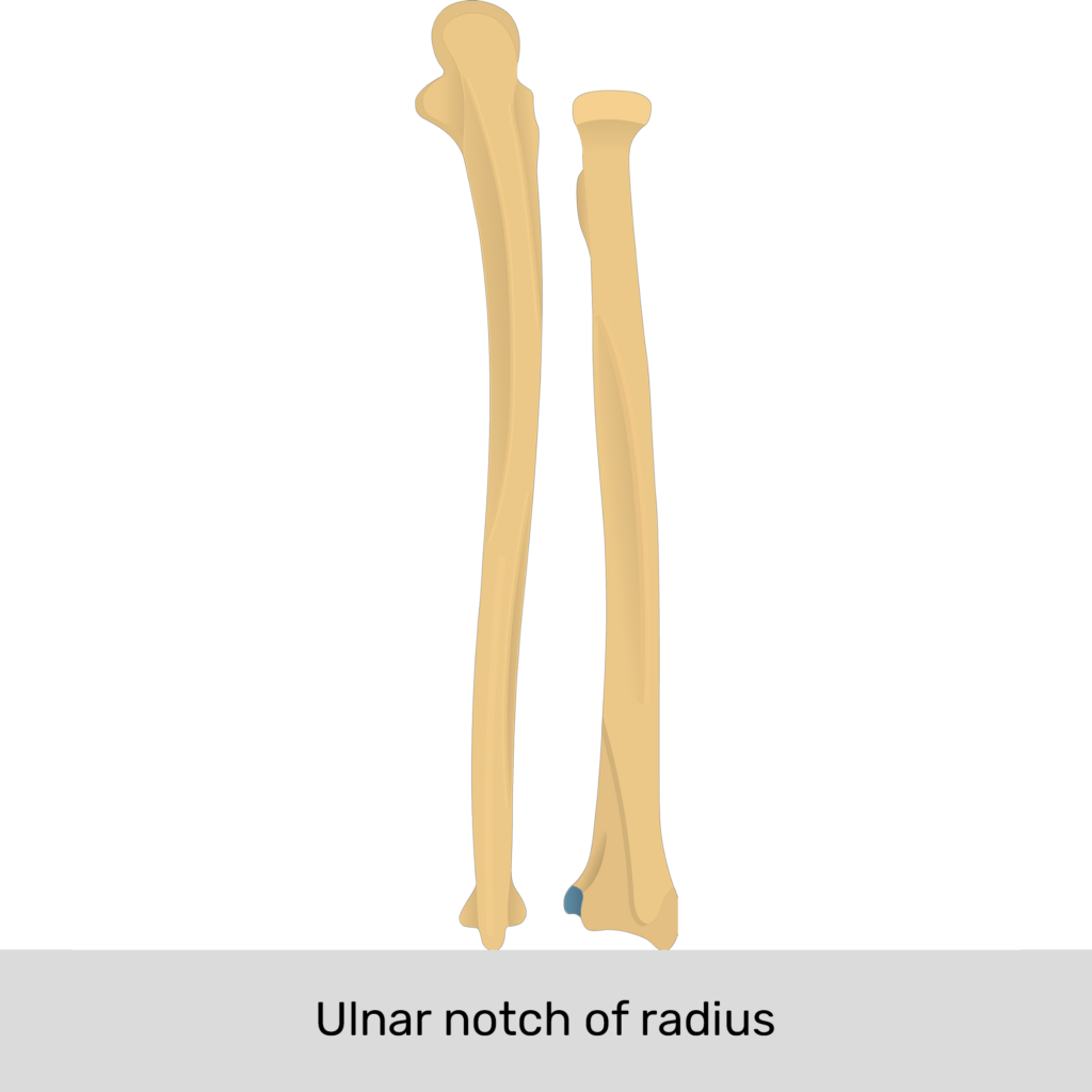
Ulnar Notch
The distal depression of the radius has a narrow concavity that articulates with the ulna at the distal radioulnar joint.
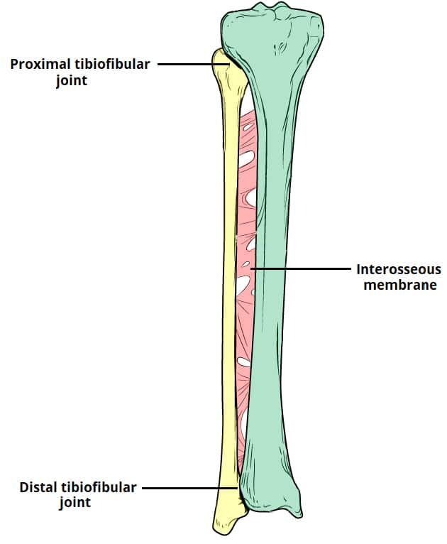
Interosseous Membrane
The first attachment site between the radius and ulna at a broad, flat, fibrous connective tissue that joins the two bones together to attach deep skeletal muscle.

Proximal Radioulnar Joint
A joint between the head of the radius and radial notch of the ulna, just below the elbow for forearm movement.

Distal Radioulnar Joint
A joint between the head of the ulna and the ulnar notch of the radius
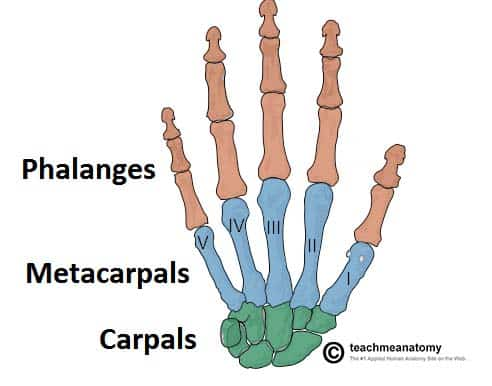
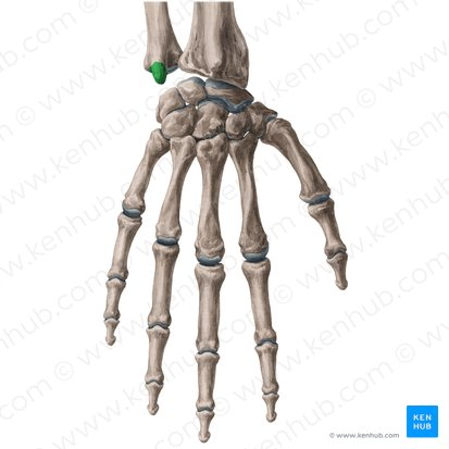
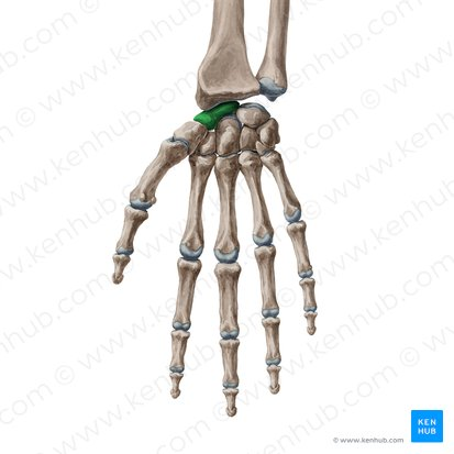
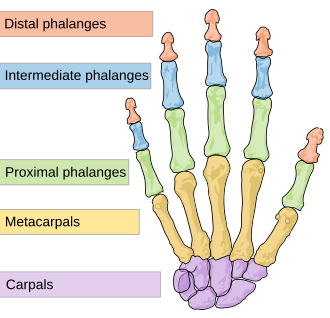
Carpals
Eight wrist bones arranged in two rows (carpus) all joined into one through ligaments
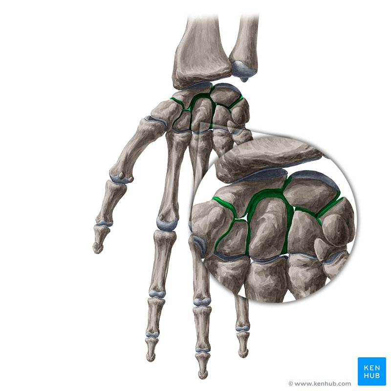
Intercarpal Joints
A complex series of synovial plane (gliding) joints that connect the eight carpal bones of the wrist together into two transverse rows of four.

Carpals: Proximal Rows
Articulates with the distal radius and ulna to form radiocarpal joint

Scaphoid
Most commonly fractured carpal bone.

Lunate
Moon-shaped carpal bone.

Triquetrum
Three-cornered carpal bone.

Pisiform
Pea-shaped carpal bone.
Radiocarpal (Wrist) Joint
A major synovial joint where the radius and ulna meet by the two carpus bone: lunate and scaphoid
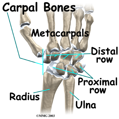
Carpals: Distal Rows
Articulates with the metacarpals

Trapezium
Distal row carpal bone that articulates with the thumb.

Trapezoid
Distal row carpal bone between trapezium and capitate.
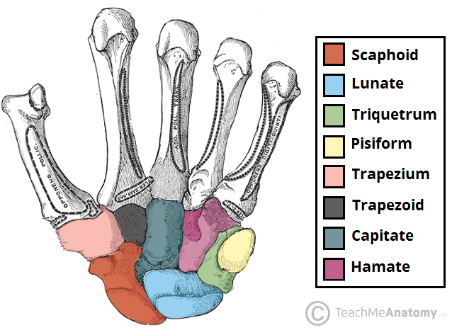
Capitate
Largest carpal bone.

Hamate
Carpal bone with a hook-shaped projection.

Carpal Tunnel
A narrow passageway containing flexor tendons and the median nerve in the wrist formed by carpal bones and the flexor retinaculum.
Ulnar Side of the Carpal Tunnel
The pisiform and hamate
Radial Side of the Carpal Tunnel
The scaphoid and trapezium.
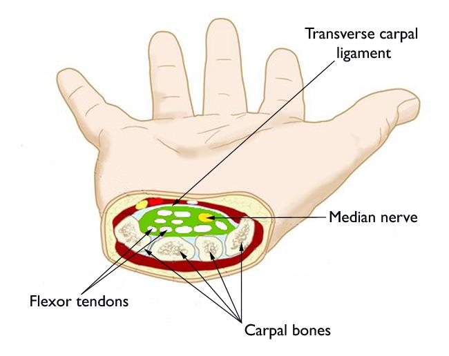
Flexor Retinaculum
A strong fibrous band of connective tissue that forms the roof of the carpal tunnel.

Metacarpals
Five bones (metacarpus) form the palm, with bones numbered 1–5 from the thumb to pinky, and they are made up of three parts: a proximal base, an intermediate body, and a distal head

Metacarpals: Base
Articulates with the distal row of carpal bones through carpometacarpal joints
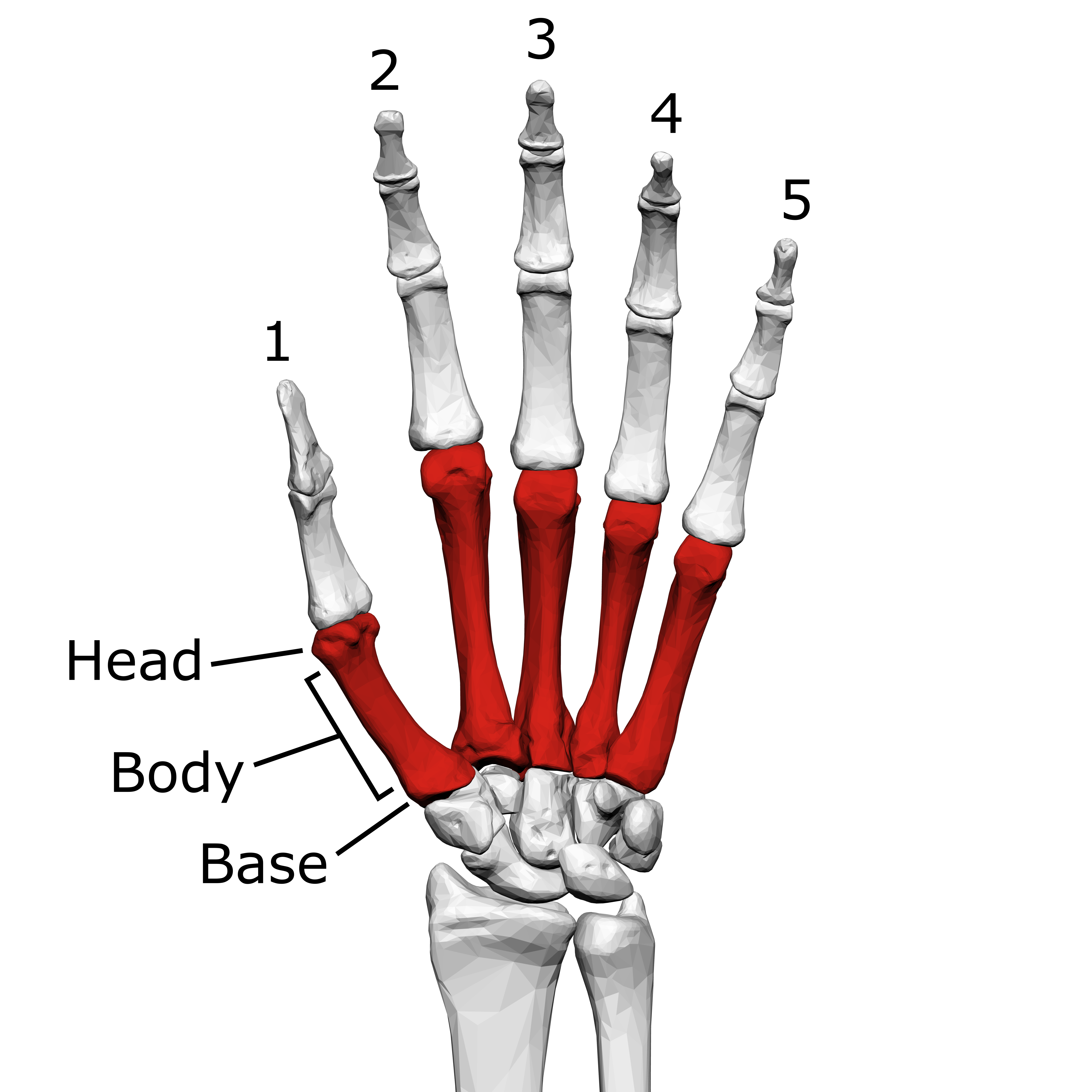
Metacarpals: Head
Articulates with the proximal phalanges through the metacarpophalangeal joints
Knuckles
Common name for the heads of the metacarpals.

Phalanges
Bones of the fingers and toes, with 14 bones (phalanx) in each hand and foot, are made up of the base, body, and head.

Phalanges: Digit Numbering
Digits are numbered 1–5, beginning with the thumb (lateral to medial).
Thumb (Pollex)
Thumb (Pollex) Has two phalanges: proximal and distal.

Digits 2–5
Have three phalanges: proximal, middle, and distal.
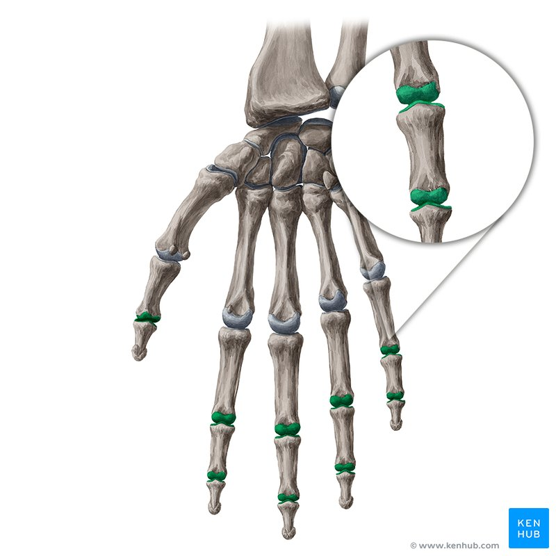
Interphalangeal Joints
Joints between phalanges.
Pelvic Girdle
Consists of the two hip bones, and connects lower limbs to the axial skeleton.
Pelvic Girdle: Function
Weight Transfer: Supports the upper body’s weight and transfers it through the hip joints to the lower limbs
Protection: Forms a pelvic cavity that houses and shields visceral organs
Movement: Serves as an anchoring point for major muscle groups for walking, running, and balancing
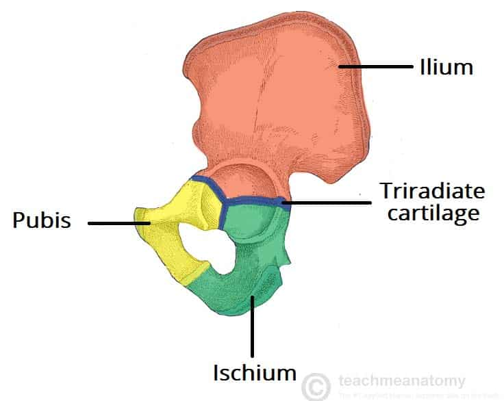
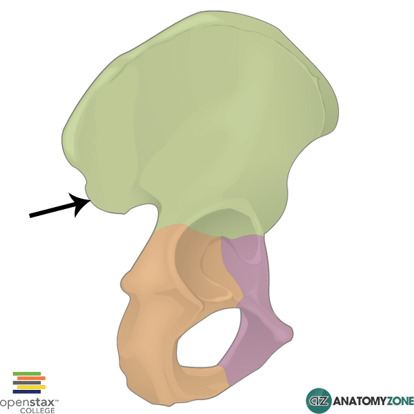
Hip Bone
Coxal bone formed by fusion of the ilium, ischium, and pubis.
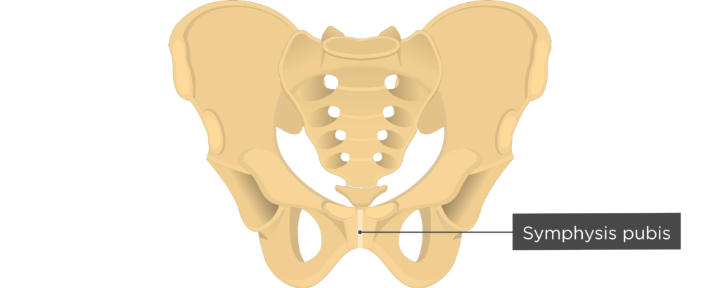
Pubic Symphysis
Fibrocartilaginous joint between the hip bones.

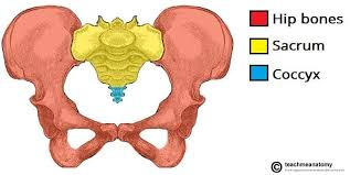
Bony Pelvis
A complete basin-shaped structure composed of the hip bones, pubic symphysis, sacrum, and coccyx that connect and support the spine to the lower limbs
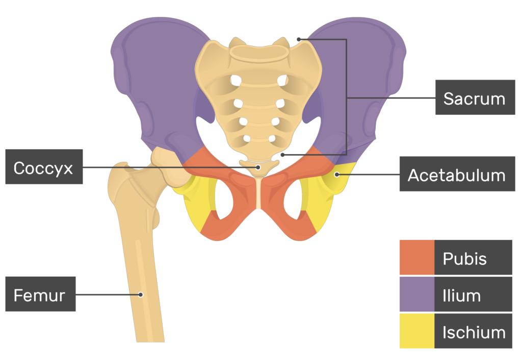
Ilium
Largest and superior portion of the hip bone, composed of a superior ala and an inferior body.
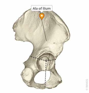
Ala of the Ilium
The broad, wing-like superior portion of the ilium.
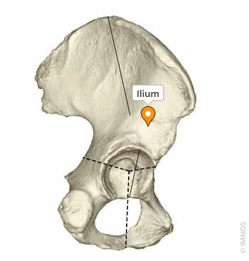
Body of the Ilium
The inferior portion of the ilium that contributes to the acetabulum.
Acetabulum
The socket of the hip bone that receives the head of the femur.
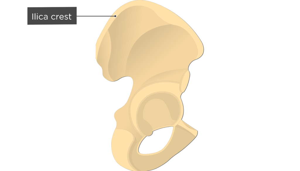
Iliac Crest
Superior border of the ilium.
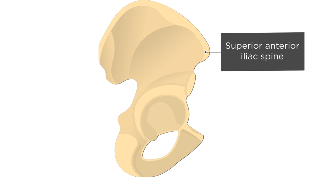
Anterior Superior Iliac Spine (ASIS)
Anterior projection of the iliac crest.
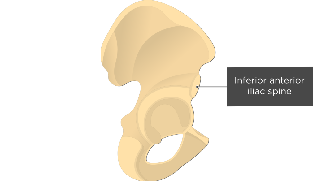
Anterior Inferior Iliac Spine (AIIS)
Projection inferior to the ASIS.
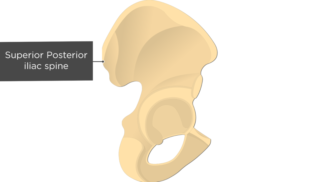
Posterior Superior Iliac Spine (PSIS)
Posterior projection of the iliac crest.
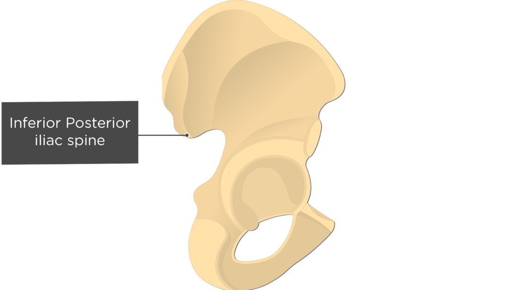
Posterior Inferior Iliac Spine (PIIS)
Projection inferior to the PSIS.
Iliac Spines
Serve as attachment sites for tendons of the trunk, hip, and thigh muscles.
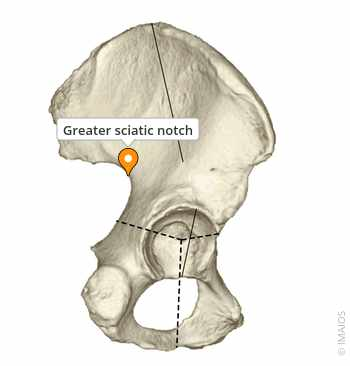
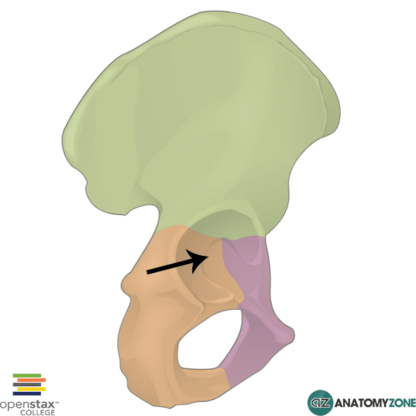
Greater Sciatic Notch
Passageway for the sciatic nerve, other nerves, blood vessels, and muscles