Mediastinum and Heart
1/67
There's no tags or description
Looks like no tags are added yet.
Name | Mastery | Learn | Test | Matching | Spaced | Call with Kai |
|---|
No analytics yet
Send a link to your students to track their progress
68 Terms
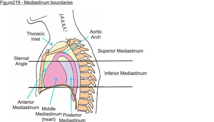
Central division of the thoracic cavity located between the two pleural sacs (lungs)
Mediastinum- the space that contains the heart, great vessels, trachea, esophagus, and other structures.
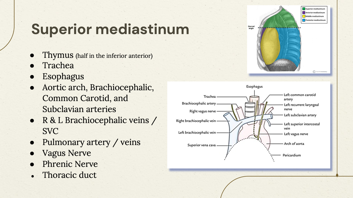
What are the contents of Superior mediastinum?
The superior mediastinum contains the thymus, great vessels (such as the aorta and its branches), trachea, esophagus, and nerves such as the vagus and phrenic nerves.
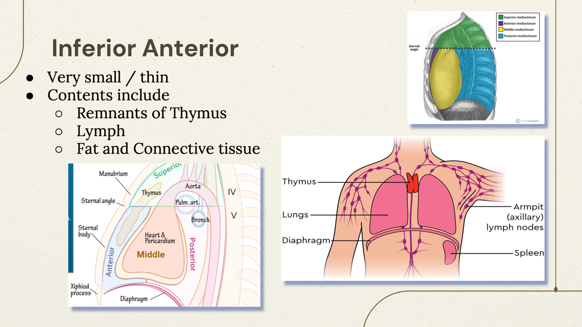
What are the contents of Inferior Anterior Mediastinum?
The inferior anterior mediastinum contains the remnants of thymus, connective and fat tissue, and lymph nodes.
What are the organization of Mediastinum?
Superior, Inferior anterior, Inferior Middle, Inferior Posterior.

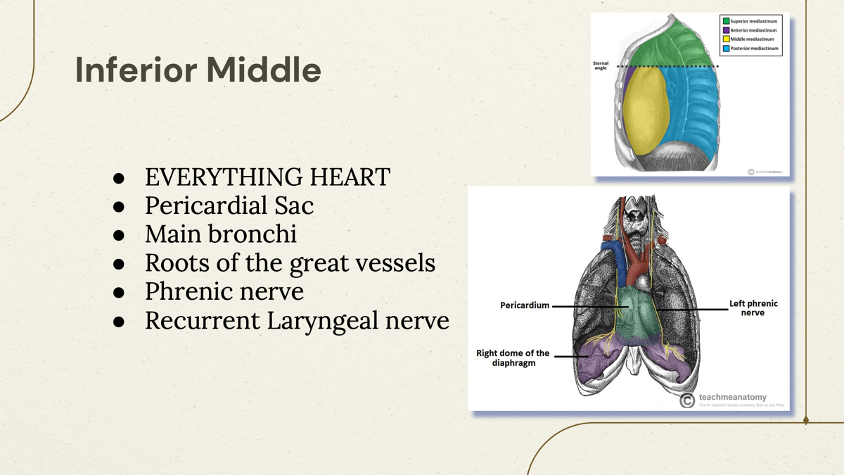
What are the contents of Inferior Middle of Mediastinum?
Everything heart, Pericardial sac, Main Bronchi, Roots of great vessels, Phrenic nerve, Recurrent Laryngel nerve.
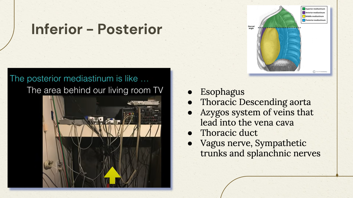
What are the contents of Inferior Posterior of mediastinum?
The inferior posterior mediastinum contains the esophagus, thoracic descending aorta, azygos and hemiazygos veins, thoracic duct, Vagus nerve, Splanchnic nerve and sympathetic trunk.
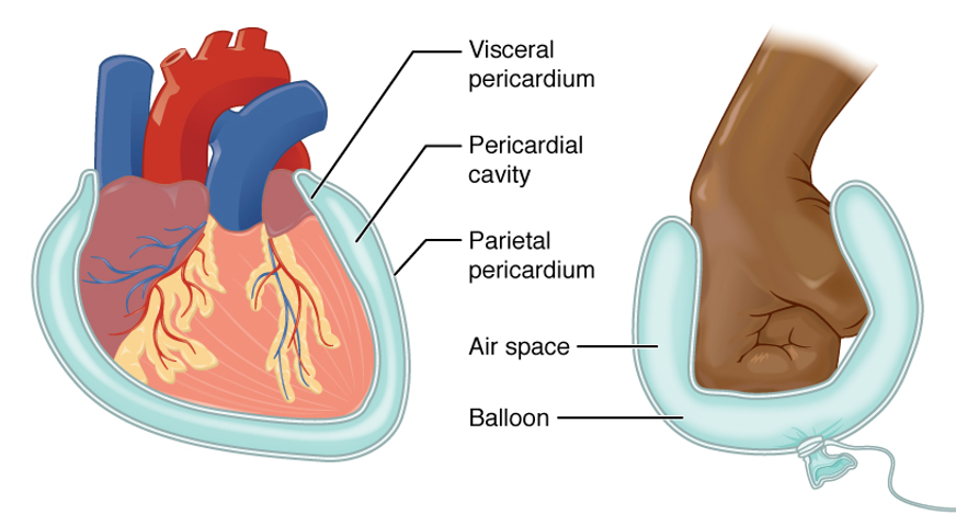
What is the sac surrounding the heart and its function?
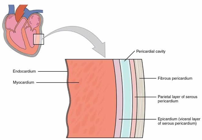
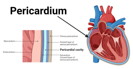
The pericardial sac- is a double-walled structure that surrounds the heart, providing protection, lubrication, and reducing friction during heartbeats.

What are the layers of Pericardium sac?
Fibrous Layer, Parietal Layer, Serosa, Visceral Layer.
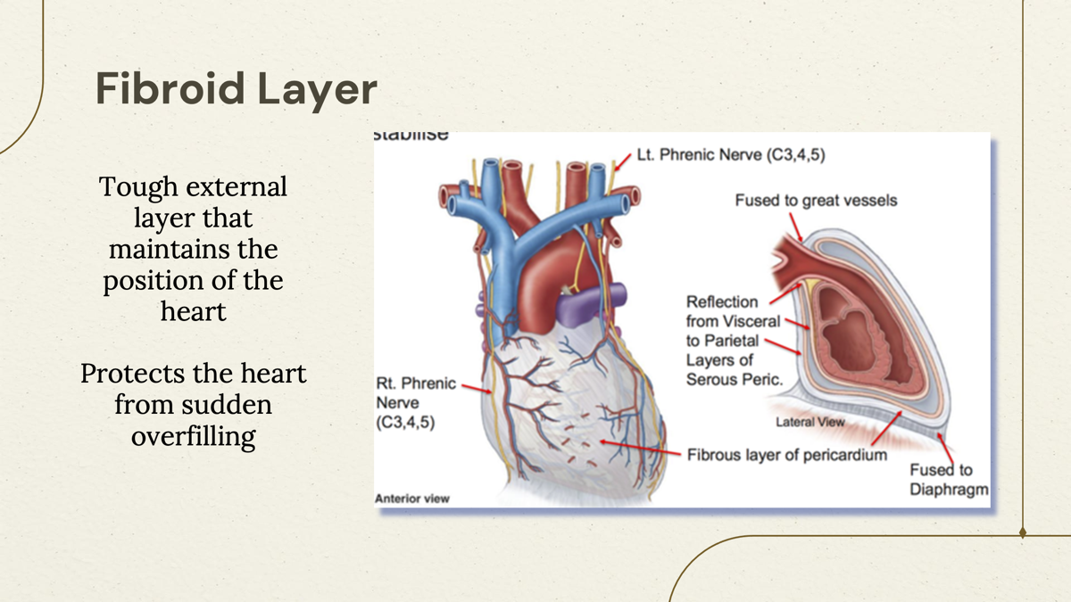
What is the toughest layer of pericardium sac?
Fibrous Layer- the outermost layer that provides structural support and protection for the heart.
Tough external layer that maintains the position of the hear Protects the heart from sudden overfilling
What is the Outer serous pericardium and the lining of the inner aspect of the fibrous pericardium
Parietal Layer
a smooth serous membrane that forms the outer layer of the pericardium, continuing onto the surface of the heart.

The layer that surrounds the VISCERA aka the heart?
Visceral Layer - the inner serous membrane that directly covers the heart, providing lubrication and protection.
Layer that contains serous fluids (Space)
Pericardial Cavity - the space between the parietal and visceral layers filled with serous fluid, allowing for smooth heart movement.

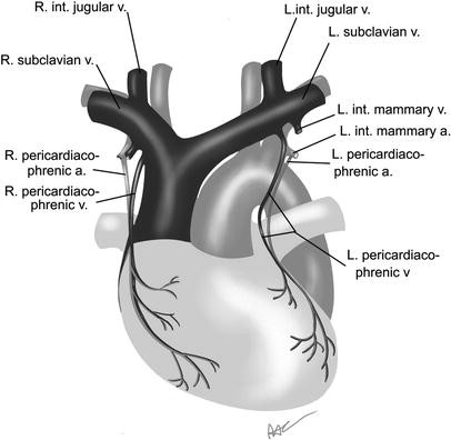
Pericardiacophrenic artery
a branch of the internal thoracic artery that supplies blood to the pericardium and diaphragm.

Pericardiophrenic veins
Empty into the brachiocephalic or internal thoracic veins.
Thick wall of the heart
Myocardium - the muscular middle layer of the heart responsible for contraction and pumping blood.
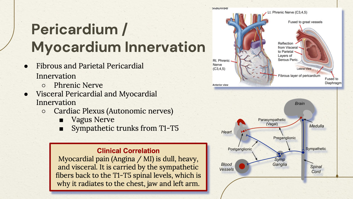
What innervates the Fibrous and Parietal Pericardial?
The fibrous and parietal pericardium are innervated primarily by the phrenic nerve, which provides sensory fibers.
What innervates the visceral pericardial and Myocardial?
Cardiac Plexus (Autonomic nerve)
Vagus Nerve
Sympathetic trunk from T1-T5
In Anterior surface, heart position looks like?
Is formed mainly by the right ventricle
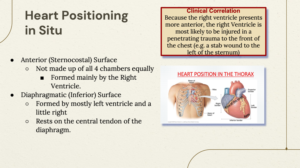
In diaphragmatic (inferior) surface it is formed mostly by:
○Formed by mostly left ventricle and a little right
○Rests on the central tendon of the diaphragm.
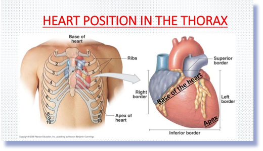
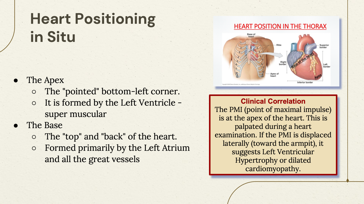
what is the Apex of the heart?
The “pointed” end of the heart, primarily formed by the left ventricle (muscular part of the heart)
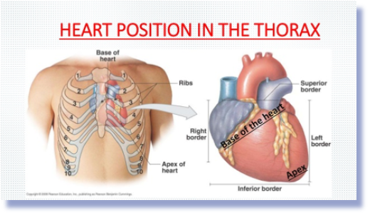
What Is the “top" and “back” of the heart?
The Base- the broad, flat part of the heart, predominantly formed by the left atrium and all great vessel.
In fetal, what organ carries highly oxygenated, nutrient-rich blood from placenta toward the fetal heart.
The umbilical vein, which transports oxygen and nutrients from the placenta to the fetus.
What are the three fetal shunts?
Shunt 1: Ductus Venous,
Shunt 2:Forearm Ovale,
Shunt 3: Ductus Arteriosus
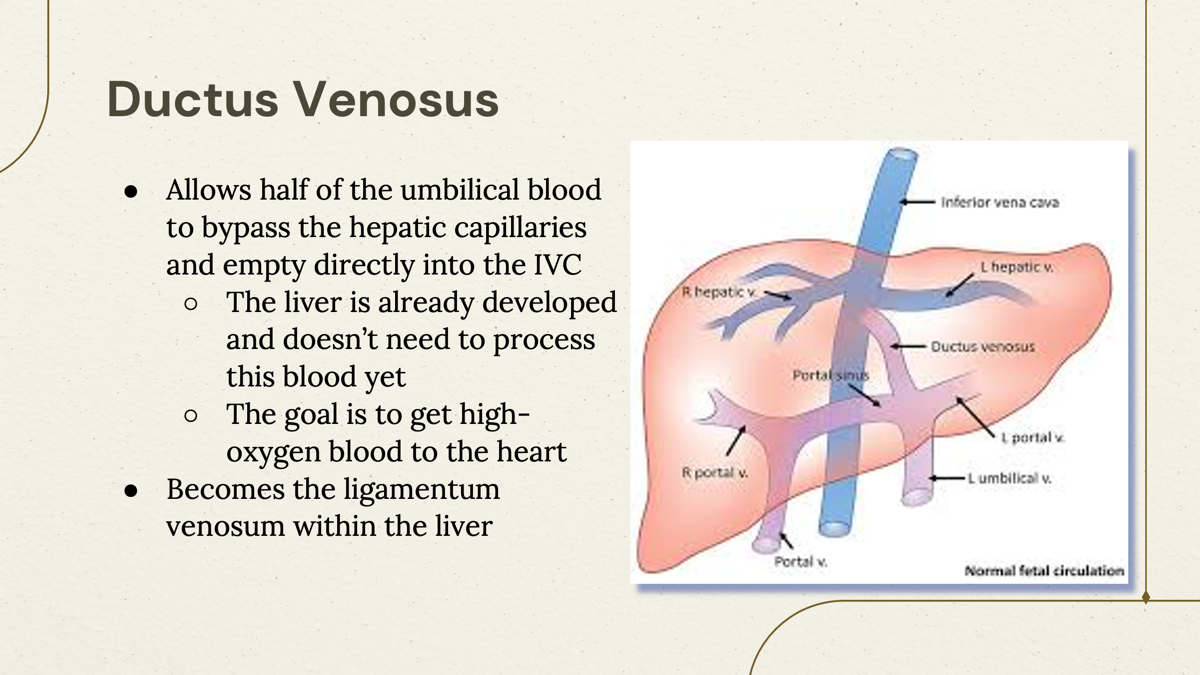
What shunt bypasses the liver?
The Ductus Venosus, which allows blood to bypass the hepatic capillaries of liver.
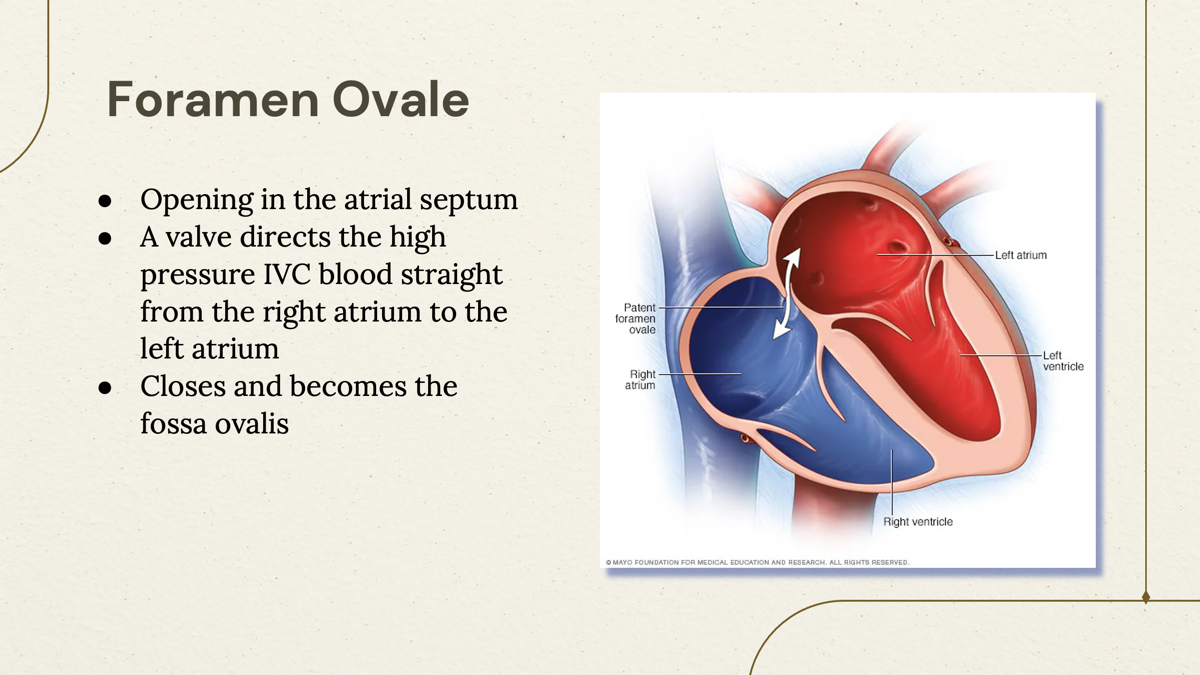
What is Forearm Ovale?
The foramen ovale is an opening in the fetal heart (atrial septum) that allows blood to flow directly from the right atrium to the left atrium, bypassing the lungs.
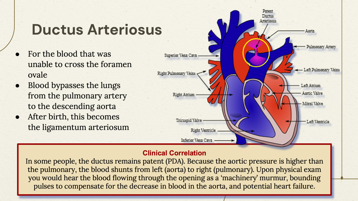
What “Shant” allows the blood to bypass the pulmonary artery to the descending Aorta in Fetal stage?
The Ductus Arteriosus, which connects the pulmonary artery to the descending aorta, allowing blood to bypass the non-functioning fetal lungs.
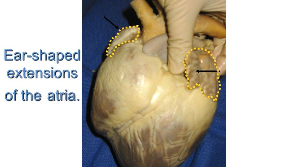
Fetal remnats aka “ears” and often thought as elephant ears:
Auricles, which are small, ear-shaped structures on the top of the heart that are remnants of the fetal heart structure.
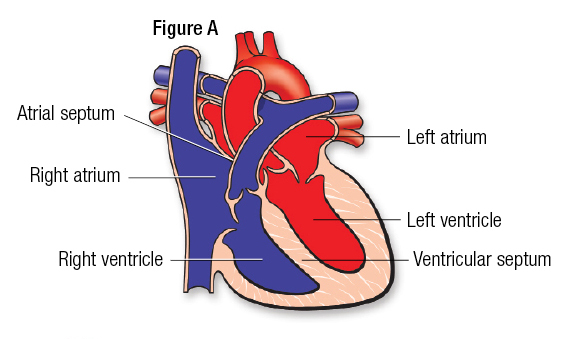
Wall between right and left atrium?
Atrial Septum:
The landmark for the atrial septum
Fossa Ovalis
is the remnant of the foramen ovale, an important fetal structure that allowed blood flow between the right and left atria.

Specialized, parallel bands of muscle tissue located in the walls of the right and left atria.
Pectinate muscles-
Increase the contractile force of the atria—allowing for efficient blood filling and pumping—while also increasing the surface area of the atrial chambers
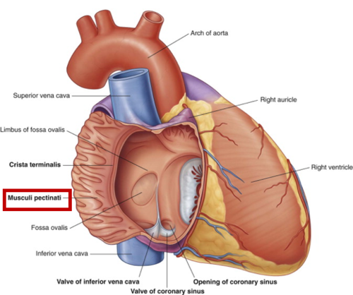
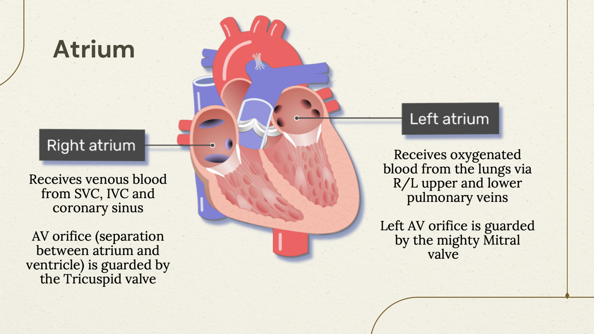
Receives venous blood from SVC, IVC, and coronary sinus
Right Atrium- one of the four chambers of the heart, located on the right side, playing a crucial role in the circulatory system by collecting deoxygenated blood from the body before it is sent to the right ventricle.

Receives oxygenated blood from the lungs via R/L upper and lower pulmonary veins
Left Atrium - one of the four chambers of the heart, located on the left side, it is responsible for receiving oxygen-rich blood from the lungs and transferring it to the left ventricle.
Ventricle that doesn't have to work as hard, receives blood form right atria via tricupsid valve.
Right Ventricle
one of the four chambers of the heart, it pumps deoxygenated blood to the lungs for oxygenation and is less muscular than the left ventricle.

The apex of part of the heart, hard worker and thick.
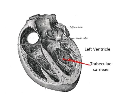
Left Ventricle - one of the four chambers of the heart, it is the strongest and most muscular chamber, responsible for pumping oxygenated blood to the whole body.

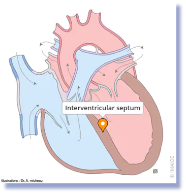
Ventricles are separated by?
Interventricular septum. A muscular wall that divides the right and left ventricles of the heart, preventing the mixing of oxygenated and deoxygenated blood.
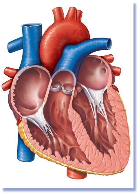
Iregular, muscular ridges and columns that project from the inner surfaces of both ventricles of the heart
Trabeculae carneae - structures that help to support the heart muscle and assist in the heart's contraction mechanism.
What is the tissue between the two ventricular, and contain the bundle if His
Interventricular septum
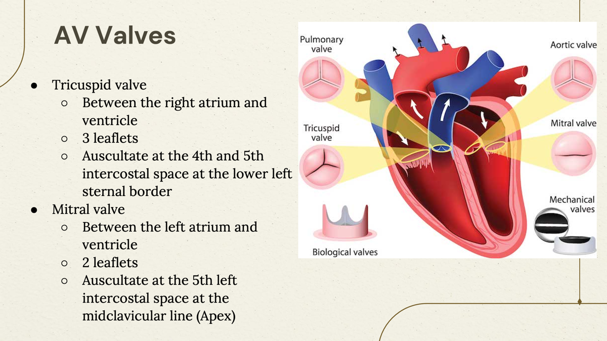
Two types of valves of the heart?
Tricuspid and Mitral valves
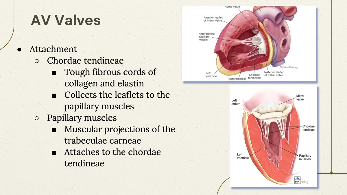
Two attachment point of AV valves?
Chordae tendineae- tough fibrous cords.
Papillary muscles- muscular projections.
Two types of Semilunar Valve and their functions?
Pulmonic valve- Between the right ventricle and the pulmonary trunk Auscultate at the 2nd left intercostal space on the left sternal border.
Aortic valve- Between the left ventricle and ascending aorta Auscultate at the 2nd right intercostal space on the right sternal border.
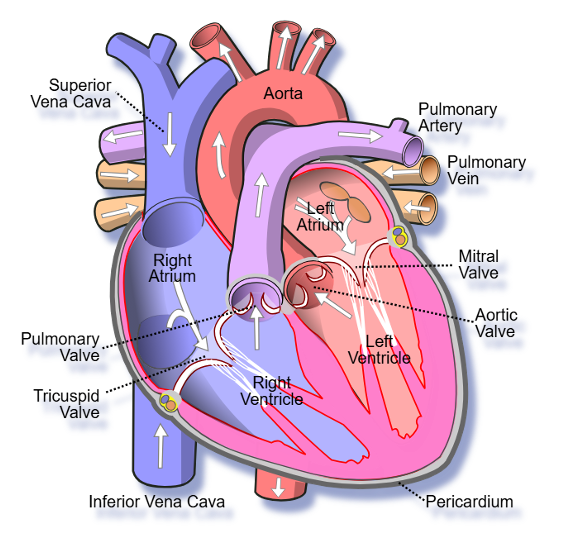
Deoxygenated blood Flow:
○From the SVC, IVC, and coronary sinus
○Into the right atrium
○Through the tricuspid valve during diastole
○Into the right ventricle
○Through the pulmonary valve into systole
○Into the pulmonary trunk
○Into the right and left pulmonary arteries
○Into the capillary beds for gas exchange
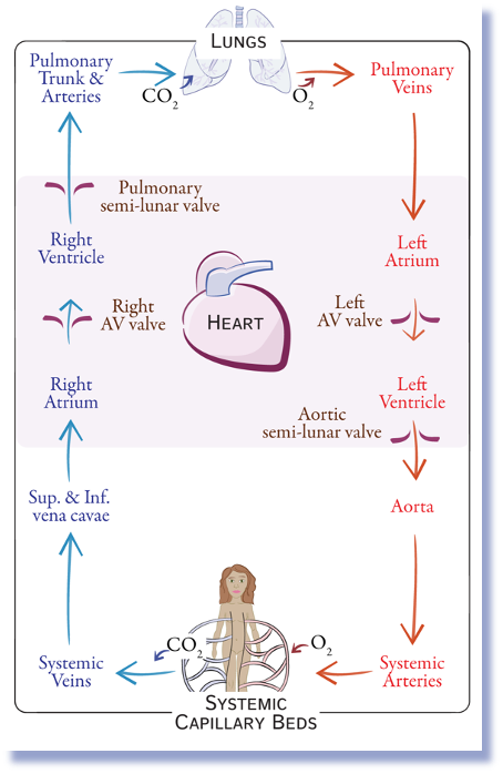
Oxygenated blood flow
○Into the 4 pulmonary veins
○Into the left atrium
○Through the mitral valve during diastole
○Into the left ventricle
○Through the aortic valve into systole
○Into the Aorta
○Through the systemic circulation for gas exchange
Structure | Blood Type |
|---|---|
SVC/IVC | Deoxygenated |
Right Atrium | Deoxygenated |
Right Ventricle | Deoxygenated |
Pulmonary Arteries | Deoxygenated 🚨 |
Pulmonary Veins | Oxygenated 🚨 |
Left Atrium | Oxygenated |
Left Ventricle | Oxygenated |
Aorta | Oxygenated |
'
Right side of the heart = sends blood to the lungs
Left side of the heart = sends blood to the body

Blood Flow:
SVC/IVC/Coronary Sinus
→ Right Atrium
→ Tricuspid Valve
→ Right Ventricle
→ Pulmonary Valve
→ Pulmonary Trunk
→ Pulmonary Arteries
→ Lungs
→ Pulmonary Veins
→ Left Atrium
→ Mitral Valve
→ Left Ventricle
→ Aortic Valve
→ Aorta
→ Body
→ SVC/IVC
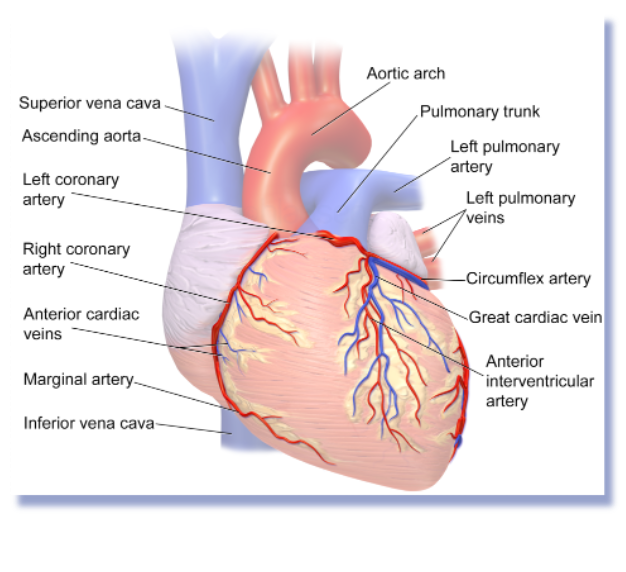
What artery supplies blood (oxygen rich) to the myocardium (heart muscle)
The coronary arteries are the blood vessels that bring oxygen-rich blood to the heart muscle.
Coronary arteries = feed the heart muscle (myocardium)
Where Do Coronary Arteries Come From?
Right and left coronary arteries originate from the aortic sinuses.
What is coronary venous system?
The coronary venous system is simply the network of veins that drains deoxygenated blood from the heart muscle (myocardium) and returns it to the heart.
where does coronary venous system ends?
Coronary venous system ends in the coronary sinus.
Where are coronary arteries and veins located?
Arteries and veins are located in coronary sulcus (tunnels for the vessels)
Sulcus = groove
The coronary sulcus is a groove on the surface of the heart.
Blood vessels travel inside this groove.
Think of it like a roadway where coronary vessels run.
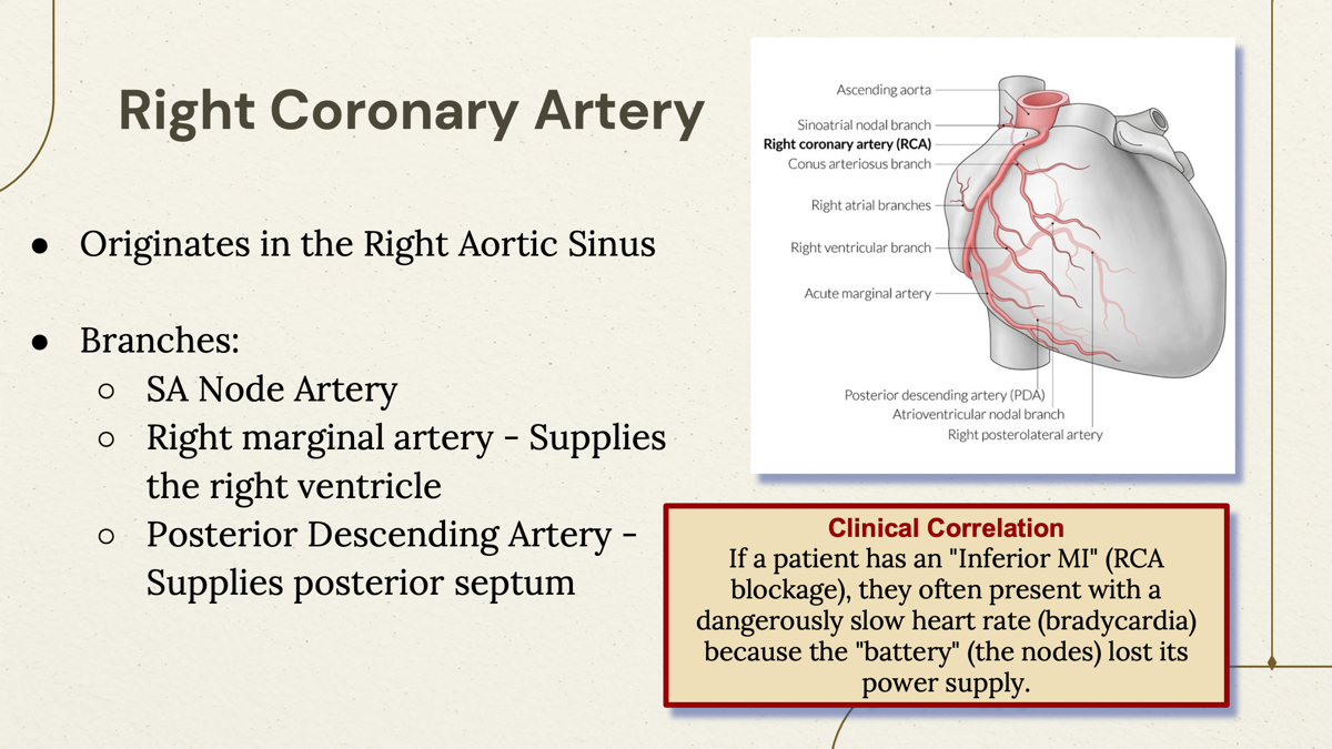
where does Right Coronary Artery originates?
Right Aortic Sinus-
Right Aortic Sinus → Right Coronary Artery
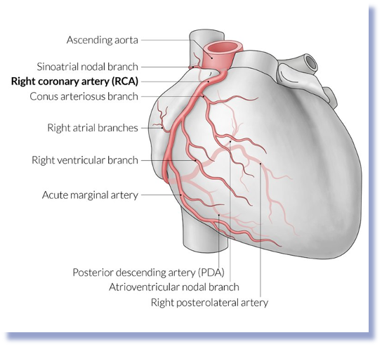
What are the branches of Right Coronary Artery (RCA)
1. SA Nodal Artery
🫀 Supplies the SA nod
Right Marginal Artery
🫀 Supplies the right ventricle
3. Posterior Descending Artery (PDA)
Also called:
Posterior interventricular artery
🫀 Supplies:
Inferior wall of the heart
Posterior 1/3 of interventricular septum
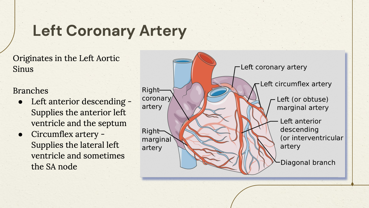
Where does Left Coronary Artery originates?
Originates in the Left Aortic Sinus
Supplies most of the left side of the heart, especially the left ventricle, which does the majority of the heart's pumping work.
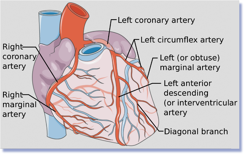
Where does LCA branches?
1. Left Anterior Descending (LAD)
Supplies the anterior (front) left ventricle
Supplies the interventricular septum
Circumflex Artery (LCX)
Supplies the lateral (side) left ventricle
May supply the SA node in some people
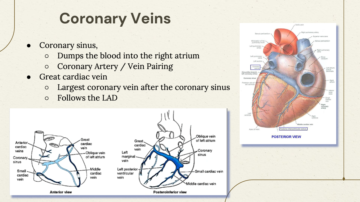
What is the function of coronary Vein?
Coronary veins collect deoxygenated blood from the heart muscle, drain into the coronary sinus, and the coronary sinus empties into the right atrium.
Where does corona sinus drain its blood?
right Atrium

What is great cardiac vein?
Largest coronary vein after the coronary sinus.
Runs alongside the LAD (Left Anterior Descending artery).
What is Sulcus?
Sulcus = groove (a groove on the surface of the heart where blood vessels travel).
What is coronary sulcus?
Sulci between the atrium and ventricles
○Coronary Sulcus (Also known as the atrioventricular groove)
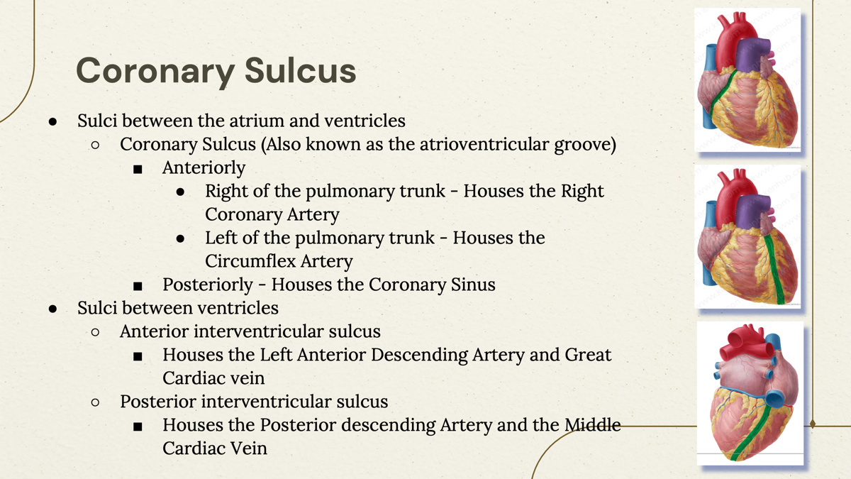
What is found in the Anterior Sulcus?
●Right of the pulmonary trunk - Houses the Right Coronary Artery
●Left of the pulmonary trunk - Houses the Circumflex Artery
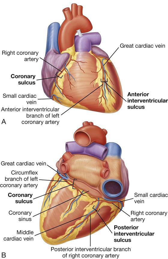
What is found in the Posterior of coronary sulcus?
Posteriorly - Houses the Coronary Sinus
What are the two Sulci between ventricles?
Anterior and Posterior Interventricular Sulcus.
-Anterior interventricular sulcus- Houses the Left Anterior Descending Artery and Great Cardiac vein.
-Posterior interventricular sulcus- Houses the Right Posterior Descending Artery and Middle Cardiac vein.

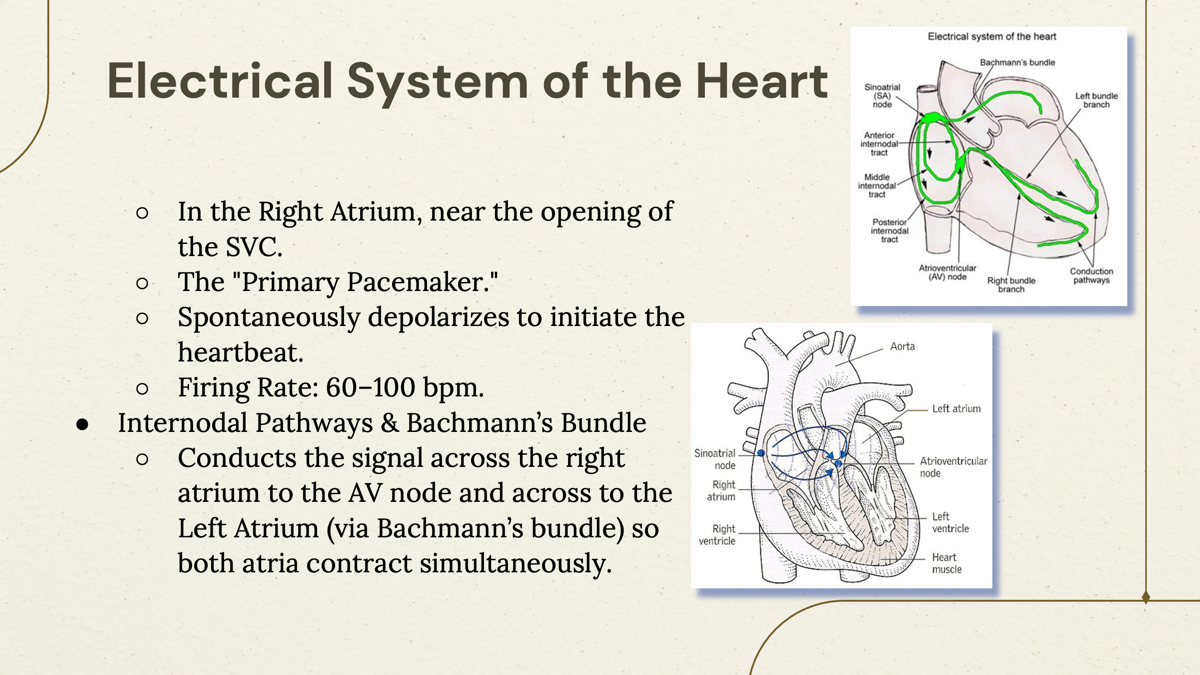
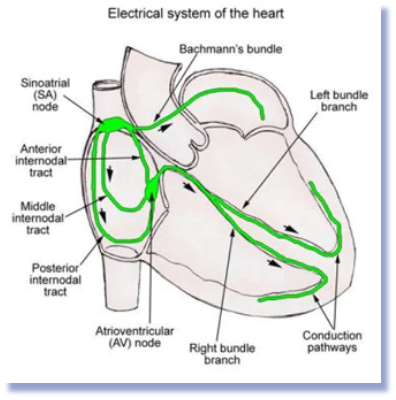
What is the “primary peace maker” of the heart?
Sinoatrial (SA) Node- “starts the heartbeat”
location: In the Right Atrium, near the opening of the SVC.
Firing: 60-100bpm
What does Internodal Pathways & Bachmann’s Bundle do?
Conducts the signal across the right atrium to the AV node and across to the Left Atrium (via Bachmann’s bundle) so both atria contract simultaneously
Bachmann's Bundle- A special pathway that carries the signal from the right atrium to the left atrium.
What does Atrioventricular (AV) node?
It functions as the “gatekeeper” of electrical signals.
Delays the signal for 0.1 seconds to allow ventricles to fill completely with blood before they countract.
SA node: Starter
AV node: gatekeeper
What happens when SA side fails?
The AV node takes over with intrinsic rate of 40-60 bpm.
Where is AV node located?
In the intertribal septum, near the opening of the coronary sinus.
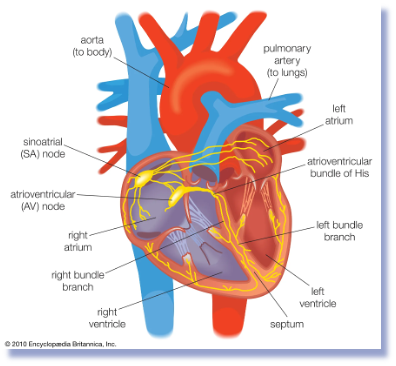
What is the bundle of His?
Small electrical connection between the atria and ventricles.
Think of the Bundle of His as the electrical wire that carries the signal from the AV node to the ventricles.
Passes through the fibrous skeleton of the heart.
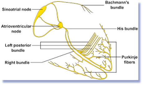
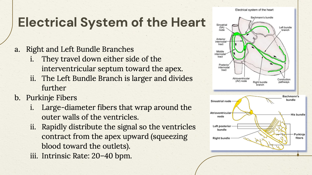
Where does the right and left bundle branches?
These branches run down the interventricular septum (the wall between the ventricles) toward the apex (tip) of the heart.
The left bundle branch is larger because the left ventricle is bigger and has more muscle and decides further.
What is Purkinje fibers and their function?
Large-diameter fibers that wrap around the outer walls of the ventricles.
Rapidly distribute the signal so the ventricles contract from the apex upward (squeezing blood toward the outlets) with the intrinsic rate of 20-40 bpm.
SA Node ⚡
↓
AV Node 🚦
↓
Bundle of His 🔌
↓
Right & Left Bundle Branches 🌳
↓
Purkinje Fibers 🌿
↓
Ventricles Contract 💥