OAT Boot day 5 - Cell Cycle
 Call Kai
 Learn
Learn Practice Test
 Spaced Repetition
 Match
Match Flashcards
 Knowt Play
1/75

There's no tags or description
Looks like no tags are added yet.
Name | Mastery | Learn | Test | Matching | Spaced | Call with Kai |
|---|
No analytics yet
Send a link to your students to track their progress
76 Terms
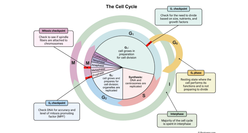
What are the two phases the Cell Cycle is divided into?
The Interphase and M Phase
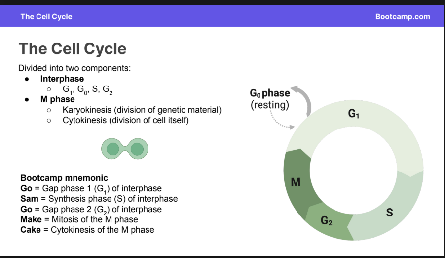
What makes up interphase?
G1, G0, S, G2
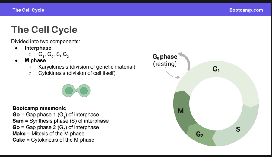
What makes up the M phase?
Karyokinesis and Cytokinesis
What is Karyokinesis?
Division of Genetic Material
What is Cytokinesis
Division of the cell itself
Pneumonic of the phases of the cell cycle (5)
Or just name the 5 stages.
Go = Gap phase 1 (G1) of interphase
Sam = Synthesis phase (S) of interphase
Go = Gap phase 2 (G2) of interphase
Make = Mitosis of the M phase
Cake = Cytokinesis of the M phase
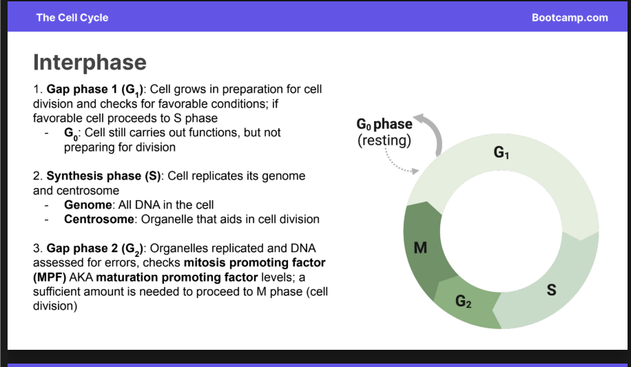
Interphase (what are the phases here)
Consists of:
G1
G0
G2
G1
What is also in tandem in this spot
1. Gap phase 1 (G1): Cell grows in preparation for cell division and checks for favorable conditions; if favorable cell proceeds to S phase
G0 can also happen at this point
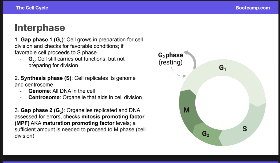
G0
Cell still carries out functions, but not preparing for division
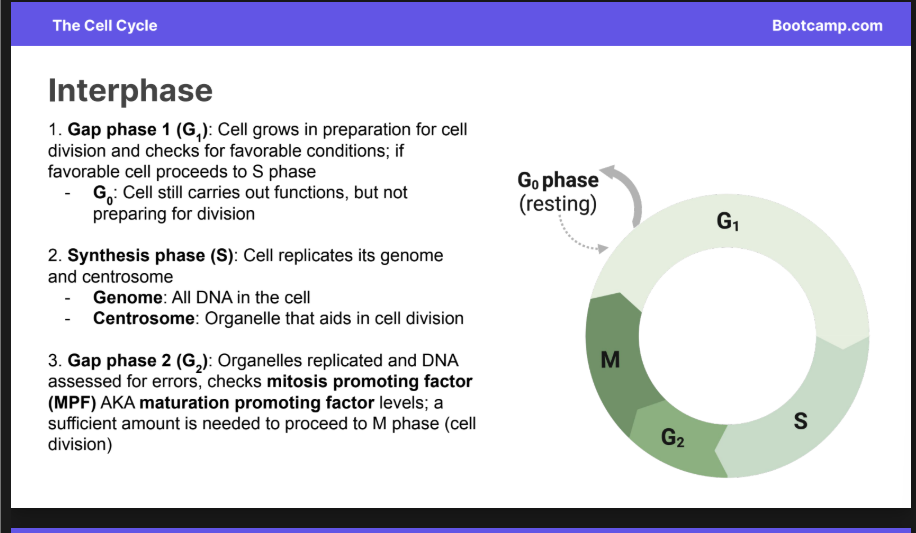
Synthesis Phase
Cell replicates its genome and centrosome
Genome
All the DNA in the cell
Centrosome
An organelle that aids in cell division
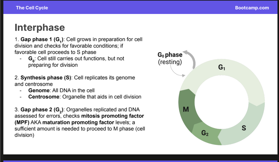
Gap Phase 2
Organelles replicated and DNA is assessed for errors, checks mitosis promoting factor (MPF) AKA maturation promoting factor levels;
a sufficient amount is needed to proceed to M phase (cell division)
M Phase
Stage in the cell cycle where karyokinesis and cytokinesis
occur
Types of Karyokinesis in Eukaryotes
Eukaryotes
Mitosis + Meiosis
Utilize microtubule organizing centers
Types of Karyokinesis in Prokaryotes
binary fission:
Genome replicates while cell division occurs (no S phase)
No microtubule organizing center
Microtubule Organizing Centers (MTOCs)
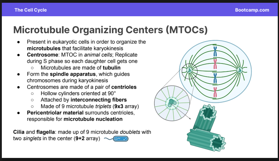
Present in eukaryotic cells in order to organize the microtubules that facilitate karyokinesis
Form the spindle apparatus, which guides chromosomes during karyokinesis
MTOC’s in Animal Cells
Centrosomes:
Replicate during S phase so each daughter cell gets one
Microtubules are made of tubulin
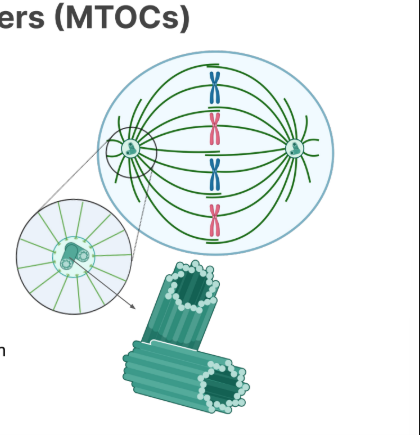
They are made of a pair of centrioles
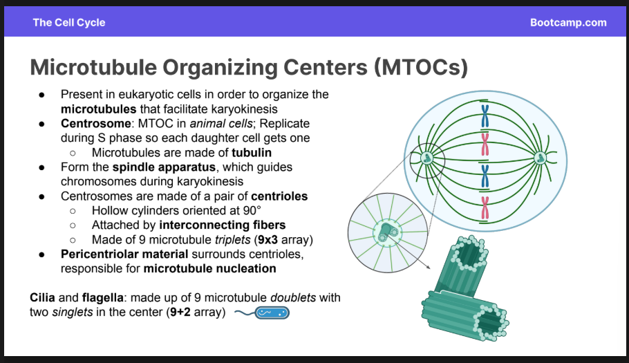
Centrioles
Hollow cylinders oriented at 90 degrees
Attatched by interconnecting fibers
made of 9 microtubule truplets (9×3)
Cillia and Flagella are made of what?
9 microtubule doublets
There is 2 singlets in the middle.

Pericentriolar Material + what is it used for?
Surrounds Centrioles, responsible for microtubule nucleation.
Microtubule Nucleation: (chat answer) process by which new microtubules begin to form inside a cell. It is the first step in building the microtubule cytoskeleton,
Microtubule Types
Polar
Astral
Kinetichore
Polar Microtubules
Connect centrosomes and push them to opposite sides of the cell.
Astral
Attach to cell membrane to orient chromosome
Kinetochore Microtubules
Attach to the kinetochore of the chromosome
Functional Limitations in the cell cycle
Physical reasons why cells need to divide once reaching a certain size
Surface to Volume Ratio:
Cell volume becomes too large for the surface area of their plasma membrane to support; Decrease in S/V leads to division
Genome to Volume Ratio
Cell Volume becomes too large for the genome to support; decrease in G/V leads to cell division.
Cell Specific Regulations: List all 5 off
Cell Cycle Checkpoints
Cyclin dependent Kinases
Growth Factors
Anchorage dependence
Density Dependent inhibition
Cell Cycle Checkpoints (3 of em)
G1 Restriction Point:
Conditions are favorable to grow and eventually divide.
End of G2
Accurately replicated genome
Sufficient MPF Levels (maturation promoting factor)
MPF is the protein complex that controls the cells entry into mitosis
M Checkpoint
Microtubules properly attached to chromosomes.
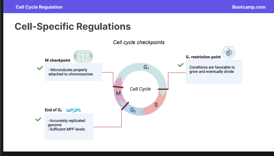
G1 Restriction Point
G1 Restriction Point:
Conditions are favorable to grow and eventually divide.
End of G2
M Checkpoint
Microtubules properly attached to chromosomes.
M Checkpoint
M Checkpoint
Microtubules properly attached to chromosomes.
Cyclin Dependent Kinases
Cyclin-dependent kinases (CDKs): phosphorylate certain molecules in order to signal cell cycle progression
Activated by cyclin, a protein that cycles through stages of synthesis and degradation
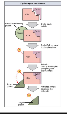
Growth Factors
Growth factors: bind plasma membrane receptors to signal growth and cell division
Anchorage Dependenance
Cells divide only when attached to an external surface
Density dependent inhibition:
halting of cell division when cell density is too high
GENERAL CHECKPOINTS IN THE CELL CYCLE
Genome
All DNA in the cell
Chromatin
Chromatin: Loosely winded DNA + protein complex (histones)
Chromosome
Separate DNA molecules that make up the genome
Sister Chromatids
Identical copies of the same chromosome
Dyad
Identical sister chromatids joined to form an X shape
Centromere
Region connecting sister chromatids in dyad
Kinetochore:
Proteins that associate with microtubules in cell division
Mitosis Phases
Prophase
Metaphase
Anaphase
Telophase
Cytokinesis
Prophase
● Chromatin condenses into chromosomes
● Nucleolus and nuclear envelope disappear
● Spindle apparatus forms
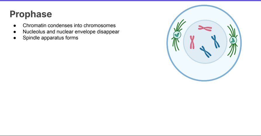
Metaphase
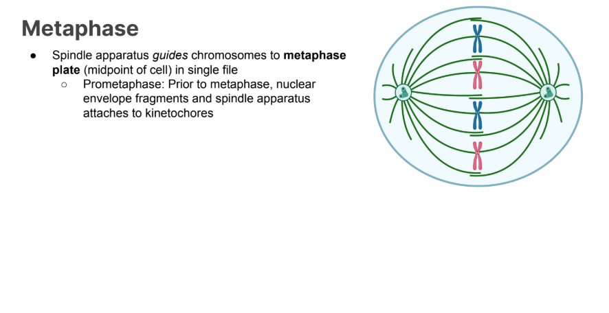
Anaphase
Kinetochore microtubules shorten to pull sister chromatids apart
Sister chromatids are now considered individual chromosomes - chromosome count doubles
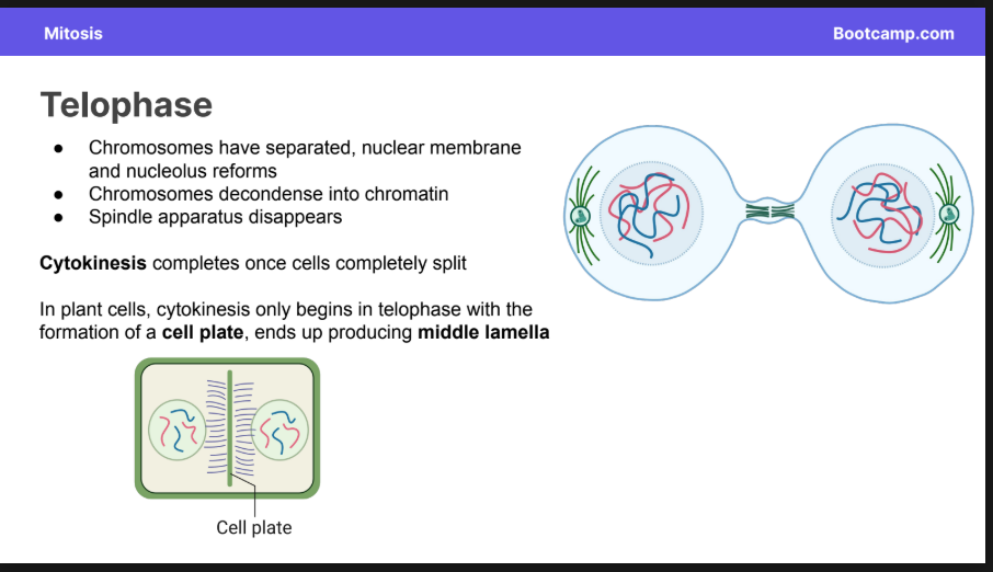
Telophase
Chromosomes have separated, nuclear membrane and nucleolus reforms
Chromosomes decondense into chromatin
Spindle apparatus disappears
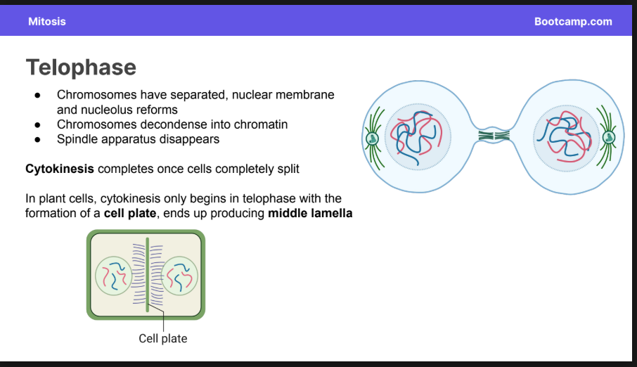
Cytokinesis in animal cells
In animal cells, cytokinesis begins in late anaphase with the formation of a cleavage furrow
Contractile ring of actin microfilaments and myosin motors that pinches cell into two
Cytokinesis ends when the cells are completley split
Cytokinesis in plant cells
Cytokinesis completes once cells completely split
In plant cells, cytokinesis only begins in telophase with the formation of a cell plate, ends up producing middle lamella
Ploidy Def
The number of chromosome sets in the body
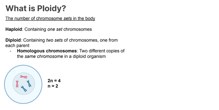
Haploid
Containing one set of chromosomes
Diploid
containing 2 sets of chromosomes
Two different copies of the same chromosome in a diploid organism.
Mitosis Vs Meiosis
Mitosis involves the division of a diploid cell into two genetically identical diploid cells
Meiosis involves two divisions of a diploid cell to produce four genetically distinct haploid daughter cells
Main two phases of meiosis and what it means.
Meiosis is divided into:
Meiosis I: homologous chromosomes separate
Reductional division
Meiosis II: sister chromatids separate
Somatic Cells in the human ploidy
Diploid
2n = 46
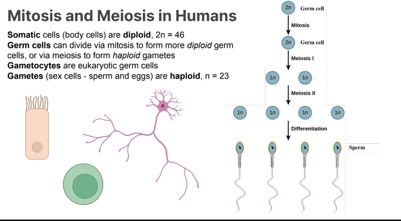
Germ Cells in the human body:
can divide via mitosis to form more diploid germ cells, or via meiosis to form haploid gametes
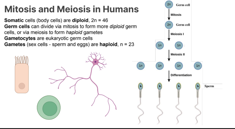
Gametocytes
Eukaryotic Germ Cells
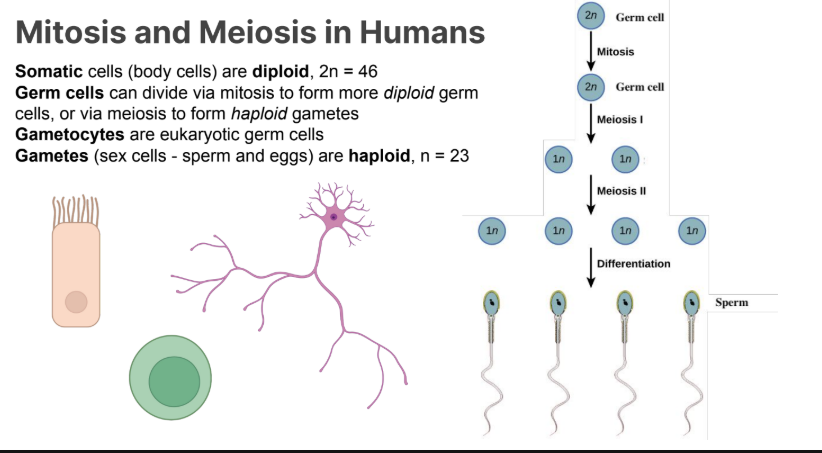
Gametes
(sex cells - sperm and eggs) are haploid n=23
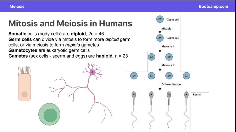
Synapsis
Homologous chromosomes pair up to form tetrads
(AKA bivalents)
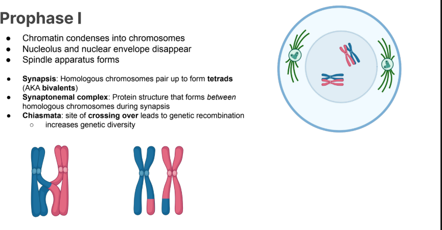
Synaptonemal Complex
Protein structure that forms between homologous chromosomes during synapsis
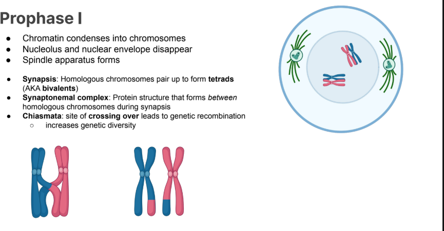
Chiasmata
Crossing over site that leads to genetic recombination
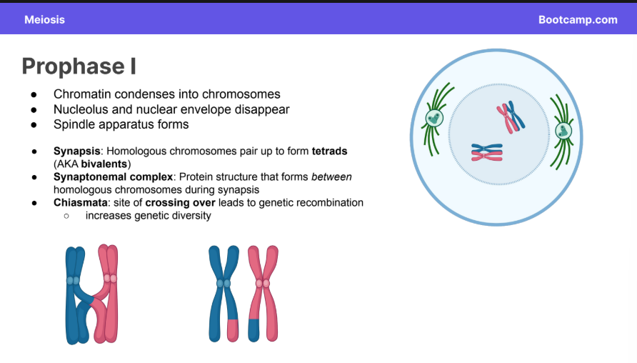
Prophase I
Chromatin condenses into chromosomes
Nucleolus and nuclear envelope disappear
Spindle apparatus forms
Synapsis:
Synaptonemal complex:
Chiasmata:
SITES of the above things.
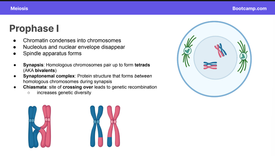
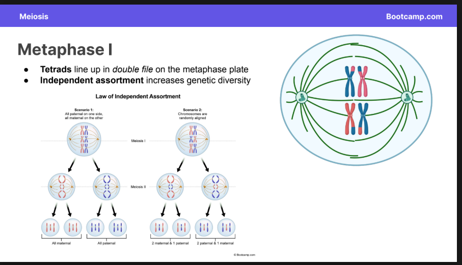
Metaphase I
Tetrads line up in double file on the metaphase plate
Independent assortment increases genetic diversity
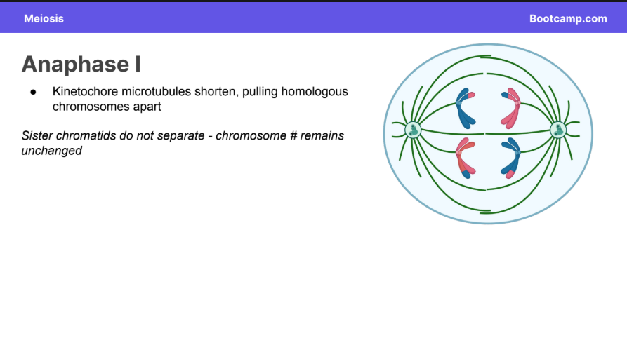
Anaphase I
Kinetochore microtubules shorten, pulling homologous chromosomes apart
Sister chromatids do not separate - chromosome # remains unchanged
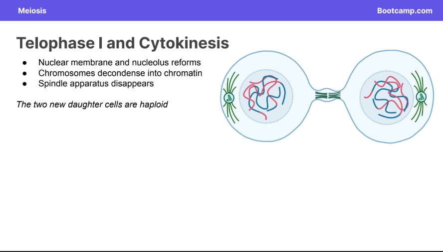
Telophase I and Cytokinesis
● Nuclear membrane and nucleolus reforms
● Chromosomes decondense into chromatin
● Spindle apparatus disappears
The two new daughter cells are haploid
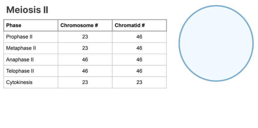
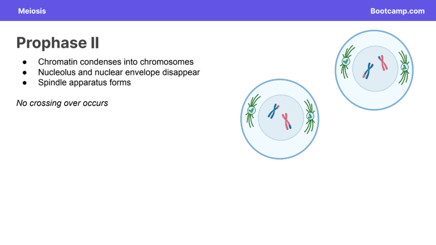
Prophase II
● Chromatin condenses into chromosomes
● Nucleolus and nuclear envelope disappear
● Spindle apparatus forms
No crossing over occurs
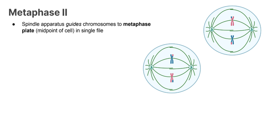
Metaphase II
● Spindle apparatus guides chromosomes to metaphase plate (midpoint of cell) in single file
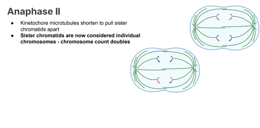
Anaphase II
Telophase II and Cytokinesis
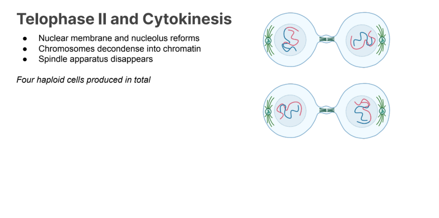
● Nuclear membrane and nucleolus reforms
● Chromosomes decondense into chromatin
● Spindle apparatus disappears
Four haploid Cells produced in total
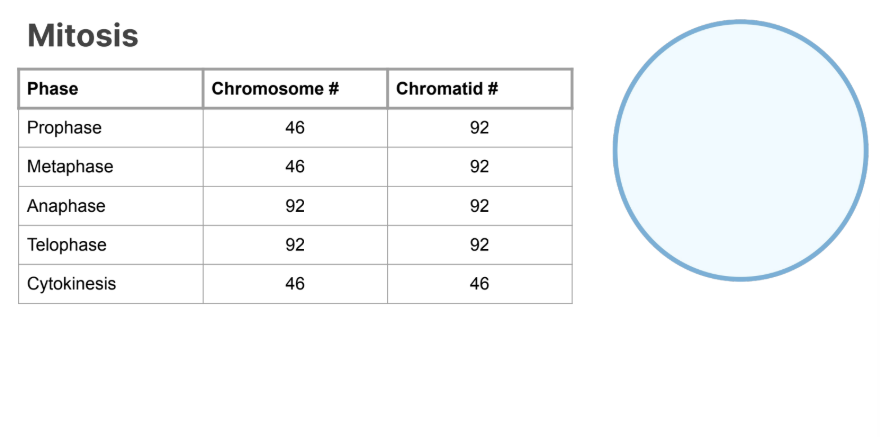
Mitosis Chromosome and Chromatid Counts across each phase
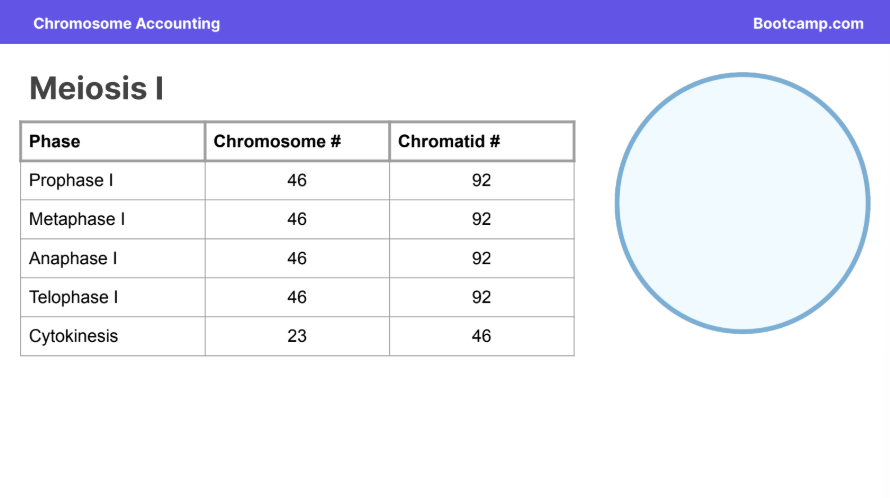
Meiosis 1 Chromosome and Chromatid Counts
Meiosis II chromosome and Chromatid Counts