BSCI201 LAB PRACTICAL 3
1/54
There's no tags or description
Looks like no tags are added yet.
Name | Mastery | Learn | Test | Matching | Spaced | Call with Kai |
|---|
No analytics yet
Send a link to your students to track their progress
55 Terms
Nervous System
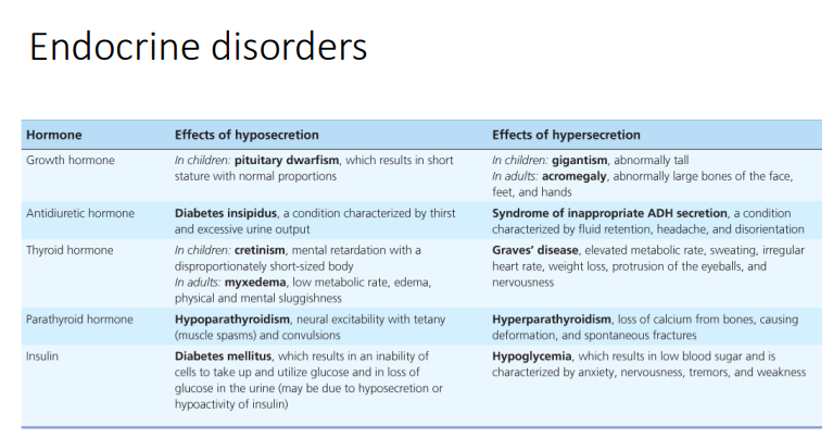
Function: Controls body, voluntary and involuntary control, initiates voluntary skeletal movements, moderates heart rate and breathing rate, processes sensory inputs, controls speech, etc
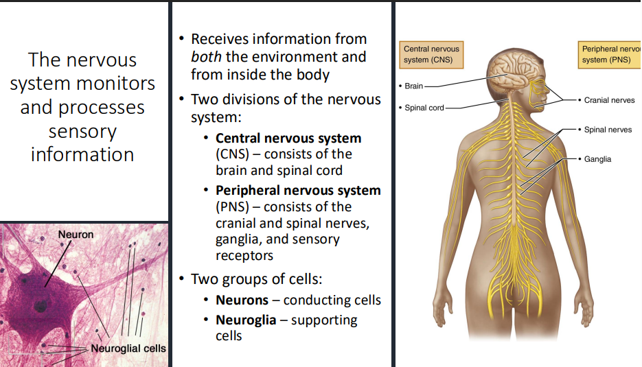
Monitors and Processes Sensory Information:
receives information from BOTH environment and inside body
Nervous system divisions:
Central Nervous System (CNS) → consists of brain and spinal cord
Spinal cord → receives information and processes lower level involuntary responses (reflexes)
Brain → more complex information, higher order processing (processing sound information, forming speech, response, etc)
Peripheral Nervous System (PNS) → consists of cranial and spinal nerves, ganglia, and sensory receptors
Types of Cells:
Neurons → conducting cells
send electrical and chemical signals that deal with communication
Neuroglia → supporting cells
support and protect neurons
Types of Neuroglial Cells
Means nerve glue; also known as glial cells
Functions: Brace, protect, and myelinate neurons
not capable of transmitting nerve impulses
Myelinating Cells → significantly high lipid content, making them appear white
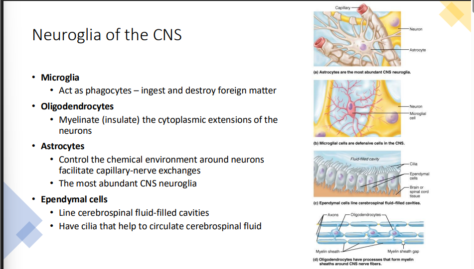
CNS Supporting Cells:
Astrocytes → control chemical environment of CNS, create blood-brain barrier, have extensions that wrap around neurons and blood vessels, etc
Microglia → phagocytic cells, engulf and destroy pathogens and debris from dead cells, protects cells of brain and spinal cord, etc
Ependymal Cells → ciliated columnar cells that line cavities of CNS filled with cerebrospinal fluid, cilia are motile and can beat, moving fluid and circulating CSF through cavities, etc
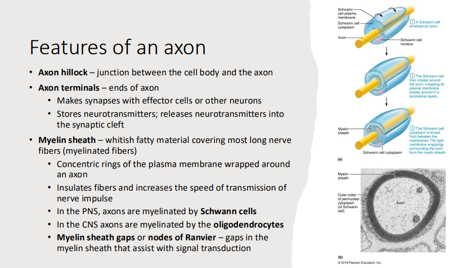
Oligodendrocytes → wrap around neurons, speed electrical transmissions that pass through neurons, etc
extend multiple processes away from main cell body and each processes wrap around a neuronal process
one oligodendrocyte can myelinate multiple neuronal processes
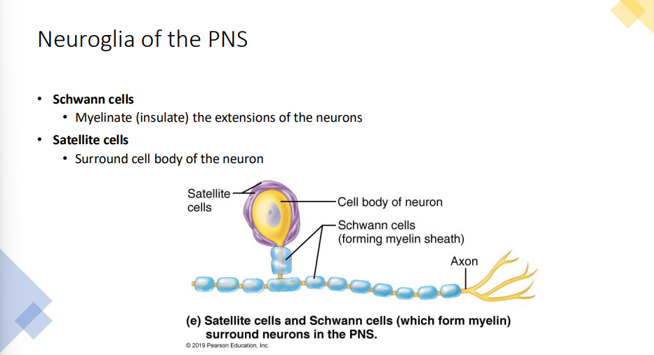
PNS Supporting Cells:
Schwann Cells → myelinating cells of PNS, myelinate neuronal processes to insulate them and speed up conduction of electrical signals, etc
entire schwann cell will flatten and wrap its entire body around a neuron
can only myelinate one neuron
Satellite Cells
wrap around neurons to control chemical environment the neuron is exposed to
multiple cells surround the cell body of the neuron
Neuron Anatomy
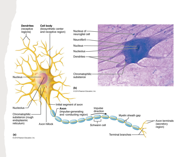
Cell Body → composed of large, round nucleus surrounded by cytoplasm, which contains:
Nucleus → large and round
Nucleolus → ribosome assembly
Neurofibrils → intermediate filaments of cytoskeleton
built from neurofilament proteins
form network throughout cytoplasm that allows cells to maintain shape
Chromatophilic substance → specialized rough ER of neurons
facilitates protein production and processing
main biosynthetic center of cell, where transcription and translation occur
receives information from various dendrites and processes information
sends signals via axon
Dendrites and Axons → processes that extend out from the cell body and transmit nerve impulses
Dendrites → receive signals and sends them towards the cell body
contains many dendrites
Axons → transmit action potentials to other cells
carry electrical signals (action potentials) away from cell body and towards axon terminal
one neuron has one axon, but there can be many branching axon terminals
axon hillock → place of origin of axon near cell body
axon terminals meet with other cells and neurons via synapses, where electrical signals are converted into chemical signals
myeline sheath gaps → gaps between myelinating cells on axon
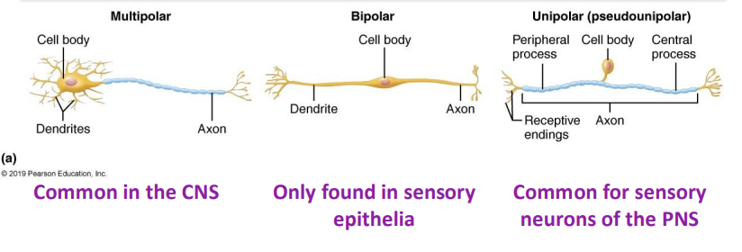
Structural Classification of Neurons
Based on how many processes extend from cell body
Multipolar: neurons have many dendrites and one axon
Bipolar: neurons have one axon and one dendrite
Unipolar (pseuounipolar): neurons have one process that branches; no dendrites
singular process is short and divides into proximal and distal branches
distal peripheral process → associated with sensory receptor
proximal central process → enter CNS and synapse onto neuron of CNS
unipolar neurons are derived from bipolar neurons
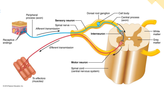
Functional Classification of Neurons
Based on the direction of impulse transmission in relation to CNS
Sensory (afferent) neurons → carry impulses from sensory receptors in skin, internal organs, muscles, and special sense organs TOWARDS CNS
Association neurons (interneurons) → conduct impulses within CNS
Motor (efferent) neurons: carry impulses AWAY from CNS to organs, muscles, and glands
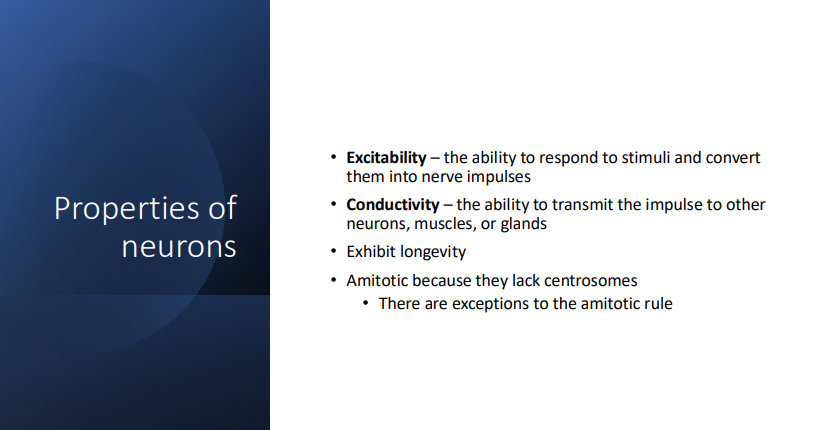
Neuron Properties
Tracts and Nerves
Tract: bundle of axons in CNS supported only by neuroglia
Nerve: bundle of axons in PNS wrapped in CT
Sensory (afferent) nerves: contains only sensory processes
Motor (efferent) nerves: contain only motor processes
Mixed nerves: contain both sensory and motor neurons
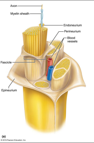
Connective Tissue Sheathes of Nerves:
Endoneurium → encloses each axon and its myelin sheath
Perineurium → encloses groups of axons called fascicles
Epineurium → encloses nerve (group of fascicles)
Human Brain Gross Anatomy
Weight: 3.5 lbs (1.6 kg)
Four Major Regions:
Cerebrum
Diencephalon
Brain Stem
Cerebellum
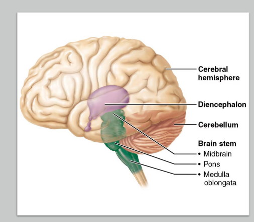
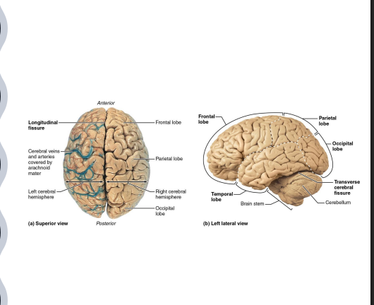
Anatomy of Cerebrum
composed of two cerebral hemispheres (left and right)
83% of brain’s total mass
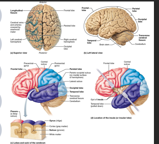
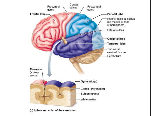
surface of cerebrum is marked by gyri (elevated ridges)
sulcus → furrow between gyri
fissure → deeper sulcus
each cerebral hemisphere is divided into five lobes
Cerebral Hemispheres
carries out higher mental functions (memory and reason)
separated by longitudinal fissure → median fissure dividing cerebrum into right and left hemispheres
held together medially by corpus callosum
lateral ventricles separated by membrane (septum pellucidum)
Cerebrum 5 Lobes
frontal, temporal, parietal, and occipital lobes:
can be viewed externally
named for the overlaying cranial bones
Insula
cannot be viewed externally
located deep to the lateral sulcus (covered by frontal, temporal, and parietal lobes)
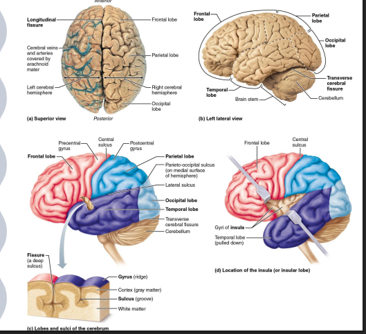
Cerebrum Major Sulci
central sulcus → separates frontal and parietal lobes
precentral gyrus of frontal lobe contains primary motor cortex
postcentral gyrus of parietal lobe contains primary somatosensory cortex
lateral sulcus → separates temporal lobe from parietal and frontal lobes
parieto-occipital sulcus → separates parietal lobe from occipital lobe
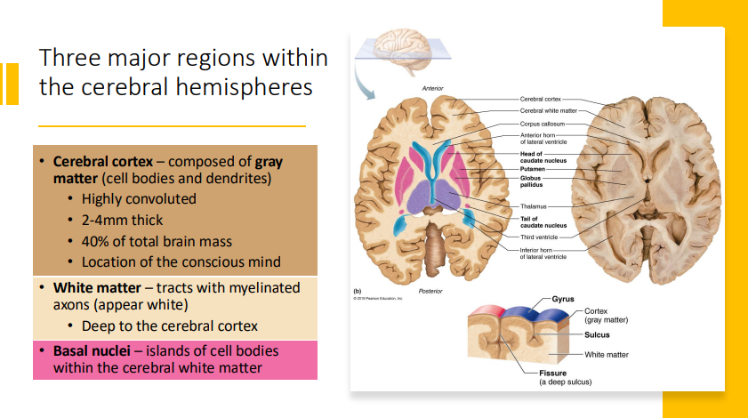
Three Major Regions in Cerebral Hemispheres
cerebral cortex → composed of grey matter (cell bodies and dendrites)
highly convoluted
2-4 mm thick
40% of total brain mass
location of conscious mind
white matter → tracts with myelinated axons (appear white)
deep to cerebral cortex
basal nuclei → islands of cell bodies within cerebral white matter
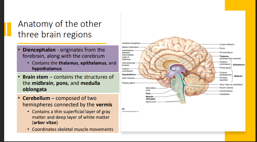
Anatomy of Other Brain Regions
Diencephalon → originates from forebrain, along with cerebrum
contains thalamus, epithalamus, and hypothalamus
Brain stem → contains structures of midbrain, pons, and medulla oblongata
Cerebellum → composed of two hemispheres connected by the vermis
contains thin superficial layer of gray matter and deep layer of white matter (arbor vitae)
coordinates skeletal muscle movements
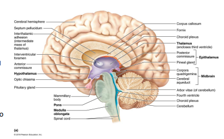
Diencephalon
Thalamus → relay center for all sensory information (except smell) on its way to cerebral cortex
Hypothalamus → regulation of visceral activities and body functions, emotions, instincts, secretes hormones
Epithalamus → contains pineal gland, which secretes the hormone melanin
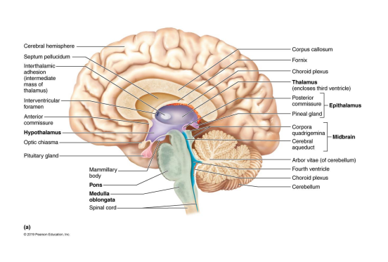
Brain Stem
Midbrain
corpora quadrigemina → pair of superior colliculi (visual reflex) and pair of inferior colliculi (auditory reflex)
cerebral peduncles → tracts in midbrain, connects pons to cerebrum
cerebral aqueduct → pathway for CSF
Pons → contains fiber tracts connecting cerebrum to cerebellum
Medulla oblongata → regulates autonomic functions (heart rate, blood pressure, breathing, etc) and it blends in with the spinal cord at the level of the foramen magnum
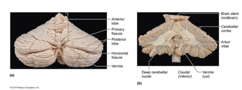
Cerebellum
two hemispheres separated by a longitudinal fissure called the vermis
thin superficial layer of gray matter and deep layer of white matter (arbor vitae)
coordinates skeletal muscle contractions
specializes fine movements
helps lean new movements (walking)
brain region most affected by alcohol intoxication
Structures Protecting Brain
cranium → body helmet composed of 8 cranial bones
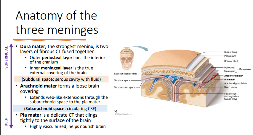
Meninges → three CT membranes surrounding the brain
CSF → fluid cushion in the subarachnoid space and ventricles
Blood-brain barrier → selective barrier that prevents harmful substances in blood from crossing into the brain
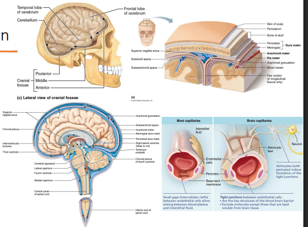
Meninges Anatomy
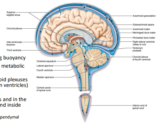
Cerebrospinal Fluid
acts as fluid cushion, providing buoyancy
provides nutrients and removes metabolic wastes
filtered from blood by the choroid plexuses (small collections of capillaries in ventricles)
supported by astrocytes
circulating through ventricles and in subarachnoid space → CSF found inside and outside brain
circulation facilitated by ciliated ependymal cells
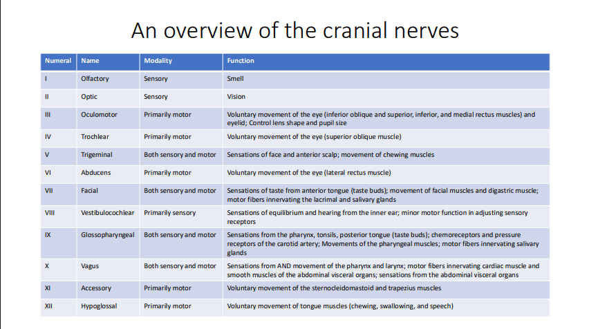
12 Pairs Cranial Nerves
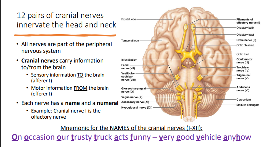
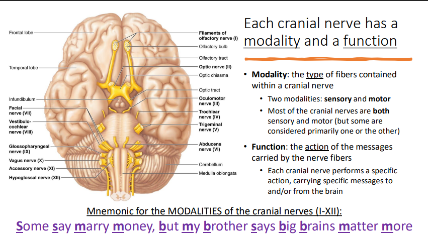
Cranial Nerve Modality + Function
Accessory Structures of Eye
palpebrae → eyelids
medial and lateral commissures
lacrimal caruncle → fleshy elevation at medial commissure
lacrimal apparatus → produces and drains tears to lubricate eye
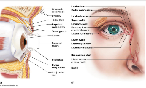
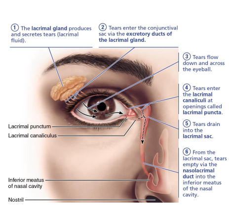
Lacrimal Apparatus
produces and drains lacrimal fluid (tears) to lubricate the eye
contains mucus, antibodies, lysozyme
lacrimal fluid is continuously released and flows across the eye:
lacrimal gland
lacrimal puncta
lacrimal canaliculi
lacrimal sac
nasolacrimal duct
Extrinsic Muscles of Eye
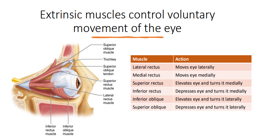
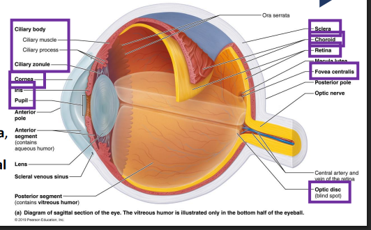
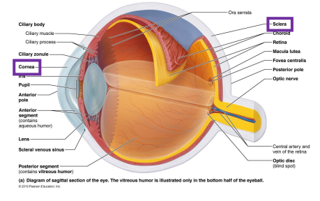
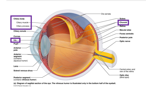
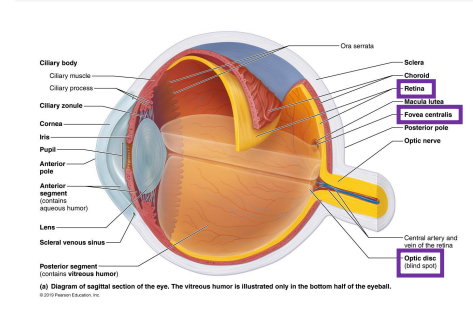
Eye Wall Layers
Fibrous Layer → composed of sclera and cornea
Vascular layer → contains choroid, ciliary body, and iris
Sensory layer → retina, composed of the pigmented and neural layers
Fibrous Layer (Outermost Eye Wall Layer)
composed of dense avascular connective tissue
sclera → opaque region occupying the posterior 5/6 of the fibrous layer
cornea → anterior transparent region, modified to let light in
major light-bending medium of eye (refracts light to focus on the retina)
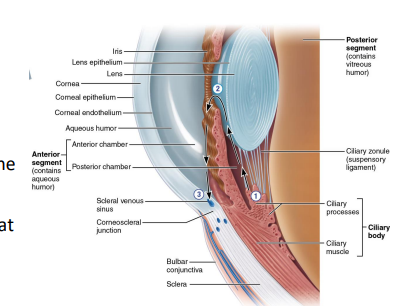
Vascular Layer (Middle Eye Wall Layer)
choroid → highly vascularized posterior region, contains melanin to prevent the scattering of light
ciliary body → encircles lens
ciliary muscle → intrinsic muscle that controls lens shape
ciliary processes → contain capillaries from which aqueous humor is filtered
zonules → fibers extended from ciliary process to hold the lens upright
Iris → pigmented smooth muscles that regulate diameter of pupil
sphincter pupillae → contract to constrict the pupil (close vision and bright light)
dilator pupillae → contracts to dilate the pupil (distant vision and dim light)
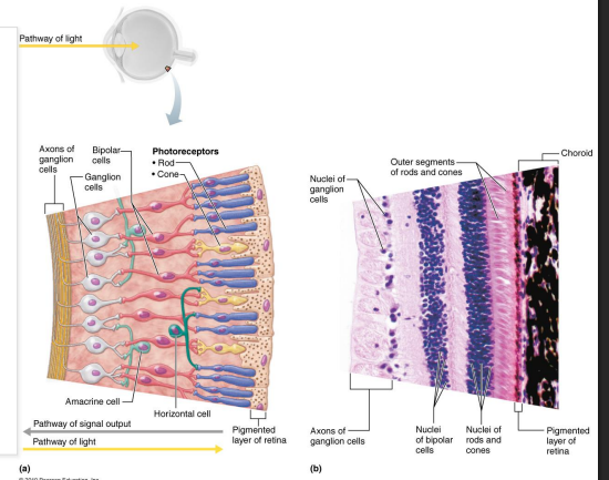
Retina (Innermost Eye Wall Layer)
pigmented layer (outer layer) → composed of melanocytes, absorbs light and prevents it from scattering
neural layer (transparent inner layer) → converts light energy into nerve impulses that then travel to brain via optic nerve
fovea centralis → area of neural layer that contains only cones
optic disc (blind spot) → region where optic nerve fibers exit the eye, lacks photoreceptors
Cells of Neural Layer of Retina
Two types of photoreceptors sense light:
rods → dim light vision
cones → high light and color vision
Bipolar cells
Ganglion cells → axons leave the retina in the optic nerve
Anterior vs Posterior Eye Segments
anterior segment → between cornea and lens (includes lens)
contains aqueous humor, fluid formed and drained throughout life
lens → biconvex structure that helps focus light on retina
posterior segment → everything posterior to lens
contains vitreous humor, a gel-like fluid formed during embryonic development that lasts a lifetime
like lens, aqueous humor and vitreous humor help refract light
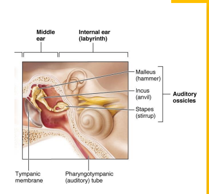
External Ear
Pinna → elastic cartilage covered with skin
external acoustic meatus → canal in temporal bone, lined with ceruminous glands
tympanic membrane → vibrates in response to soundwaves; separates external and middle ear
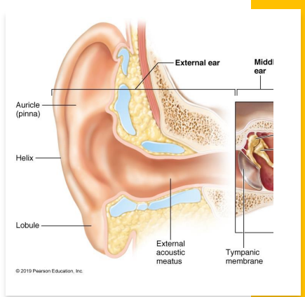
Middle Ear
three ossicles transmit and amplify vibrations
malleus → attached to tympanic membrane and incus
incus → articulates with malleus and stapes
stapes → articulates with incus and attaches to oval window
oval window → transmits vibrations from stapes to Scala vestibuli
pharyngotympanic tube → connects middle ear to throat
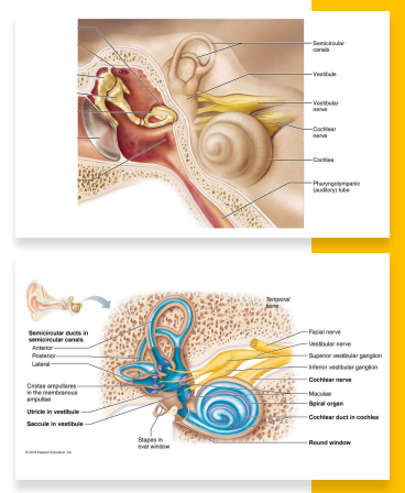
Internal Ear
bony labyrinth → cavity in temporal bone, filled with perilymph
semicircular canals
vestibule
cochlea
membranous labyrinth → ducts and sacs within body labyrinth, filled with endolymph
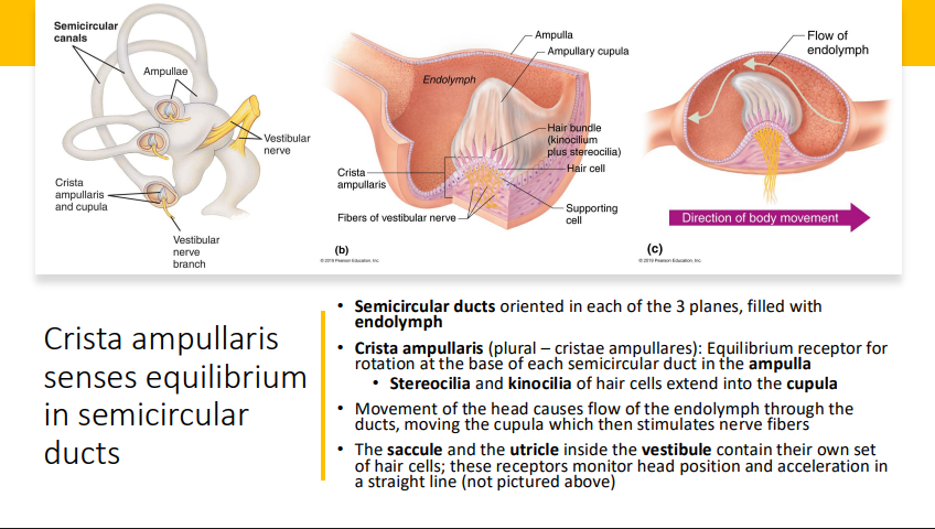
Equilibrium:
semicircular ducts
saccule
utricle
Hearing
cochlear duct
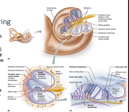
Spiral Organ - Responsible for Hearing
Spiral organ (organ of corti) is within cochlear duct
hair cells (auditory receptors) rest on basilar membrane and project stereocilia into tectorial membrane
movement of basilar membrane bends the stereocilia, depolarizing hair cell membrane, starting nerve impulse in the cochlear nerve
Crista Ampullaris
Olfaction and Gustation
chemical senses
olfaction: sense of smell
gustation: sense of taste
receptors for olfaction and gustation are called chemoreceptors:
chemoreceptors respond to chemicals dissolved in aqueous solution
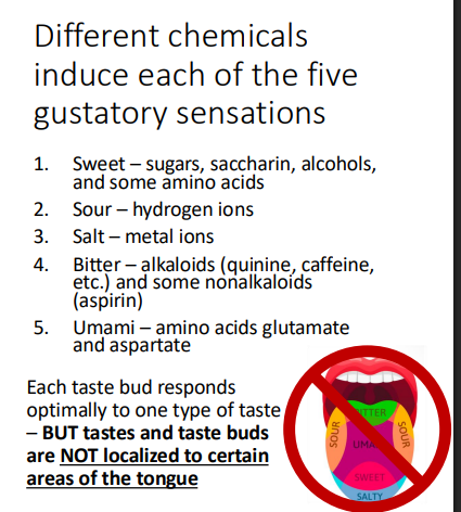
gustation chemoreceptors are divided into 5 subtypes that sense diff tastes
olfaction chemoreceptors are sensitive to wide range of chemical sensations
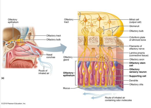
Olfaction + Olfactory Epithelium
cell types of olfactory epithelium:
olfactory sensory neurons → bipolar neurons with radiating olfactory cilia
bundles of axons of olfactory receptor cells form the filaments of olfactory nerve
replaced every 30-60 days by olfactory stem cells
supporting cells → surround and cushion the olfactory system neurons
olfactory stem cells → lie at the basal surface (superior side of epithelium) and divide to replace olfactory sensory neurons
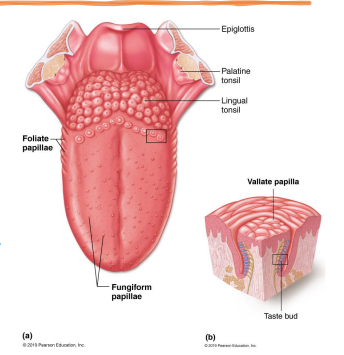
Sense of Taste
receptor organs are taste buds
most located on tongue in papillae
few found on soft palate, epiglottis, pharynx, and inner surface of cheeks
types of papillae:
fungiform → most numerous papillae, taste buds are on superior surface
foliate → on lateral edges of tongue, taste buds located in the side walls
vallate → large papillae in a V formation on posterior surface of tongue, taste buds are located in the side walls
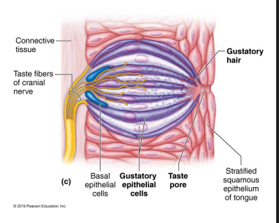
Taste Bud Structure + Chemicals
flask-shaped
all taste buds response to all 5 classes of chemical stimuli
each taste bud responds optimally to one type of taste
contain 50-100 epithelial cells
gustatory epithelial cells → receptor cells
microvilli (gustatory hairs) project through a taste pore to the surface of the epithelium bathed in saliva
basal epithelial cells → stem cells that replace the gustatory epithelial cells every 7 days
Endocrine System
endocrine glands → ductless glands that release chemical regulators called hormones directly into ECF (interstitial fluid and blood)
Endocrinology → study of biological effects of hormones released by endocrine glands and diseases caused by dysfunction
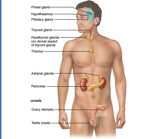
Major Endocrine Glands
Hormone Release
released in response to homeostatic imbalances referred to as stimuli
hormones usually work via negative feedback to maintain homeostasis
biological effects of hormones negate/eliminate stimuli that caused release of hormones
exception: hormone prolactin stimulates milk production through positive feedback
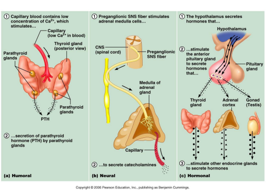
Stimuli Types
Humoral stimuli → changes in levels of chemicals in body’s humors (bodily fluids) stimulate endocrine glands to release hormones
Neural stimuli → activation of nervous system stimulates endocrine glands to release hormones
hormonal stimuli → released hormones stimulate endocrine glands to release other hormones
Hormone Nomenclature
organ of origin → endocrine gland releasing hormone
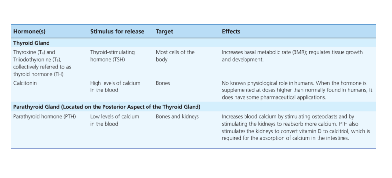
example: parathyroid hormone
function → major biological effect of hormone
example: follicle stimulating hormone
chemical structure → unique feature of chemical structure
example: triiodothyronine (t3)
Chemical Structure of Hormones Classes
biogenic amine hormones → derived from amino acid tyrosine
peptide/protein/glycoprotein hormones → composed of amino acid sequences
steroid hormones → derived from cholesterol
Target Cells/Tissues of Hormones
express accessible, functional receptors that hormones bind to
multiple hormones can target same cell
target cell can bind multiple hormones simultaneously
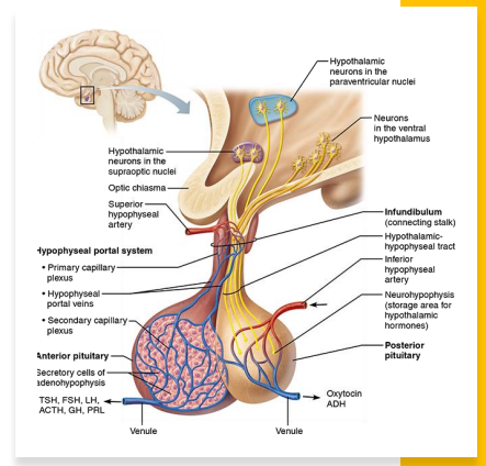
Pituitary Gland (Hypophysis)
two functional lobes attached to the hypothalamus by infundibulum:
adenohypophysis (anterior)
controlled by neurosecretions produced by neurons of ventral hypothalamus then liberated into hypophyseal portal system and carried to cells of adenohypophysis where they control hormone release
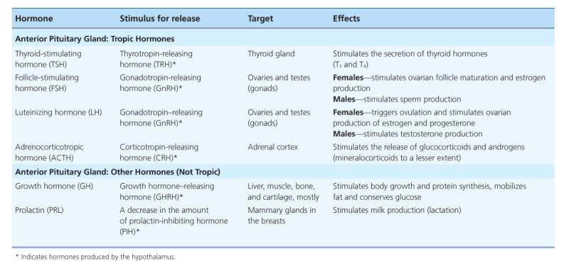
produces 4 tropic hormones that stimulate target organs that are endocrine glands
produces 2 other main hormones not directly involved in regulation of other endocrine glands
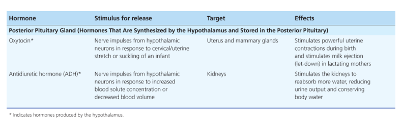
neurohypophysis (posterior)
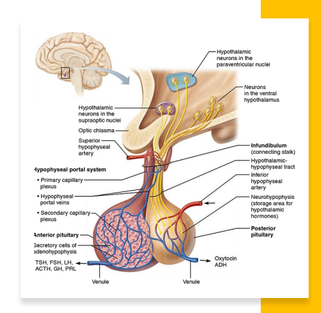
Pituitary Gland (Hypophysis)
Neurohypophysis: not an endocrine gland but storage for 2 hormones transported to it via the axons of neurons in the paraventricular and supraoptic nuclei of the hypothalamus
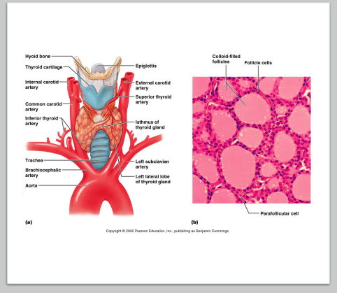
Thyroid Gland
located in throat just inferior to larynx
two lobes joined by central mass or isthmus
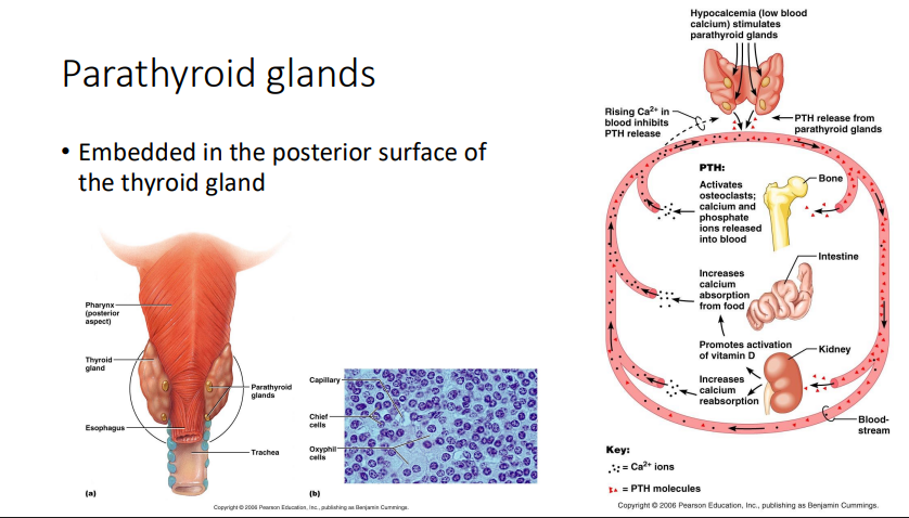
Parathyroid Glands
embedded in posterior surface of thyroid gland
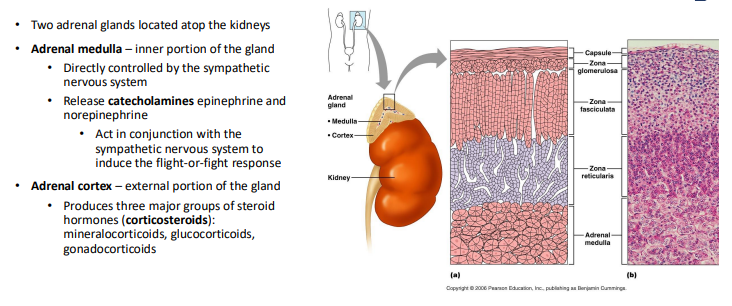
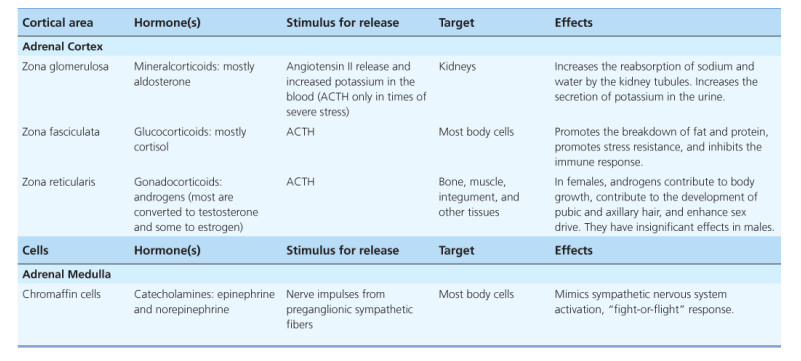
Adrenal Glands
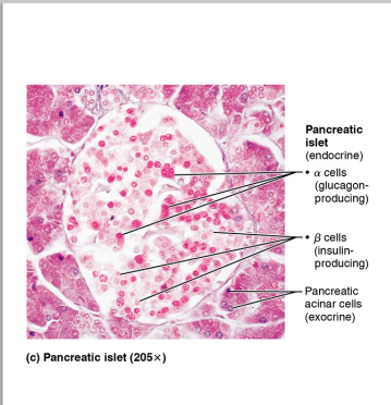
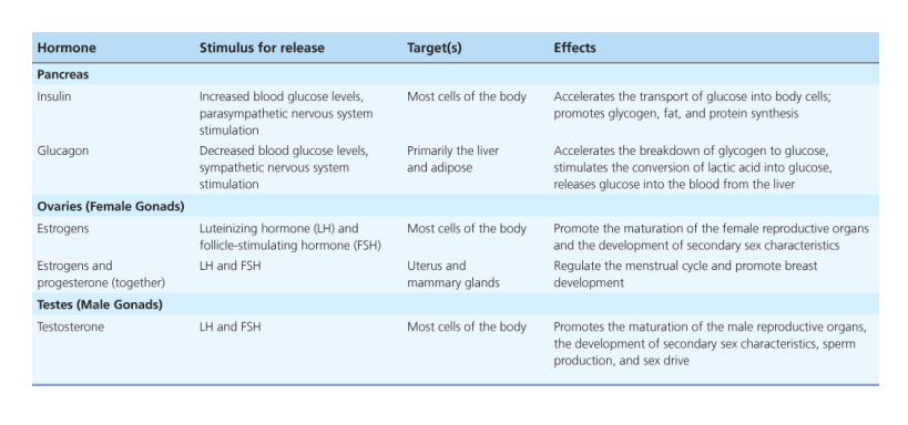
Pancreas
located behind stomach
functions as endocrine and exocrine gland
exocrine: produces digestive enzymes
endocrine: produces insulin and glucagon, hormones that act to regulate blood sugar levels
Gonads
two tropic hormones released by anterior pituitary induce the gonads to produce steroidal hormones
follicle stimulating hormone (FSH)
luteinizing hormone (LH)
Ovaries (females) produce estrogens and progesterone
Testes (males) produce testosterone
Endocrine Disorders